. 2023 Nov;29(11):1658-1672.
doi: 10.1261/rna.079427.122. Epub 2023 Jul 7.

A transient intermediate RNA structure underlies the regulatory function of the E. coli thiB TPP translational riboswitch

Affiliations

A transient intermediate RNA structure underlies the regulatory function of the E. coli thiB TPP translational riboswitch

Katherine E Berman et al. RNA. 2023 Nov.

Abstract

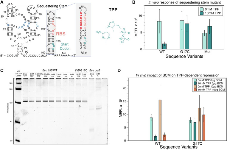
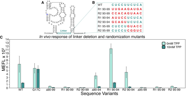
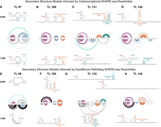
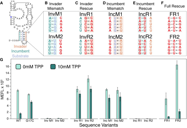
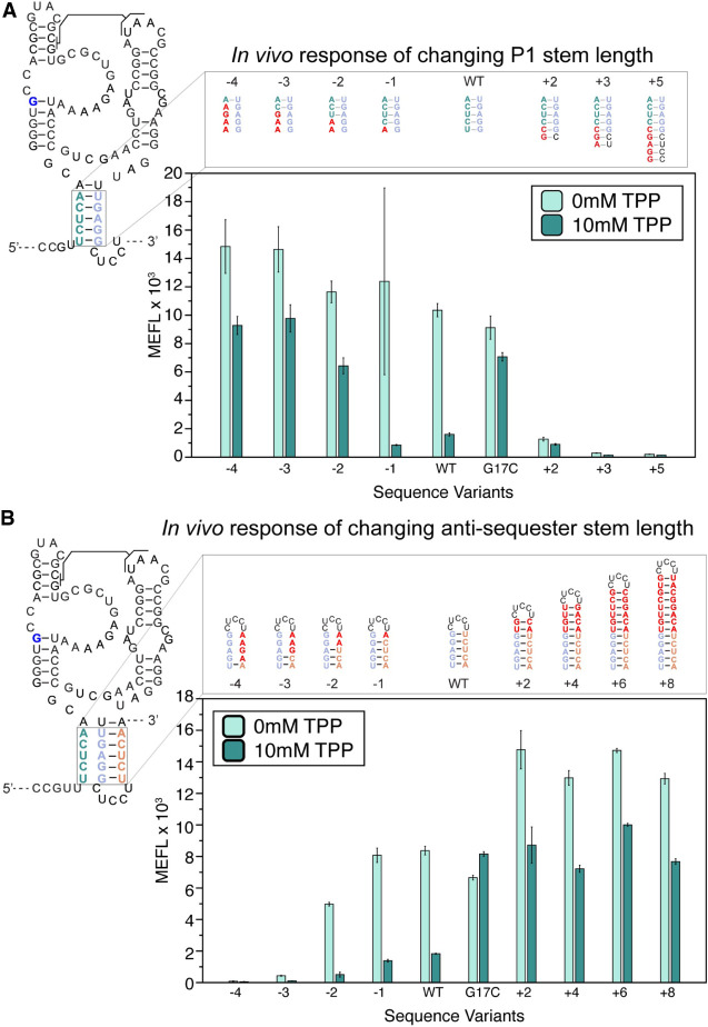
Riboswitches are cis-regulatory RNA elements that regulate gene expression in response to ligand binding through the coordinated action of a ligand-binding aptamer domain (AD) and a downstream expression platform (EP). Previous studies of transcriptional riboswitches have uncovered diverse examples that utilize structural intermediates that compete with the AD and EP folds to mediate the switching mechanism on the timescale of transcription. Here we investigate whether similar intermediates are important for riboswitches that control translation by studying the Escherichia coli thiB thiamin pyrophosphate (TPP) riboswitch. Using cellular gene expression assays, we first confirmed that the riboswitch acts at the level of translational regulation. Deletion mutagenesis showed the importance of the AD-EP linker sequence for riboswitch function. Sequence complementarity between the linker region and the AD P1 stem suggested the possibility of an intermediate nascent RNA structure called the antisequestering stem that could mediate the thiB switching mechanism. Experimentally informed secondary structure models of the thiB folding pathway generated from chemical probing of nascent thiB structures in stalled transcription elongation complexes confirmed the presence of the antisequestering stem, and showed it may form cotranscriptionally. Additional mutational analysis showed that mutations to the antisequestering stem break or bias thiB function according to whether the antisequestering stem or P1 is favored. This work provides an important example of intermediate structures that compete with AD and EP folds to implement riboswitch mechanisms.

Keywords: RNA folding; SHAPE-Seq; riboswitches; translation regulation.


Figures

FIGURE 1.
FIGURE 1.
The thiB E. coli TPP riboswitch represses translation through a sequestering stem that includes the RBS. (A) Proposed secondary structure of the ligand-bound E. coli thiB TPP riboswitch based on the published crystal structure (Serganov et al. 2006), Rfam sequence alignment (Kalvari et al. 2017), and the sequestering stem secondary structure modeled using RNAstructure (Reuter and Mathews 2010). Ligand-binding nucleotides within the ligand-binding pocket are colored blue. The RBS and the start codon for the naturally encoded downstream thiB gene are colored in salmon and teal, respectively. The chemical structure of TPP is drawn next to the RNA, and a known tertiary interaction between the P2 and P3 stems is highlighted. Inset shows secondary structure model of a mutant sequestering stem designed to weaken base-pairing with the RBS. Mutated nucleotides in red. Secondary structures determined by using RNAstructure version 6.4 (Reuter and Mathews 2010). (B) Wild-type (WT) and mutant thiB riboswitch-regulated super folder green fluorescent protein (sfGFP) expression in E. coli cells measured by flow cytometry. Units shown are molecules of equivalent fluorescein (MEFL) determined by flow cytometry. (C) Single-round in vitro E. coli RNA polymerase transcription assay measuring transcription products on a 10% urea poly-acrylamide gel. The Bce crcB F riboswitch, used as a positive control, terminates in the absence of F at an expected length of 82 nt, and antiterminates in the presence of F at an expected length of 124 nt. The expected length of the full thiB transcription product is 173 nt. (D) WT and mutant thiB riboswitch-regulated sfGFP expression in E. coli cells in the presence and absence of the Rho-inhibitor bicyclomycin (BCM) measured by flow cytometry. Bar graphs in B and D represent mean values across three biological replicates, each performed in technical triplicate for nine total data points (n = 9). Error bars represent the standard deviation from the mean. Data in C are n = 2 representative gels (Supplemental Fig. S7).
FIGURE 2.
FIGURE 2.
A specific portion of the linker region in the E. coli thiB TPP riboswitch is essential for TPP-dependent repression. (A) Secondary structure model of the thiB E. coli TPP riboswitch after Figure 1A, with linker sequence (nts 90–99) outlined in teal. (B) A table of sequence randomizations (R) tested within the linker sequence, labeled according to which nucleotides were randomized. (C) Flow cytometry assay of plasmid sfGFP expression regulated by the ThiB riboswitch sequence variants. X-axis labels indicate the riboswitch sequence variant tested ([WT] wild-type, [G17C] ligand unresponsive mutant, [Δ] deletion). Bar graphs represent mean values across three biological replicates, each performed in technical triplicate for nine total data points (n = 9). Error bars represent the standard deviation from the mean.
FIGURE 3.
FIGURE 3.
Experimentally informed models of the cotranscriptional and equilibrium-refolded RNA folding pathways for E. coli thiB riboswitch in the presence and absence of TPP. R2D2 models of secondary structure intermediate in the folding pathway. One hundred secondary structure models were generated from the experimental data for each intermediate RNA length. Models are depicted as RNA bow plots (0 mM TPP top, 1 mM TPP bottom), where arcs between nucleotides represent base pairs in a particular structure, arc thickness is proportional to the number of times that base pair is present in the one hundred structure set, and arcs are color coded according to portions of the riboswitch models. Secondary structure depictions are the consensus structure over the 100 iterations. Models and consensus structures from cotranscriptional SHAPE-Seq data sets at TLs (A) 87 nt, (B) 100 nt, (C) 131 nt, (D) 142 nt, and from equilibrium-refolded SHAPE-Seq data sets at TL (E) 89 nt, (F) 105 nt, (G) 133 nt, (H) 142 nt. Representative lengths for cotranscriptional and refolded conditions were chosen to be similar but do not exactly match due to differences in read coverage (see Materials and Methods).
FIGURE 4.
FIGURE 4.
Complementary base pairs within P1 and within the antisequestering stem are required for E. coli thiB TPP riboswitch TPP-dependent repression. (A) Secondary structure model of the thiB E. coli TPP riboswitch after Figure 1A including the proposed top portion of the antisequestering stem drawn as invading the P1 helix. Invader, incumbent and substrate strands are labeled according to a mechanism in which the incumbent:substrate duplex forms the P1 stem of the aptamer and the substrate:invader duplex forms the top of the antisequestering stem. Arrows indicate a potential mechanism where the invader displaces the incumbent during antisequestering stem folding. (BF) Mutant nucleotides are depicted in red, while WT nucleotides are teal (incumbent), orange (invader), and blue (substrate). Mutations were designed to break the invasion of the antisequestering stem (B, invader mismatch, InvM), rescue the invasion and break P1 (C, invader rescue, InvR), break P1 and allow invasion by the antisequestering stem (D, incumbent mismatch, IncM), rescue P1 and break the invasion (E, incumbent rescue, IncR), and rescue all base-pairing interactions with sequences different than the WT sequence (F, full rescue, FR). (G) Flow cytometry assay of plasmid sfGFP expression regulated by the thiB riboswitch sequence variants. X-axis labels indicate the riboswitch sequence variant tested ([WT] wild-type, [G17C] ligand unresponsive mutant). Bar graphs represent mean values across three biological replicates, each performed in technical triplicate for nine total data points (n = 9). Error bars represent the standard deviation from the mean.
FIGURE 5.
FIGURE 5.
Changing the length of P1 and the antisequestering stem have the opposite effects on E. coli thiB TPP riboswitch function. (A) P1 stem mutants that lengthen and shorten the P1 stem. Lengthening mutants change nucleotides in the leader sequence before the riboswitch to extend P1. Mismatches on the 5′ end shorten the P1 helix from the bottom. Secondary structure depictions of the mutations are depicted above flow cytometry assay data of plasmid sfGFP expression regulated by the thiB riboswitch P1 sequence variants. X-axis labels indicate the riboswitch sequence variant tested ([WT] wild-type, [G17C] ligand unresponsive mutant). (B) Sequence mutants for the antisequestering stem. Mutants to the left of the WT hairpin are mismatch mutations which shorten the antisequestering helix, while mutants to the right extend the helix. Secondary structure depictions and flow cytometry assay data as in (A). X-axis labels indicate the riboswitch sequence variant tested ([WT] wild-type, [G17C] ligand unresponsive mutant). Bar graphs represent mean values across one biological replicate, each performed in technical triplicate for three total data points (n = 3). Error bars represent the standard deviation from the mean.

Similar articles

Cited by

References

    1. Aalberts DP, Jannen WK. 2013. Visualizing RNA base-pairing probabilities with RNAbow diagrams. RNA 19: 475–478. 10.1261/rna.033365.112 - DOI - PMC - PubMed
    1. Antunes D, Jorge NAN, Garcia de Souza Costa M, Passetti F, Caffarena ER. 2019. Unraveling RNA dynamical behavior of TPP riboswitches: a comparison between Escherichia coli and Arabidopsis thaliana. Sci Rep 9: 4197. 10.1038/s41598-019-40875-1 - DOI - PMC - PubMed
    1. Baker JL, Sudarsan N, Weinberg Z, Roth A, Stockbridge RB, Breaker RR. 2012. Widespread genetic switches and toxicity resistance proteins for fluoride. Science 335: 233–235. 10.1126/science.1215063 - DOI - PMC - PubMed
    1. Bastet L, Chauvier A, Singh N, Lussier A, Lamontagne A-M, Prévost K, Masśe E, Wade JT, Lafontaine DA. 2017. Translational control and Rho-dependent transcription termination are intimately linked in riboswitch regulation. Nucleic Acids Res 45: 7474–7486. 10.1093/nar/gkx434 - DOI - PMC - PubMed
    1. Blaha GM, Wade JT. 2022. Transcription-translation coupling in bacteria. Annu Rev Genet 56: 187–205. 10.1146/annurev-genet-072220-033342 - DOI - PubMed

Publication types

LinkOut - more resources