. 2023 Jul 7;8(1):16.
doi: 10.1038/s41525-023-00362-z.

Atypical splicing variants in PKD1 explain most undiagnosed typical familial ADPKD

Affiliations

Atypical splicing variants in PKD1 explain most undiagnosed typical familial ADPKD

Yvonne Hort et al. NPJ Genom Med. .

Abstract

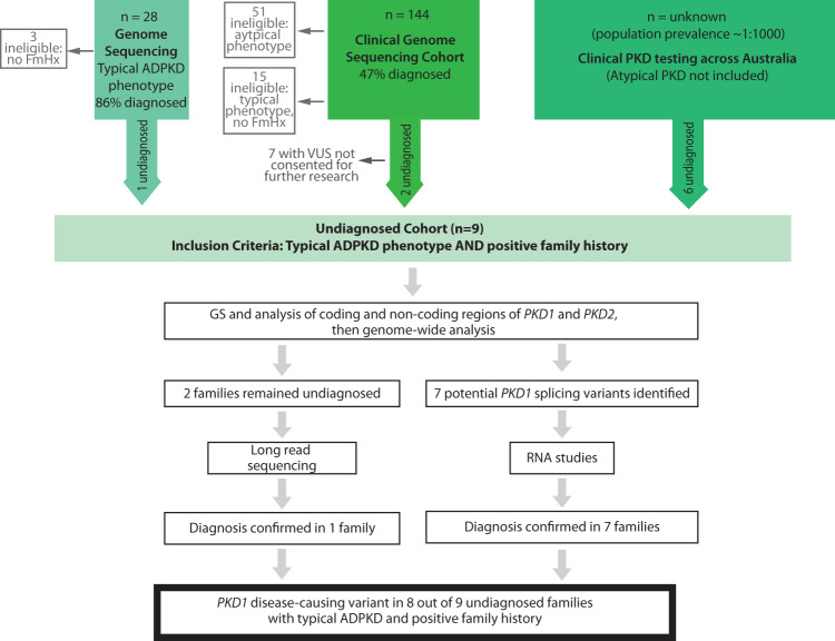
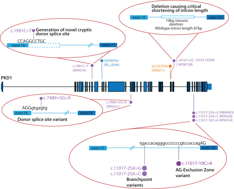
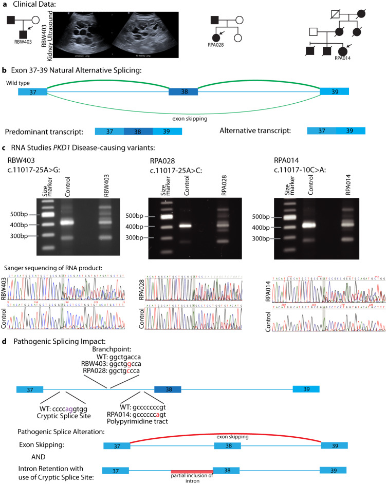
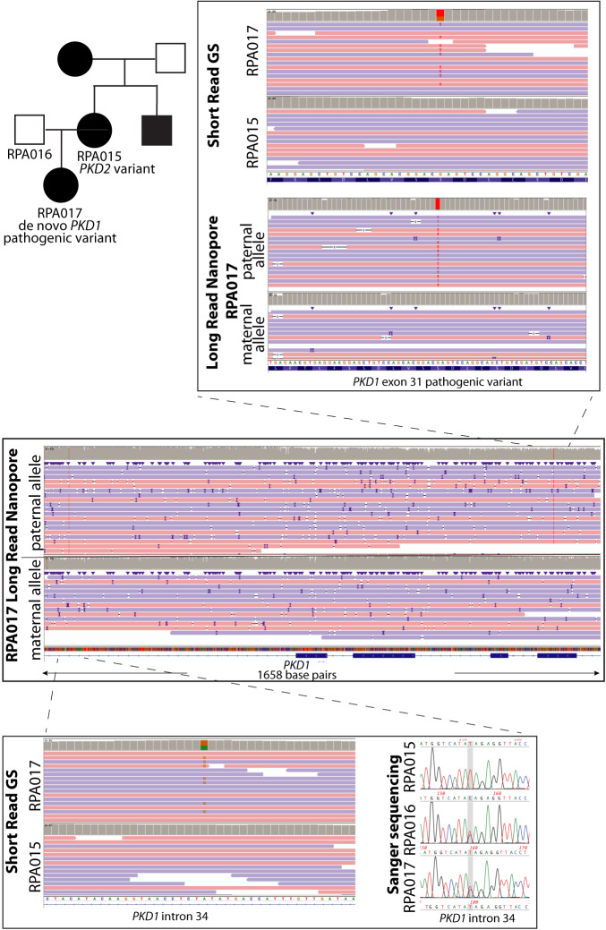
Autosomal dominant polycystic kidney disease (ADPKD) is the most common monogenic cause of kidney failure and is primarily associated with PKD1 or PKD2. Approximately 10% of patients remain undiagnosed after standard genetic testing. We aimed to utilise short and long-read genome sequencing and RNA studies to investigate undiagnosed families. Patients with typical ADPKD phenotype and undiagnosed after genetic diagnostics were recruited. Probands underwent short-read genome sequencing, PKD1 and PKD2 coding and non-coding analyses and then genome-wide analysis. Targeted RNA studies investigated variants suspected to impact splicing. Those undiagnosed then underwent Oxford Nanopore Technologies long-read genome sequencing. From over 172 probands, 9 met inclusion criteria and consented. A genetic diagnosis was made in 8 of 9 (89%) families undiagnosed on prior genetic testing. Six had variants impacting splicing, five in non-coding regions of PKD1. Short-read genome sequencing identified novel branchpoint, AG-exclusion zone and missense variants generating cryptic splice sites and a deletion causing critical intron shortening. Long-read sequencing confirmed the diagnosis in one family. Most undiagnosed families with typical ADPKD have splice-impacting variants in PKD1. We describe a pragmatic method for diagnostic laboratories to assess PKD1 and PKD2 non-coding regions and validate suspected splicing variants through targeted RNA studies.


Conflict of interest statement

The authors declare no competing interests.

Figures

Fig. 1
Fig. 1. Study design.
Overview of patients assessed for study suitability and study method. VUS variant of uncertain significance, GS genome sequencing; FmHx family history.
Fig. 2
Fig. 2. Overview of variants types identified in PKD1.
Variants identified in the PKD1 gene in the study, including a range of different splicing variants. Gene illustration developed using Protein Paint. PKD1 NM_001009944.3.
Fig. 3
Fig. 3. Disease-causing splicing variants in PKD1 intron 37.
A Pedigrees and renal ultrasound images from RBW403 demonstrating bilateral kidney cysts. B Natural splicing of exons 37, 38, and 39 of PKD1, depicting that skipping of exon 38 naturally occurs at a low level. C RT-PCR studies and Sanger sequencing of RT-PCR product in RBW403, RPA028, and RPA014. Additional bands are demonstrated in the affected individuals compared with controls, consistent in size with skipping of exon 38 and partial retention of exon 37. This is also reflected in Sanger sequencing of the RT-PCR product. Low-level skipping of exon 38 is evident in the controls. D Illustration of the splicing impact of the different variants identified in exon 37 across the cohort. All three variants result in the skipping of exon 38 and, less frequently, partial retention of exon 37 due to the use of an upstream cryptic splice site.
Fig. 4
Fig. 4. Long read nanopore sequencing confirms de novo PKD1 variant occurred on the paternal allele.
The middle panel shows long-read genome sequencing data from RPA017 over PKD1 exons 31–35, separated into maternal and paternal alleles. The top panel ‘zooms in’ over the region that includes the pathogenic PKD1 exon 31 variant identified in RPA017. The variant is absent in RPA015 (affected mother). The bottom panel ‘zooms in’ over an intronic single nucleotide variant identified in RPA017 that short-read GS and Sanger sequencing shows is present in RPA016 (unaffected father) and absent in RPA015 (affected mother). In RPA017, this variant is seen on the same long-read sequencing as the pathogenic PKD1 variant, demonstrating that the de novo disease-causing variant in RPA017 has occurred on her paternally inherited PKD1 allele. GS genome sequencing.

References

    1. Mallawaarachchi AC, et al. Whole-genome sequencing overcomes pseudogene homology to diagnose autosomal dominant polycystic kidney disease. Eur. J. Hum. Genet. 2016;24:1584–1590. doi: 10.1038/ejhg.2016.48. - DOI - PMC - PubMed
    1. Audrézet M-P, et al. Autosomal dominant polycystic kidney disease: comprehensive mutation analysis of PKD1 and PKD2 in 700 unrelated patients. Hum. Mutat. 2012;33:1239–1250. doi: 10.1002/humu.22103. - DOI - PubMed
    1. Rossetti S, et al. Identification of gene mutations in autosomal dominant polycystic kidney disease through targeted resequencing. J. Am. Soc. Nephrol. 2012;23:915–933. doi: 10.1681/ASN.2011101032. - DOI - PMC - PubMed
    1. Mallawaarachchi A, et al. Genomic diagnostics in polycystic kidney disease: an assessment of real-world use of whole-genome sequencing. Eur. J. Hum. Genet. 2021;29:760–770. doi: 10.1038/s41431-020-00796-4. - DOI - PMC - PubMed
    1. Senum SR, et al. Monoallelic IFT140 pathogenic variants are an important cause of the autosomal dominant polycystic kidney-spectrum phenotype. Am. J. Hum. Genet. 2022;109:136–156. doi: 10.1016/j.ajhg.2021.11.016. - DOI - PMC - PubMed