. 2023 Jul 7;14(1):4035.
doi: 10.1038/s41467-023-39665-1.

Amphetamine disrupts dopamine axon growth in adolescence by a sex-specific mechanism in mice

Affiliations

Amphetamine disrupts dopamine axon growth in adolescence by a sex-specific mechanism in mice

Lauren M Reynolds et al. Nat Commun. .

Abstract

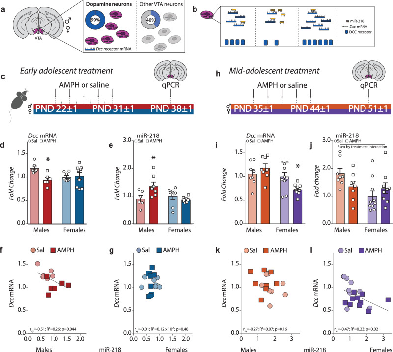
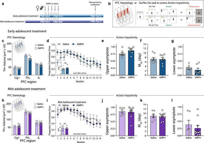
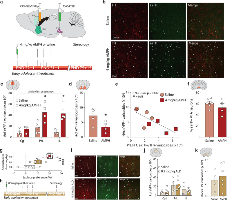
Initiating drug use during adolescence increases the risk of developing addiction or other psychopathologies later in life, with long-term outcomes varying according to sex and exact timing of use. The cellular and molecular underpinnings explaining this differential sensitivity to detrimental drug effects remain unexplained. The Netrin-1/DCC guidance cue system segregates cortical and limbic dopamine pathways in adolescence. Here we show that amphetamine, by dysregulating Netrin-1/DCC signaling, triggers ectopic growth of mesolimbic dopamine axons to the prefrontal cortex, only in early-adolescent male mice, underlying a male-specific vulnerability to enduring cognitive deficits. In adolescent females, compensatory changes in Netrin-1 protect against the deleterious consequences of amphetamine on dopamine connectivity and cognitive outcomes. Netrin-1/DCC signaling functions as a molecular switch which can be differentially regulated by the same drug experience as function of an individual's sex and adolescent age, and lead to divergent long-term outcomes associated with vulnerable or resilient phenotypes.


Conflict of interest statement

The authors declare no competing interests.

Figures

Fig. 1
Fig. 1. Regulation of Dcc expression in the VTA by AMPH in adolescence is sexually dimorphic.
a Dcc mRNA is expressed by 99% of dopamine neurons in the VTA of both male and female mice. b The microRNA miR-218 represses Dcc mRNA expression,. c Timeline of experiments in early adolescence. Male and female mice were exposed to a recreational-like amphetamine (AMPH, 4 mg/kg) regimen from P21 ± 1 to P31 ± 1. One week later, Dcc mRNA and miR-218 expression was quantified in the VTA using qPCR. dj AMPH in early adolescence downregulated Dcc expression in males, but not females (d) and increased miR-218 only in males (e) (Table 1A, B). In early adolescence, VTA miR-218 and Dcc mRNA levels correlated negatively in male, (f) but not female mice (g) (Table 1C, D). h Timeline of experiments in mid-adolescence. Male and female mice were exposed to the same recreational-like AMPH regimen, but from P35 ± 1 to P44 ± 1 and VTA transcripts were quantified one week later. il In mid-adolescence, AMPH no longer altered Dcc mRNA in males but downregulated levels in females (i), and it did not significantly alter miR-218 expression in either group (j) (Table 1E, F). In mid-adolescence, VTA miR-218 and Dcc mRNA levels did not correlate in males (k) but were negatively correlated in females (l) (Table 1G, H). All bar graphs are presented as mean values ± SEM, and were normalized to the saline condition in female mice (d, e, i, j). Source data are provided as a Source Data file. *p < 0.05.
Fig. 2
Fig. 2. Females are protected against the detrimental effects of AMPH in adolescence on the maturation of PFC dopamine connectivity and impulse control.
a Experimental timeline. b In adulthood, mice were randomly assigned to have their brains processed for stereological quantification of PFC dopamine innervation (left schematic) or were tested for impulse control in the Go/No-Go task (right schematic). c AMPH in early adolescence does not augment the span of the dopamine input to the cingulate (Cg1), prelimbic (PrL), and infralimbic (IL) subregions of the medial PFC in female mice, in contrast to our previous results in males,. Instead, a decrease in the volume of dopamine input to the PrL is evident (Table 2A). d AMPH in early adolescence does not alter action impulsivity in adulthood in female mice, unlike our previous observations in males. Area under the curve (AUC) analysis indicates that the proportion of commission errors is similar between the AMPH-treated and saline groups (Table 2B, C). eg Sigmoidal curve fit analysis (Table 2D–F) further revealed that there is no difference in the number of commission errors at the beginning of the task (e, upper asymptote), that both groups began to improve their inhibitory control performance around day 7-8 (f, M50), and that both groups show similar proportion of commission errors during the last sessions (g, lower asymptote). h AMPH in mid-adolescence does not alter the extent of the dopamine input to the Cg1, PrL and IL subregions of the PFC in female mice (Table 2G), despite downregulating Dcc in dopamine neurons (Fig. 1i). il In mid-adolescence, females continue to be insensitive to AMPH-induced deficits in action impulsivity, with no differences in the proportion of commission errors in the task (Table 2H–I). i Sigmoidal curve fit analysis (Table 2J–L) revealed that AMPH and saline groups perform equally at the beginning of the task (j, upper asymptote), start showing improvement around day 7-8 (j, M50), and have similar low proportion of commission errors during the last session (l, lower asymptote). All graphs are presented as mean values ± SEM. Source data are provided as a Source Data file.
Fig. 3
Fig. 3. AMPH upregulates Netrin-1 in the NAc of mid-adolescent females, counteracting its downregulation of Dcc levels in the VTA.
a Netrin-1 is highly expressed in the PFC, with lower expression in the NAc. DCC is expressed in a complementary pattern, with DCC-expressing dopamine axons segregated to the NAc. b When Dcc is reduced in dopamine neurons of adolescent male mice, their axons fail to recognize the NAc as their final target and instead grow ectopically to the PFC. c Reducing Netrin-1 expression in the NAc during adolescence also results in ectopic growth of Dcc-expressing dopamine axons to the PFC in male mice. d Experimental timeline for early adolescent treatment. e Experimental timeline for mid-adolescent treatment. f AMPH in early adolescence downregulates NAc Netrin-1 levels in males, but not in females (Table 3A, B). g AMPH in mid-adolescencence no longer downregulates Netrin-1 in the NAc, however mid-adolescent females show significant Netrin-1 upregulation in response to AMPH (Fig. 1i) (Table 3C, D). All graphs of Western blots are normalized to the saline condition for each age and sex. h Dcc mRNA expression in the VTA and Netrin-1 protein levels in the NAc of female mice treated with AMPH in mid-adolescence show a strong and significant negative correlation (Table 3E). i Top, Netrin-1 protein in the NAc of female mice was downregulated using an shRNA approach before treatment with AMPH or saline in mid-adolescence. Left, Netrin-1 shRNA virus was well expressed in the NAc of female mice. Right, Adult female mice treated with AMPH in mid-adolescence were not different from their saline-treated counterparts when receiving a scrambled control virus, in agreement with the results in Fig. 2h. However, adult females had an increased expanse of dopamine input to the PFC as adults when Netrin-1 in the NAc was downregulated with shRNA before treatment with AMPH in mid-adolescence (Table 3F). All bar graphs are presented as mean values ± SEM. Source data are provided as a Source Data file. *p < 0.05, **p < 0.01.
Fig. 4
Fig. 4. Recreational AMPH in adolescence induces ectopic growth of mesolimbic dopamine axons to the PFC in male mice.
a Experimental design. b Photomicrographs showing the prelimbic PFC (PrL) of adult mice injected with tracer viruses in adolescence. Top Dopamine axons continued to grow from the NAc to the PFC in adolescence in the saline condition (closed arrowheads). Bottom The number of axons that grew to the PFC in adolescence is dramatically increased in adult mice that were exposed to AMPH early in adolescence. c Stereological quantification reveals a significant increase in fluorescent axon terminals across the cingulate 1 (Cg1), PrL, and infralimbic (IL) subregions of the medial PFC and a pronounced dorsal-to-ventral gradient (Table 4A), paired with a significant decrease of fluorescent terminals in the NAc (d, Table 4B). e The number of labeled terminals in the PFC and in the NAc are negatively correlated (Table 4C). f The percentage of VTA dopamine neuron infection is similar between treatment groups (Table 4D). g Exposure to AMPH induces robust conditioned place preference (CPP) (Table 1E). This is not the case in male mice exposed to a therapeutic-like amphetamine regimen (ALD, 0.5 mg/kg). h Experimental design. i Photomicrographs showing the PrL of adult mice injected with tracer viruses in adolescence. The number of labeled axons that continued to grow from the NAc to the PFC in adolescence (closed arrowheads) is not different between adult mice that were exposed to saline (Top) or to ALD in adolescence (Bottom). ALD in adolescence does not alter the number of labeled dopamine terminals in the PFC (j, Table 1F) or the in the NAc (k, Table 1G), indicating that this AMPH dose does not interfere with dopamine axon targeting. All bar graphs are presented as mean values ± SEM. Box plots include a box extending from the 25th to 75th percentiles, with the median indicated by a line and with whiskers extending from the minima to the maxima. Source data are provided as a Source Data file. *p < 0.05, **p < 0.01, ***p < 0.01.
Fig. 5
Fig. 5. CRISPRa-mediated upregulation of VTA Dcc transcription prevents the harmful effect of recreational AMPH in adolescence on impulse control in male mice.
a The murine Dcc gene and mRNA. b CRISPR activation (CRISPRa) system. c Co-immunofluorescence of the mCherry tag for the Dcc sgRNA and TH (arrowheads) in cultured dopamine neurons (2 coverslips per guide combination). d All sgRNA sequences augmented Dcc mRNA expression (Table 5A; minimum 1.3983 ± 0.04752, maximum 1.93135 ± 0.22296 fold change). e The multiplex of all 4 sgRNAs gave the most robust increase in Dcc mRNA, with a maximal fold change of 3.81668 ± 1.03421 over LacZ control (Table 5B). f In vivo experimental design. g Low and h high magnification images of dopamine neurons (TH+) expressing the sgRNA viruses (mCherry+; arrowhead; n = 4 mice). i Dcc sgRNAs upregulated Dcc mRNA in the VTA compared to those receiving the LacZ sgRNA (Table 5C). j DCC protein expression in the NAc, where only dopamine axons express DCC receptors, was also significantly increased (Table 5D). k NAc DCC protein upregulation was strongly correlated with VTA Dcc mRNA upregulation (Table 5E). l Experimental design. m Mice with LacZ sgRNA treated with AMPH in adolescence showed a greater rate of commission errors compared to mice with LacZ sgRNA and treated with saline. This effect of AMPH was not observed in mice that received Dcc sgRNA (Table 5G). Area under the curve (AUC, inset) indicates that Dcc CRISPRa protects against AMPH-induced action impulsivity (Table 5H). n Illustration of sigmoidal curve fit analysis. o All groups showed a similar number of commission errors at the beginning of the task (upper asymptote, Table 5I). p LacZ sgRNA AMPH-treated mice took longer to improve their task performance (M50) in comparison to the LacZ sgRNA saline group (Table 5J), with some mice never improving (an M50 of 14 days), an effect rescued by Dcc CRISPRa treatment. q During the last trials, only LacZ sgRNA AMPH-treated mice showed significant impulse control deficits (Table 2K). All bar and line graphs are presented as mean values ± SEM. Source data are provided as a Source Data file. *p < 0.05, **p < 0.01.

Similar articles

Cited by

References

    1. Sawyer SM, Azzopardi PS, Wickremarathne D, Patton GC. The age of adolescence. Lancet Child Adolesc Health. 2018;2:223–228. doi: 10.1016/S2352-4642(18)30022-1. - DOI - PubMed
    1. Schneider, M. Adolescence as a vulnerable period to alter rodent behavior. Cell Tissue Res.354, 99–106 (2013). - PubMed
    1. Granata L, Gildawie KR, Ismail N, Brenhouse HC, Kopec AM. Immune signaling as a node of interaction between systems that sex-specifically develop during puberty and adolescence. Dev. Cogn. Neurosci. 2022;57:101143. doi: 10.1016/j.dcn.2022.101143. - DOI - PMC - PubMed
    1. Reynolds LM, Flores C. Mesocorticolimbic dopamine pathways across adolescence: diversity in development. Front. Neural Circuit. 2021;15:735625. doi: 10.3389/fncir.2021.735625. - DOI - PMC - PubMed
    1. Spear LP. The adolescent brain and age-related behavioral manifestations. Neurosci. Biobehav. Rev. 2000;24:417 463. doi: 10.1016/S0149-7634(00)00014-2. - DOI - PubMed

Publication types