

Evaluation of nimotuzumab Fab2 as an optical imaging agent in EGFR positive cancers
- PMID: 37419997
- PMCID: PMC10328982
- DOI: 10.1038/s41598-023-37873-9
Evaluation of nimotuzumab Fab2 as an optical imaging agent in EGFR positive cancers
Abstract
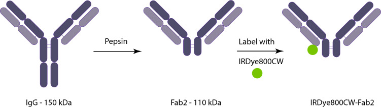
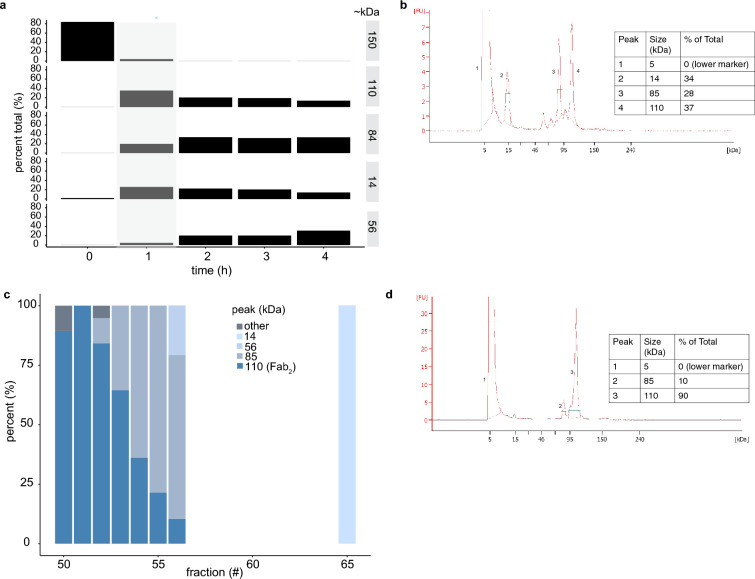
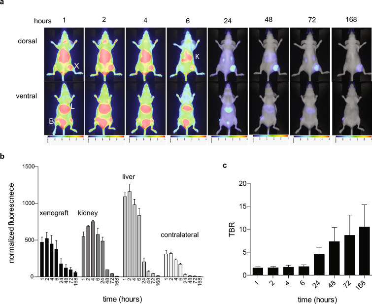
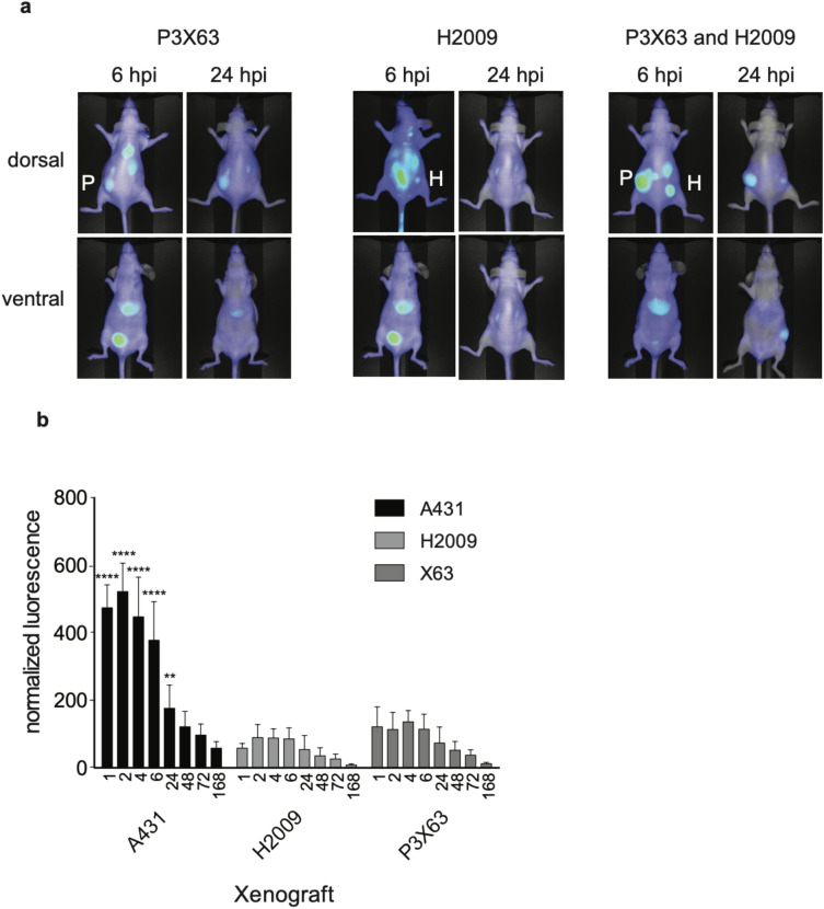
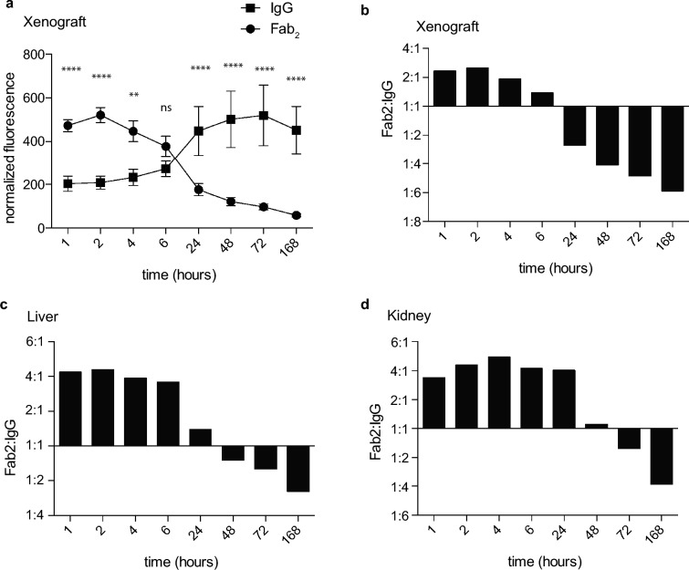
Molecular-targeted imaging probes can be used with a variety of imaging modalities to detect diseased tissues and guide their removal. EGFR is a useful biomarker for a variety of cancers, because it is expressed at high levels relative to normal tissues. Previously, we showed the anti-EGFR antibody nimotuzumab can be used as a positron emission tomography and fluorescent imaging probe for EGFR positive cancers in mice. These imaging probes are currently in clinical trials for PET imaging and image-guided surgery, respectively. One issue with using antibody probes for imaging is their long circulation time and slow tissue penetration, which requires patients to wait a few days after injection before imaging or surgery, multiple visits and longer radiation exposure. Here, we generated a Fab2 fragment of nimotuzumab, by pepsin digestion and labeled it with IRDye800CW to evaluate its optical imaging properties. The Fab2 had faster tumor accumulation and clearance in mice relative to the nimotuzumab IgG. The fluorescent signal peaked at 2 h post injection and remained high until 6 h post injection. The properties of the Fab2 allow a higher signal to background to be obtained in a shorter time frame, reducing the wait time for imaging after probe infusion.
© 2023. The Author(s).
Conflict of interest statement
The authors declare no competing interests.
Figures

Similar articles
-
Pre-clinical study of IRDye800CW-nimotuzumab formulation, stability, pharmacokinetics, and safety.BMC Cancer. 2021 Mar 12;21(1):270. doi: 10.1186/s12885-021-08003-3. BMC Cancer. 2021. PMID: 33711962 Free PMC article.
-
Site-Specific Fluorescent Labeling of Antibodies and Diabodies Using SpyTag/SpyCatcher System for In Vivo Optical Imaging.Mol Imaging Biol. 2019 Feb;21(1):54-66. doi: 10.1007/s11307-018-1222-y. Mol Imaging Biol. 2019. PMID: 29948640
-
Near infrared fluorescence imaging of EGFR expression in vivo using IRDye800CW-nimotuzumab.Oncotarget. 2017 Dec 21;9(5):6213-6227. doi: 10.18632/oncotarget.23557. eCollection 2018 Jan 19. Oncotarget. 2017. PMID: 29464066 Free PMC article.
-
Near infrared imaging of epidermal growth factor receptor positive xenografts in mice with domain I/II specific antibody fragments.Theranostics. 2019 Jan 30;9(4):974-985. doi: 10.7150/thno.30835. eCollection 2019. Theranostics. 2019. PMID: 30867810 Free PMC article.
-
Preclinical Evaluation of 111In-Labeled PEGylated Maytansine Nimotuzumab Drug Conjugates in EGFR-Positive Cancer Models.J Nucl Med. 2019 Aug;60(8):1103-1110. doi: 10.2967/jnumed.118.220095. Epub 2019 Jan 17. J Nucl Med. 2019. PMID: 30655327
References
-
- clinicaltrials.gov (NCT04459065, NCT04235114).
MeSH terms
Substances
LinkOut - more resources
Full Text Sources
Medical
Research Materials
Miscellaneous
