

Protective Effect of Platelet-Rich Plasma on Cisplatin-Induced Nephrotoxicity in Adult Male Albino Rats: Histological and Immunohistochemical Study
- PMID: 37420147
- PMCID: PMC10803452
- DOI: 10.1007/s12011-023-03742-9
Protective Effect of Platelet-Rich Plasma on Cisplatin-Induced Nephrotoxicity in Adult Male Albino Rats: Histological and Immunohistochemical Study
Abstract
Cisplatin is a potent antineoplastic drug that is used for treatment of many solid tumors. It has a wide range of adverse effects. Nephrotoxicity is the most common one of them. Platelet-rich plasma (PRP) is an autologous human plasma that activates the tissue regeneration through cell proliferation and differentiation. Study the role of PRP in amelioration of cisplatin-induced nephrotoxicity on the kidney of adult male albino rats by biochemical, morphometric, histological, and immunohistochemical studies. Thirty-five adult male albino rats were used. Thirty rats were included as experimental group and five were used to obtain the PRP. The experimental group was classified into as follows: control group which received 1mL of sterile saline by intraperitoneal injection (IP), cisplatin-treated group which received cisplatin 7.5 mg/kg IP in a single dose and cisplatin and PRP-treated group rats received cisplatin 7.5 mg/kg single IP dose followed by 1ml of PRP IP after 24 h of cisplatin injection. There was a significant increase in urea and creatinine levels in cisplatin-treated group in comparison to the control and the PRP groups. The kidneys of cisplatin-treated group showed distorted renal structure, where specimens of PRP-treated group revealed restoration of the classical appearance of the renal tissue similar to the control group. PRP has protective effects on renal structure and functions and it helps to ameliorate the histological changes induced by cisplatin.
Keywords: Cisplatin; Drug-induced nephrotoxicity; Immunohistochemistry; Morphometric parameters; PRP.
© 2023. The Author(s).
Conflict of interest statement
The authors declare no competing interests.
Figures
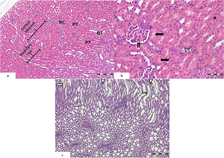
 ). Proximal convoluted tubules showing prominent brush border (
 ). The distal convoluted tubules (DT) appear with wider lumen and cuboidal cells with rounded nuclei. c The medulla is seen occupied by medullary rays (MR), collecting ducts (CD), renal interstitium (
 ), and blood vessels (
 ). (H&E. Mic.Mag a ×100, b ×400, and c ×100)
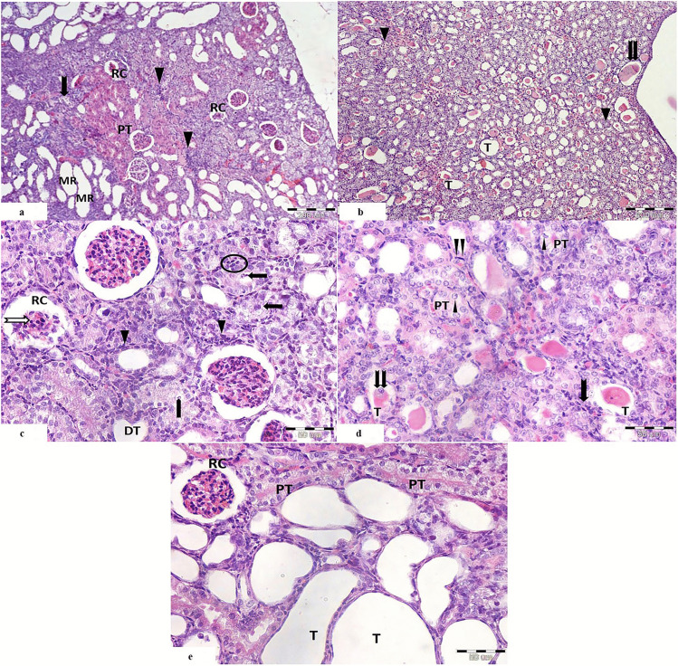
 ) among the degenerated tubules (
 ), and focal areas of proximal tubules with acidophilic cytoplasm (PT). b The medulla shows distorted tubules with multiple hyaline casts within their lumina (
 ). Some of the tubules appears dilated (T), and others show obliterated lumen (
 ). c The renal corpuscles (RC) appear with shrunken glomerular tuft and pyknotic nuclei (
 ). Peritubular cellular infiltration is noticed around the distorted tubules (
 ). The proximal convoluted tubules show vaculations (
 ), bizzare shaped nuclei of the lining cells (
 ), and extruded cells in the lumen with loss of brush border and basophilic cytoplasm (
 ). Distorted distal convoluted tubules with flattened cells are seen (DT). d The proximal tubules (PT) show karyolitic nuclei (
 ). Some tubules (T) show hyaline casts and exfoliated cells within their lumina (
 ). There are peritubular cellular infiltration (
 ) and proliferating interstitial fibroblasts (
 ). e Renal corpuscle (RC) appears with shrunken glomerular tuft and dark nuclei. Many tubules show severe ballooning (T). Cells of proximal convoluted tubules show vaculations with loss of brush boarder (PT). (H&E. Mic.Mag a, b ×100, c–e ×400)
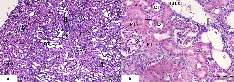
 ), and others appeared degenerated with hyalinized material in their lumen (
 ). b Distal convoluted tubules appear with cuboidal epithelial lining (DT). Widened interstitial space with evident cellular infiltration (
 ) and extravasated RBCs (RBCs) are also seen (
 ); brush border of PCT. (H&E. Mic.Mag a ×100 and b ×400)
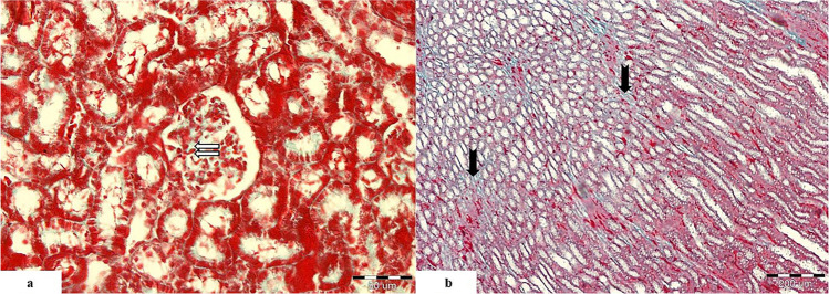
 ). b The medulla shows some collagen fibers in the peritubular interstitium (
 ). (Massonʼs trichrome. Mic.Mag a ×400 and b ×100)
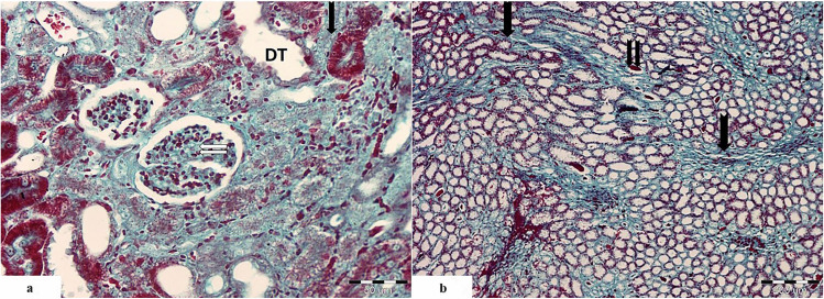
 ) and in the mesangium of the renal corpuscle (
 ). Some distal tubules show dilatation (DT). b Intense collagen fiber deposition in the medullary interstitium (
 ). Some tubules show hyaline casts deposition (
 ). (Massonʼs trichrome. Mic.Mag a ×400 and b ×100)
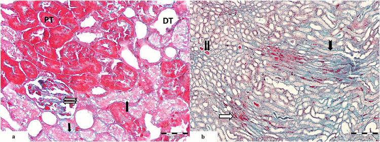
 ) and peritubular interstitium (
 ). Some proximal tubules are normal (PT), whereas others are abnormal with prominent vaculations (
 ). Some of the distal tubules are dilated (DT). b Moderate collagen fiber deposition in the medullary interstitium (
 ). Some tubules show hyaline cast deposition (
 ). There are vasa recta and extravasated RBCs in between the parallel tubules (
 ). (Masson trichrome. Mic.Mag a ×400 and b ×100)
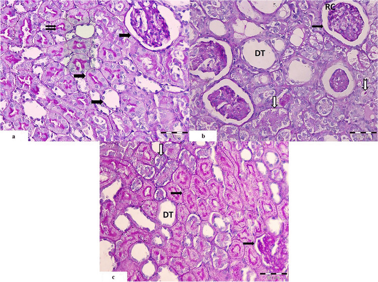
 ), and brush border of proximal convoluted tubules (
 ). It is stained magenta red with the PAS stain. b The cisplatin-treated group shows thickened basement membrane of the glomerular tuft (
 ). One of the glomeruli shows hyper cellularity (RC). Many proximal convoluted tubules appear degenerated with thickened basement membrane around them (
 ). Some tubules show hyaline casts within its lumen (
 ). Some distal tubules show dilatation (DT). c Cisplatin and PRP-treated group shows positive reaction within the basement membrane of the renal corpuscle and the proximal convoluted tubules (
 ). Few of them appear degenerated with thickened basement membrane (
 ). Some distal tubules show dilatation with thickened epithelial basement membrane (DT), and others appear nearly normal (PAS. Mic.Mag ×400)
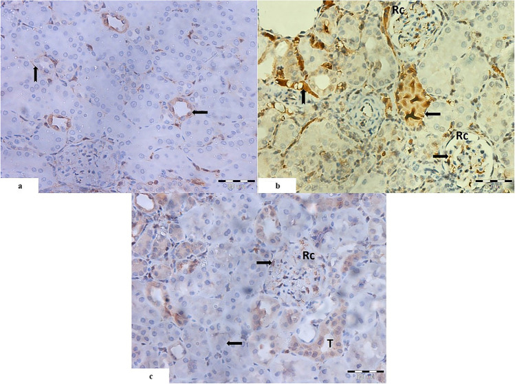
 ) and the interstitium (
 ). b Cisplatin-treated group shows intense positive reaction in the degenerated tubules with obliterated lumen (
 ). The interstitium shows strong positive reaction (
 ). Also, there is strong positive reaction within the cells of the glomerular capillary tuft (
 ) of the renal corpuscle (RC). c Cisplatin and PRP-treated group shows positive reaction within the lining cells of some tubules (T), and others show weak reaction (
 ). Few cells of the glomerular tuft show positive reaction (
 ) (caspase-3 immunohistochemical staining. Mic.Mag ×400)
References
-
- Gold J, A. R. Cisplatin. StatPearls Publishing: StatPearls Publishing; 2020.
MeSH terms
Substances
LinkOut - more resources
Full Text Sources
Research Materials
