Review
. 2023 Jul 7;22(1):106.
doi: 10.1186/s12943-023-01807-w.

The use of RNA-based treatments in the field of cancer immunotherapy

Affiliations
Review

The use of RNA-based treatments in the field of cancer immunotherapy

Mohammad Chehelgerdi et al. Mol Cancer. .

Abstract

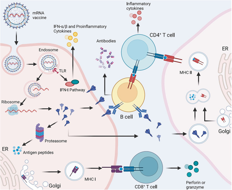
Over the past several decades, mRNA vaccines have evolved from a theoretical concept to a clinical reality. These vaccines offer several advantages over traditional vaccine techniques, including their high potency, rapid development, low-cost manufacturing, and safe administration. However, until recently, concerns over the instability and inefficient distribution of mRNA in vivo have limited their utility. Fortunately, recent technological advancements have mostly resolved these concerns, resulting in the development of numerous mRNA vaccination platforms for infectious diseases and various types of cancer. These platforms have shown promising outcomes in both animal models and humans. This study highlights the potential of mRNA vaccines as a promising alternative approach to conventional vaccine techniques and cancer treatment. This review article aims to provide a thorough and detailed examination of mRNA vaccines, including their mechanisms of action and potential applications in cancer immunotherapy. Additionally, the article will analyze the current state of mRNA vaccine technology and highlight future directions for the development and implementation of this promising vaccine platform as a mainstream therapeutic option. The review will also discuss potential challenges and limitations of mRNA vaccines, such as their stability and in vivo distribution, and suggest ways to overcome these issues. By providing a comprehensive overview and critical analysis of mRNA vaccines, this review aims to contribute to the advancement of this innovative approach to cancer treatment.

Keywords: Animal models; Cancer immunotherapy; Clinical trials; Infectious diseases; Messenger RNA; Technological advancements; Therapeutic use; mRNA vaccines.


Conflict of interest statement

The authors declare that they have no competing interests.

Figures

Fig. 1
Fig. 1
A Adjusting mRNA Medicine Dosage Pharmacokinetics. a) Crucial structural components of in vitro transcribed (IVT) mRNA and approaches for their alterations. b) Based on the individual or combined use of these elements (such as modification of caps, UTRs, or poly(A) tails), the protein product's expression duration and kinetic profile can be controlled and optimized. eIF4E represents eukaryotic translation initiation factor 4E; IRES refers to the internal ribosome entry site; and ORF denotes open reading frame. Reprinted from [51] with permission from Springer Nature. B Fundamentals of mRNA-based antigen pharmacology. a) A linear DNA plasmid containing the antigen-encoding sequence is employed for in vitro transcription. The transcribed mRNA consists of the cap, 5′ and 3′ UTRs, the open reading frame (ORF), and the poly(A) tail, which influence the mRNA's translational activity and stability once introduced into cells. b) Step 1: A portion of the foreign mRNA avoids degradation by common RNases and is taken up by cell-specific mechanisms (such as macropinocytosis in immature dendritic cells) into endosomal pathways. Step 2: The release of mRNA into the cytoplasm is not entirely understood. Step 3: Host cell protein synthesis machinery translates the mRNA. mRNA translation's rate-limiting step involves eukaryotic eIF4E binding to the cap structure. The formation of circular structures and active translation result from mRNA binding to ribosomes, eIF4E, eIF4G, and poly(A)-binding protein. Step 4: Exonucleases catalyze the termination of translation via mRNA degradation. Decapping enzymes D CP1, DCP2, and DCPS hydrolyze the cap, followed by the digestion of residual mRNA by 5′–3′ exoribonuclease 1 (XRN1). Degradation might be delayed if mRNA is silenced and located within cytoplasmic processing bodies. Alternatively, exosomal endonucleolytic cleavage of mRNA may take place. Various mechanisms control the breakdown of aberrant mRNA (such as mRNA with a premature stop codon). Step 5: The translated protein undergoes post-translational modifications based on the host cell's characteristics. The synthesized protein can then function within the cell it was produced in. Step 6: Alternatively, the protein is secreted and can function through autocrine, paracrine, or endocrine pathways. Step 7: For immunotherapeutic mRNA application, the protein must be broken down into antigenic peptide epitopes. These peptides are loaded onto major MHC molecules, which present the antigens to immune effector cells. Proteasomes degrade cytoplasmic proteins, which are then transported to the endoplasmic reticulum and loaded onto MHC class I molecules for presentation to CD8 + cytotoxic T lymphocytes. Almost all cells express MHC class I molecules. Step 8: In antigen-presenting cells, the protein must be directed to MHC class II loading compartments to obtain T cell assistance for a stronger, lasting immune response. This can be achieved by incorporating routing signal-encoding sequences into the mRNA. Additionally, DCs can process and load exogenous antigens onto MHC class I molecules through a mechanism called cross-priming. Step 9: Antigens derived from the protein can be displayed on the cell surface by both MHC class I and MHC class II molecules, enabling the immune system to recognize and respond to them accordingly. Reprinted from [51] with permission from Springer Nature
Fig. 2
Fig. 2
Essential breakthroughs and progress in mRNA-based treatment development. The creation of mRNA-based treatments can be split into three primary phases. Phase 1 (1961–1990) involves mRNA discovery, in vitro synthesis, and the construction of nucleic acid delivery systems, including mRNA identification, protamine uses for RNA delivery, in vitro mRNA translation, mRNA cap discovery, liposome-trapped mRNA delivery, commercialization of cap analogs and T7 RNA polymerases, cationic lipid-mediated mRNA delivery, and in vivo translation of naked mRNA through direct injection. Phase 2 (1990–2019) encompasses the accumulation of knowledge through numerous attempts and diverse applications, particularly protein replacement therapies and vaccination strategies for cancer and infectious diseases, such as mRNA-based cancer immunotherapy, founding of an mRNA-based company, 3′-UTR regulation of mRNA localization, antitumor T cell response triggered by mRNA, first clinical trial with mRNA using ex vivo transfected DCs, mRNA-based immunotherapy for human cancer, preclinical study with intranodally injected DC-targeted mRNA, protective mRNA vaccinations for influenza and respiratory syncytial virus, CRISPR-Cas9 mRNA for gene editing, and personalized mRNA cancer vaccines for clinical trials. Phase 3 (2019-present) sees mRNA-based therapeutics emerging as a disruptive technology, providing powerful and versatile tools for treating diseases, including clinical trials of mRNA vaccines for cancer and infectious diseases, as well as the emergency use of mRNA-1273 and BNT162b for the SARS-CoV-2 pandemic. Reprinted from [52] with permission from Springer Nature
Fig. 3
Fig. 3
PERSIST-seq summary and representative ribosome load findings. a Schematic representation of the mRNA optimization process. 5′ and 3′ UTRs sourced from literature and rational design were merged with Eterna and algorithmically created coding sequences. All sequences underwent simultaneous experimental evaluation for in-solution and in-cell stability, as well as ribosome load. Unique 6–9 nt barcodes in the 3′ UTR of the mRNA design enabled tag counting via short-read sequencing. b Experimental layout for assessing in-solution and in-cell stability and ribosome load concurrently. mRNAs were in vitro transcribed, 5′ capped, and polyadenylated in a pooled format prior to HEK293T cell transfection or in-solution degradation exposure. Cells were then collected for sucrose gradient fractionation or in-cell degradation examination. c Polysome trace from a 233-mRNA pool transfected into HEK293T cells. d 5′ UTR variations exhibit greater variability in average ribosome load per construct, as determined by polysome sequencing. The ribosome load formula is provided. Box hinges display 25% quantile, median, and 75% quantile from left to right, while whiskers indicate lower or upper hinge ± 1.5 × interquartile range. e Heatmaps of polysome profiles for top, middle, and bottom five mRNA designs (based on ribosome load) from each design category. f SARS-CoV-2 5′ UTR secondary structure model, with highlighted mutations and substitutions. g Heatmaps of SARS-CoV-2 5′ UTR variant polysome profiles, sorted by ribosome load. Reprinted from [87] with permission from Springer Nature
Fig. 4
Fig. 4
Various delivery methods and carrier molecules for mRNA vaccines, along with the typical diameters of the particulate complexes: a Naked mRNA: mRNA without any carrier or delivery system. b) Naked mRNA with in vivo electroporation: mRNA is introduced into cells by applying an electric field to facilitate uptake. c Protamine-complexed mRNA: mRNA is complexed with protamine, a cationic peptide, to improve stability and cellular uptake. d mRNA in cationic nanoemulsion: mRNA is associated with a positively charged oil-in-water cationic nanoemulsion to enhance delivery. e mRNA with dendrimer and PEG-lipid: mRNA is associated with a chemically modified dendrimer and complexed with polyethylene glycol (PEG)-lipid for improved delivery and reduced immunogenicity. f Protamine-complexed mRNA in a PEG-lipid nanoparticle: mRNA is complexed with protamine and encapsulated in a PEG-lipid nanoparticle for enhanced stability and delivery. g mRNA with polyethylenimine (PEI): mRNA is associated with a cationic polymer like PEI to improve delivery and transfection efficiency. h mRNA with PEI and lipid component: mRNA is associated with PEI and a lipid component for improved delivery and reduced immunogenicity. i mRNA in a polysaccharide particle or gel: mRNA is associated with a polysaccharide, such as chitosan, to form a particle or gel for improved stability and delivery. j mRNA in cationic lipid nanoparticle: mRNA is encapsulated in a cationic lipid nanoparticle (e.g., DOTAP or DOPE lipids) for enhanced stability and cellular uptake. k mRNA complexed with cationic lipids and cholesterol: mRNA is complexed with cationic lipids and cholesterol for improved stability and delivery. l mRNA complexed with cationic lipids, cholesterol, and PEG-lipid: mRNA is complexed with cationic lipids, cholesterol, and PEG-lipid for enhanced stability, delivery, and reduced immunogenicity. Reprinted from [126] with permission from Springer Nature
Fig. 5
Fig. 5
The impact of different intracellular delivery methods on in vitro and in vivo functionality. a In vitro colony-forming assays are used to compare the differentiation potential of electroporated and squeezed human CD34 + HSCs into CFU-GM and BFU-E colonies over a 2-week period. b The viability of mouse T cells after undergoing squeeze and electroporation is displayed. c and d The proportion of CD3 + mouse T cells expressing PD-1 or CD69 activation after squeeze, electroporation, or no treatment (control) is presented over time. e A diagram illustrates the experimental method for evaluating the effects of delivery methods on T cell activation. f and g On day 4, after re-exposure to OVA, CD45.2 + /CD8 + /IFN-γ + T cells were stained intracellularly for IFN-γ. Reprinted from [127] with permission from the Proceedings of the National Academy of Sciences
Fig. 6
Fig. 6
In an mRNA vaccine, the mRNA is taken up by cells through endocytosis and subsequently released from the endosome to be converted into proteins by ribosomes. These proteins can activate the immune system in two primary ways: i) the proteins are broken down by proteasomes into peptides that are then displayed as antigens on the cell surface by MHC class I molecules, which bind to the TCR and activate CD8 + T cells to destroy infected cells by releasing perforin and granzyme; ii) proteins secreted externally are taken up by APCs and broken down into peptides that are then displayed on the cell surface by MHC class II molecules for recognition by CD4 + T cells, which can activate both cellular immune responses by producing cytokines and humoral immune responses by co-activating B cells. Additionally, single-stranded RNA and double-stranded RNA in mRNA vaccines bind to TLR in the endosome to activate antiviral innate immune responses through the production of type-I interferon (IFN-I), which leads to the induction of numerous IFN-1-stimulated genes involved in antiviral innate immunity, a process known as the self-adjuvant effect of sequence-engineered mRNA. Reprinted from [205] with permission from Springer Nature
Fig. 7
Fig. 7
A Key elements and processes of an effective cancer vaccine. a The tumor antigen presentation process involves several steps. Initially, APCs such as DCs encounter antigens at the injection site or have antigens externally loaded onto them before injection, as in the case of DC vaccines. Antigen-loaded APCs then travel through the lymphatic system to the draining lymph nodes, where T cell activation primarily takes place. In the lymph node, mature DCs present tumor-derived peptides on MHC class I and II molecules to CD8 + and CD4 + T cells, both of naïve and memory types. The development of tumor-specific T cell responses is facilitated by delivering a costimulatory "signal 2" to T cells through interactions like CD80-CD28, CD86-CD28, CD70-CD27, and CD40-CD40 ligand (CD40L). Costimulation is enhanced by IL-12 and type I interferons (IFNs) produced by DCs. These interactions collectively support the generation and expansion of activated tumor-specific CD4 + and CD8 + T cell populations. CD4 + and CD8 + T cells migrate to the tumor site, and upon recognizing their specific antigens, they can destroy tumor cells through cytotoxicity and effector cytokine production, such as IFNγ and tumor necrosis factor (TNF). Consequently, lysed tumor cells release tumor antigens, which can be captured, processed, and presented by APCs to induce polyclonal T cell responses, thus increasing the antigenic variety of the anti-tumor immune response and leading to epitope spreading. b Cancer vaccines consist of four main components: tumor antigens, formulations, immune adjuvants, and delivery vehicles. Abbreviations: CpG ODN, CpG oligodeoxynucleotide; GM-CSF, granulocyte–macrophage colony-stimulating factor; MPL, monophosphoryl lipid A; poly-ICLC, polyinosinic–polycytidylic acid with polylysine and carboxymethylcellulose; STING, stimulator of interferon genes protein; TCR, T cell receptor; TLR, Toll-like receptor. Reprinted from [241] with permission from Springer Nature. B Key factors in the efficacy of directly injected mRNA vaccines. The effectiveness of an injected mRNA vaccine depends on several factors: the amount of antigen expression in professional APCs, which is affected by the carrier's efficiency, the presence of PAMPs such as double-stranded RNA (dsRNA) or unmodified nucleosides, and the optimization of the RNA sequence (including codon usage, G:C content, and 5' and 3' UTRs); the maturation and migration of DCs to secondary lymphoid tissues, which is enhanced by PAMPs; and the vaccine's capacity to stimulate strong T follicular helper (TFH) cell and germinal center (GC) B cell responses, an aspect that is not yet well-understood. An intradermal injection is provided as an illustration. EC refers to extracellular. Reprinted from [126] with permission from Springer Nature
Fig. 8
Fig. 8
Obstacles in the clinical application of mRNA. a In vitro production of therapeutic mRNA involves a linear DNA template and RNA polymerase (T7), followed by purification; the mRNA consists of a 5′ cap, a 5′ UTR, an ORF that encodes the target protein, a 3′ UTR, and a poly(A) tail. b) Upon local or systemic administration, mRNA encounters various extracellular hurdles such as rapid breakdown by prevalent nucleases, removal through macrophage phagocytosis, and elimination via renal filtration. c A portion of mRNA that escapes from blood vessels can be taken up by cells. The majority of these internalized mRNAs get confined in endosomes and can be identified by endosomal and cytosolic RNA sensors, which ultimately decrease the mRNA's translation and stability. Enhancing the 5′ cap can boost the binding efficiency of cytoplasmic mRNAs to ribosomes, consequently increasing mRNA translation efficiency. Although the endosomal escape of bare and unaltered mRNA is difficult, it can be facilitated by employing mRNA carriers. Reprinted from [322] with permission from Springer Nature
Fig. 9
Fig. 9
Possible therapeutic uses of IVT mRNA. Solid arrows in the right-hand column signify clinical applications, while dotted arrows indicate preclinical applications. Reprinted from [51] with permission from Springer Nature
Fig. 10
Fig. 10
Inflammatory Reactions to Artificial mRNA. In vitro transcribed (IVT) mRNA is identified by a variety of endosomal innate immune receptors, including Toll-like receptors 3, 7, and 8 (TLR3, TLR7, TLR8), as well as cytoplasmic innate immune receptors like protein kinase RNA-activated (PKR), retinoic acid-inducible gene I protein (RIG-I), melanoma differentiation-associated protein 5 (MDA5), and 2′–5′-oligoadenylate synthase (OAS). These pathways signal to produce inflammation connected with type 1 interferon (IFN), tumor necrosis factor (TNF), interleukin-6 (IL-6), IL-12, and the initiation of various transcriptional programs. Collectively, these elements generate a pro-inflammatory environment conducive to triggering specific immune responses. Furthermore, downstream consequences such as eukaryotic translation initiation factor 2α (eIF2α) phosphorylation-induced translation slowdown, increased RNA degradation due to ribonuclease L (RNASEL) overexpression, and self-amplifying mRNA replication inhibition are significant for the pharmacokinetics and pharmacodynamics of IVT mRNA. Reprinted from [51] with permission from Springer Nature
Fig. 11
Fig. 11
Natural immune detection of mRNA vaccines. DC recognizes two types of mRNA vaccines, with RNA sensors in yellow, antigens in red, DC maturation elements in green, and peptide-MHC complexes in light blue and red; a sample lipid nanoparticle carrier is depicted in the top right corner. A non-comprehensive list of major known RNA sensors responsible for identifying double-stranded and unaltered single-stranded RNAs is provided. Unaltered, unrefined (part a) and nucleoside-altered, FPLC-purified (part b) mRNAs are chosen to demonstrate two types of mRNA vaccines where known mRNA sensing mechanisms are either present or absent. The dotted arrow signifies diminished antigen expression. Ag stands for antigen; PKR for interferon-induced, double-stranded RNA-activated protein kinase; MDA5 for interferon-induced helicase C domain-containing protein 1 (also called IFIH1); IFN for interferon; m1Ψ for 1-methylpseudouridine; OAS for 2'-5'-oligoadenylate synthetase; and TLR for Toll-like receptor. Reprinted from [126] with permission from Springer Nature
Fig. 12
Fig. 12
A LNP formulation boosts protein production in vivo following intramuscular administration. BALB/c mice (4 per group) received intramuscular injections of either non-formulated (mRNA) or LNP-formulated (mRNA/LNP) PpLuc mRNA. a) Luciferase expression was observed in vivo through optical imaging at 24- and 48-h post-injection. b) Luminescence was used to quantify luciferase expression. Data points from individual mice (represented as dots) and the median (depicted as solid lines) are shown for each group. B LNP-based mRNA vaccine triggers both humoral and cellular immune reactions in mice. BALB/c mice (10 per group) received intramuscular vaccinations on day 0 and day 21 using either non-formulated RABV-G mRNA (mRNA), LNP-encapsulated RABV-G mRNA (mRNA/LNP), or buffer. Rabies Virus Neutralization Tests (VNTs) were conducted on the serum three weeks post-initial vaccination (a) and two weeks post-boost vaccination (b). Two weeks after the boost vaccination, splenocytes were exposed to an overlapping peptide library encompassing the RABV-G protein. Antigen-specific, multifunctional (IFN-γ + /TNF +) CD8 + (c) and CD4 + (d) T cells were identified using intracellular cytokine staining. Data points represent individual mice, while the solid lines indicate median values for each group. The dashed line marks the generally accepted protective titer threshold of 0.5 IU/ml for rabies VNTs. Reprinted from [540] with permission from Springer Nature
Fig. 13
Fig. 13
Obstacles in delivering and administering lipid nanoparticle-mRNA concoctions. a Natural hurdles faced by lipid nanoparticle-mRNA (LNP-mRNA) mixtures following systemic and localized distribution. b Methods of delivering LNP-mRNA compounds. Reprinted from [551] with permission from Springer Nature
Fig. 14
Fig. 14
Nanoscale particles and complexes for cancer immunotherapy delivery systems. a Lipid nanoparticles usually comprise an ionizable lipid, a supporting lipid, cholesterol, and polyethylene glycol (PEG)-lipid. Nucleic acids are integrated into the nanoparticles' hydrophilic core. b The structures of ready-made lipids explored for nucleic acid delivery and, more recently, mRNA vaccines are depicted, including the structure of DOPE (1,2-dioleoyl-sn-glycero-3-phosphoethanolamine), a helper lipid that enhances lipid nanoparticle effectiveness. c) The structures of ionizable, lipid-like substances developed using combinatorial chemistry methods for better in vivo mRNA delivery and reduced toxicity. d The structure of an amphiphilic peptide-vaccine conjugate designed to attach to albumin in the bloodstream, enhancing lymph node delivery. e A matrix-binding checkpoint inhibitor conjugate with increased retention in the area surrounding the tumor to initiate an immune response. The checkpoint inhibitor is connected to a placental growth factor 2 (PLGF2) peptide using an amine-to-sulfhydryl linker. The PLGF2 peptide facilitates binding to proteins present in the extracellular matrix (ECM). Reprinted from [555] with permission from Springer Nature
Fig. 15
Fig. 15
Identifying cancer-immunity cycle regulators through CRISPR screens. The cancer-immunity cycle outlines the step-by-step development of immune responses against tumors. CRISPR screening can be employed to examine cells at each stage of this cycle, identifying regulatory genes and the resulting phenotypic effects. A In the initial phase, tumor antigens are released by cancer cells and collected by APCs such as DCs, which may secrete cytokines when stimulated. APCs process and present the captured antigens using major histocompatibility complex proteins on their surface. APC trafficking to nearby lymph nodes enables cancer antigen presentation to naïve T cells, leading to T cell activation. Screens on APCs can reveal genes that regulate APC stimulation in response to tumor antigens and antigen presentation efficiency to T cells. B T cells exposed to antigens become primed and activated to target specific tumor antigens. T cell screens can pinpoint genes responsible for activation efficiency. C Primed T cells, including cytotoxic T lymphocytes, exit the lymph node, travel through the bloodstream, and infiltrate tumors as tumor-infiltrating lymphocytes (TILs). In vivo T cell screens can identify genes that facilitate TIL trafficking and infiltration. D Inside the tumor, T cells can finally recognize and respond to cancer-specific antigens, leading to tumor cell destruction. T cell screens can detect genes that improve tumor-killing activity. E Concurrently, screening tumor cells can uncover genes involved in resistance to T cell killing. CRISPR screens can identify positive regulators (shown in red) and negative regulators (shown in blue) at each step of the process. Reprinted from [657] with permission from Springer Nature

Similar articles

Cited by

References

    1. Lu YC, Robbins PF. Cancer immunotherapy targeting neoantigens. Semin Immunol. 2016;28(1):22–7. - PMC - PubMed
    1. Miao L, Zhang Y, Huang L. mRNA vaccine for cancer immunotherapy. Mol Cancer. 2021;20(1):1–23. - PMC - PubMed
    1. Hegde PS, Chen DS. Top 10 Challenges in Cancer Immunotherapy. Immunity. 2020;52(1):17–35. - PubMed
    1. Li W, Deng Y, Chu Q, Zhang P. Gut microbiome and cancer immunotherapy. Cancer Lett. 2019;447:41–7. - PubMed
    1. Dobosz P, Dzieciątkowski T. The Intriguing History of Cancer Immunotherapy. Front Immunol. 2019;2965. - PMC - PubMed