. 2023 Jul 7;23(1):460.
doi: 10.1186/s12903-023-03190-w.

The salivary proteome in relation to oral mucositis in autologous hematopoietic stem cell transplantation recipients: a labelled and label-free proteomics approach

Affiliations

The salivary proteome in relation to oral mucositis in autologous hematopoietic stem cell transplantation recipients: a labelled and label-free proteomics approach

S J M van Leeuwen et al. BMC Oral Health. .

Abstract

Background: Oral mucositis is a frequently seen complication in the first weeks after hematopoietic stem cell transplantation recipients which can severely affects patients quality of life. In this study, a labelled and label-free proteomics approach were used to identify differences between the salivary proteomes of autologous hematopoietic stem cell transplantation (ASCT) recipients developing ulcerative oral mucositis (ULC-OM; WHO score ≥ 2) or not (NON-OM).

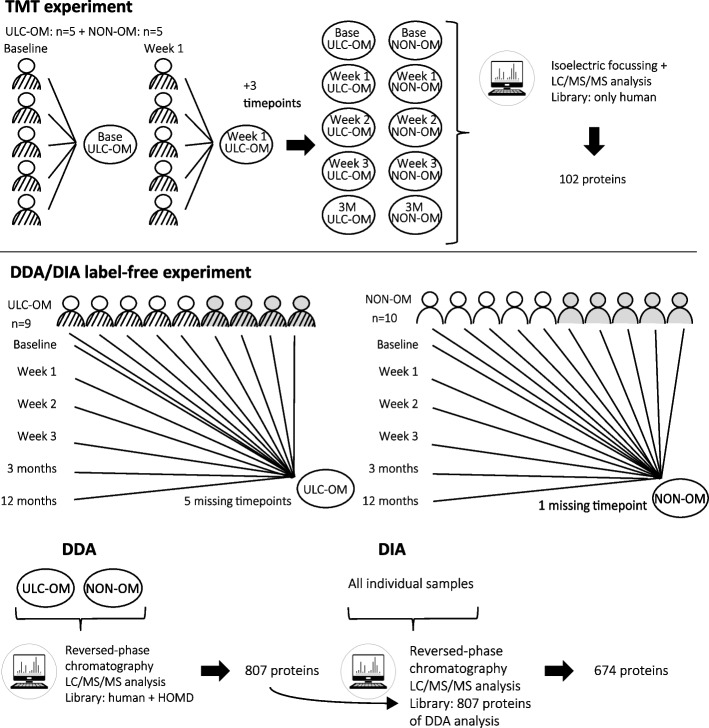
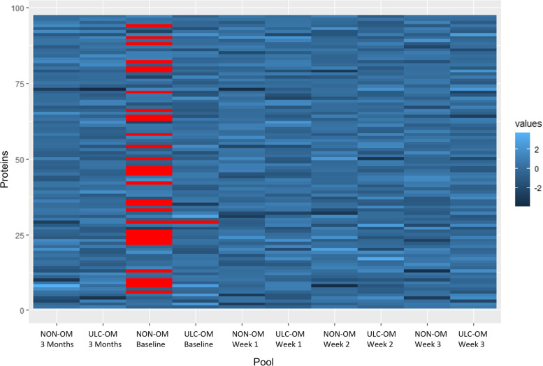
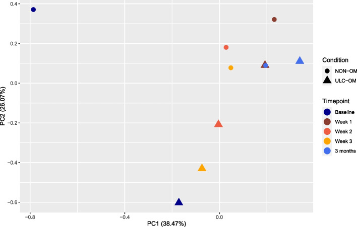
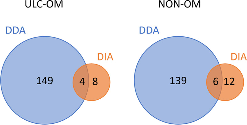
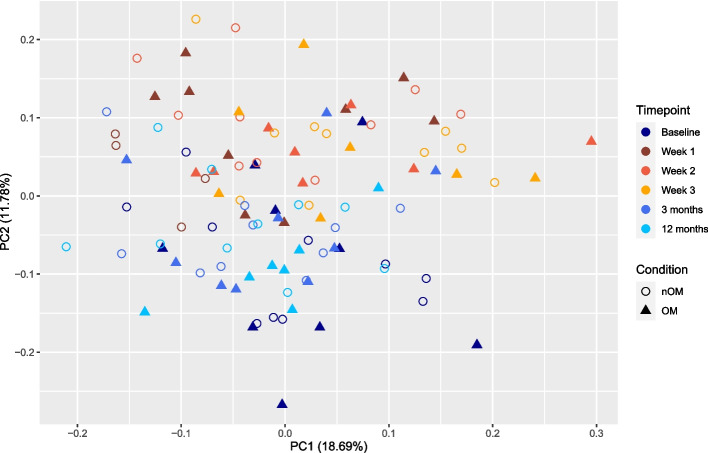
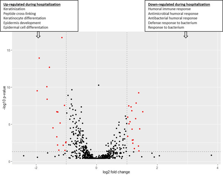
Methods: In the TMT-labelled analysis we pooled saliva samples from 5 ULC-OM patients at each of 5 timepoints: baseline, 1, 2, 3 weeks and 3 months after ASCT and compared these with pooled samples from 5 NON-OM patients. For the label-free analysis we analyzed saliva samples from 9 ULC-OM and 10 NON-OM patients at 6 different timepoints (including 12 months after ASCT) with Data-Independent Acquisition (DIA). As spectral library, all samples were grouped (ULC-OM vs NON-OM) and analyzed with Data Dependent Analysis (DDA). PCA plots and a volcano plot were generated in RStudio and differently regulated proteins were analyzed using GO analysis with g:Profiler.

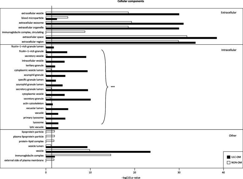
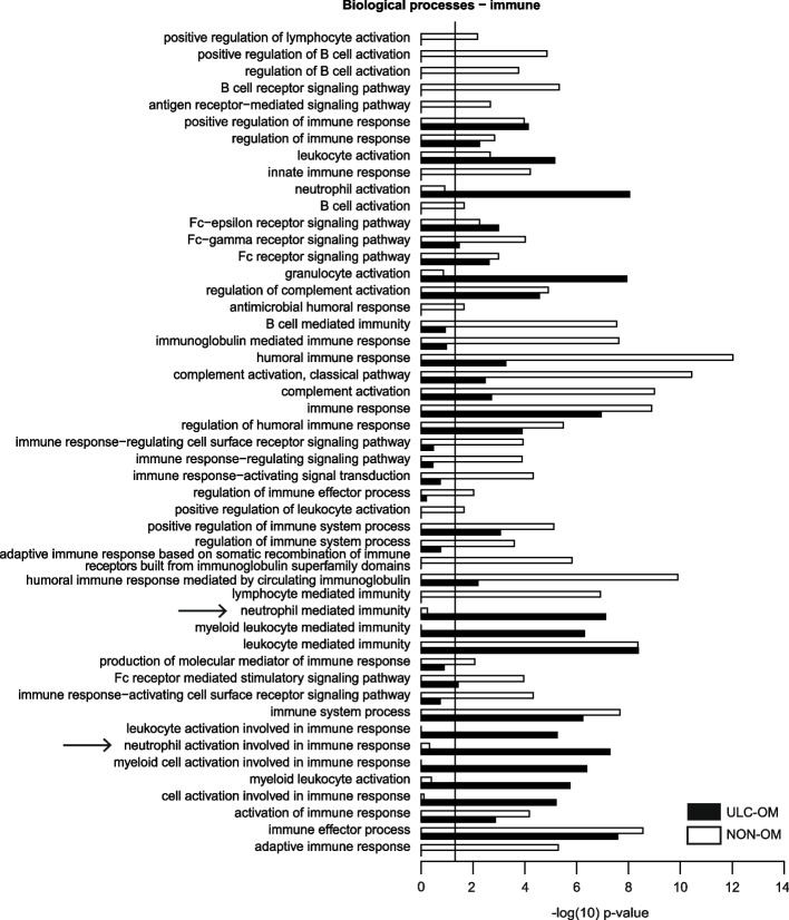
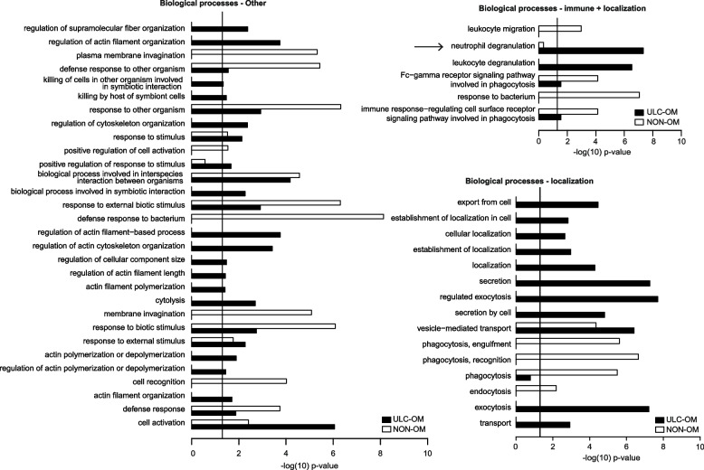
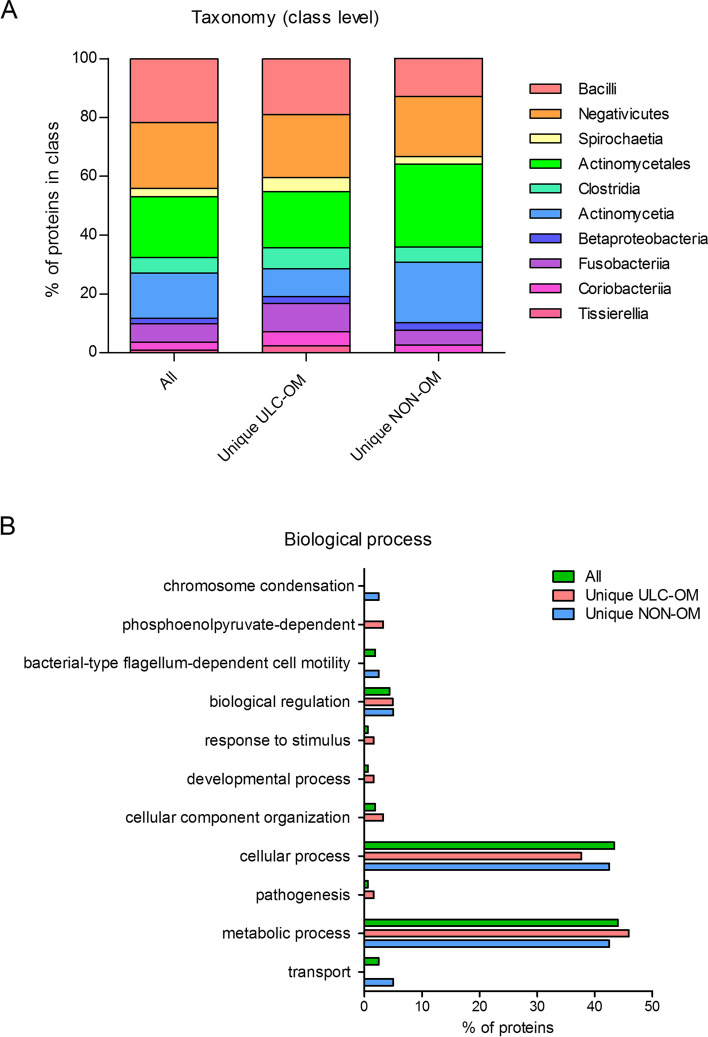
Results: A different clustering of ULC-OM pools was found at baseline, weeks 2 and 3 after ASCT with TMT-labelled analysis. Using label-free analysis, week 1-3 samples clustered distinctly from the other timepoints. Unique and up-regulated proteins in the NON-OM group (DDA analysis) were involved in immune system-related processes, while those proteins in the ULC-OM group were intracellular proteins indicating cell lysis.

Conclusions: The salivary proteome in ASCT recipients has a tissue protective or tissue-damage signature, that corresponded with the absence or presence of ulcerative oral mucositis, respectively.

Trial registration: The study is registered in the national trial register (NTR5760; automatically added to the International Clinical Trial Registry Platform).

Keywords: Autologous hematopoietic stem cell transplantation; Label-free quantification; Multiple myeloma; Oral mucositis; Saliva; TMT-labelled proteomics.


Conflict of interest statement

The authors declare no competing interests.

Figures

Fig. 1
Fig. 1
Schematic overview of the TMT-labelled, data dependent analysis (DDA) and data-independent analysis (DIA) experimental designs. TMT experiment: stimulated whole-mouth saliva samples were pooled from 5 ulcerative oral mucositis (ULC-OM) and 5 non-oral mucositis (NON-OM) patients at 5 different time points (baseline, 0–4 days (week 1), 6–11 days (week 2), 13–18 days (week 3) and 3 months). The resulting 10 pools were labelled and analyzed with LC/MS/MS. Label-free quantification (LFQ) experiment: for the DDA analysis, stimulated whole-mouth saliva samples from 9 ULC-OM and 10 NON-OM patients at 6 different time points (baseline, 0–4 days (week 1), 6–11 days (week 2), 13–18 days (week 3), 3 months and 12 months) were grouped resulting in 2 groups that were analyzed with LC/MS/MS. Of the ULC-OM patients, 5 saliva samples could not be collected, resulting in 5 missing timepoints (week 2, twice week 3 and twice 12 months). Only 1 saliva sample of a NON-OM patient at week 1 could not be collected. The oblique striped jackets represent ULC-OM patients and the empty jackets represent NON-OM patients. Patients selected for the TMT experiment were also used in the LFQ experiment in addition to 4 ULC-OM or 5 NON-OM other patients (in grey). For the DIA analysis, individual samples were analyzed with LC/MS/MS using the with DDA generated library
Fig. 2
Fig. 2
Missing values (red) of the Log2 transformed ratios (blue) of the different pools in the TMT-labelled experiment
Fig. 3
Fig. 3
Principal component analysis (PCA) plot of the TMT-labelled experiment with colors indicating the different timepoints (blue colors: baseline and 3 months; orange colors: weeks 1–3 (hospitalization)) and shapes indicating the condition: ulcerative oral mucositis (ULC-OM, filled round) or non-oral mucositis (NON-OM, filled triangle)
Fig. 4
Fig. 4
Heatmap of the TMT experiment with the different timepoints (Baseline, Week 1, Week 2, Week 3 and 3 Months) for the ulcerative oral mucositis (ULC-OM) and non-oral mucositis (NON-OM) pools. The baseline NON-OM pool is excluded, since it is an outlier in the PCA plot (Fig. 3)
Fig. 5
Fig. 5
Bar graph of significant cellular components gene ontology (GO) terms for unique and up-regulated proteins in the ulcerative oral mucositis (ULC-OM; black bars) and non-oral mucositis (NON-OM; white bars) groups of the label-free quantification Data Dependent Analysis (DDA) experiment. The vertical line represents significance threshold of p = 0.05. The intracellular CC GO terms were more significant in the ULC-OM group compared to the NON-OM group (ULC-OM: (mean ± SD) 10.77 ± 6.54, NON-OM: 0.48 ± 0.98, p < 0.001, 95% CI [7.44, 13.13] (shown with accolade and ***)
Fig. 6
Fig. 6
Bar graphs of significant gene ontology (GO) terms related to subgroup immune within biological processes for unique and up-regulated proteins in the ulcerative oral mucositis (ULC-OM; black bars) and non-oral mucositis (NON-OM; white bars) groups of the label-free quantification Data Dependent Analysis (DDA) experiment. The vertical line represents significance threshold of p = 0.05. Neutrophil degranulation and activation terms (mentioned in the Discussion section) are indicated with an arrow
Fig. 7
Fig. 7
Bar graphs of significant gene ontology (GO) terms related to subgroups other, immune + localization and localization within the biological processes for unique and up-regulated proteins in the ulcerative oral mucositis (ULC-OM; black bars) and non-oral mucositis (NON-OM; white bars) groups of the label-free quantification Data Dependent Analysis (DDA) experiment. The vertical line represents significance threshold of p = 0.05. Neutrophil degranulation and activation terms (mentioned in the Discussion section) are indicated with an arrow
Fig. 8
Fig. 8
Taxonomy at class level (A) of microbial proteins and biological processes of those proteins (B). ‘All’ represents all identified microbial proteins, in ‘unique ULC-OM’ or ‘unique NON-OM’ only the uniquely identified proteins in the ulcerative oral mucositis (ULC-OM) or non-oral mucositis (NON-OM) group are shown (Data Dependent Analysis (DDA) experiment)
Fig. 9
Fig. 9
Dot plot of the log2 transformed label-free quantification intensity (LFQ) and corresponding abundance ranking of the identified proteins in the DDA experiment. The red dots are the proteins identified in both the DDA and TMT-labelled experiment
Fig. 10
Fig. 10
Venn diagrams showing the overlap of the regulated proteins between Data Dependent Analysis (DDA) and Data Independent Analysis (DIA) of the ulcerative oral mucositis (ULC-OM) and non-oral mucositis (NON-OM) samples
Fig. 11
Fig. 11
Principal component analysis (PCA) plot of the Data Independent Acquisition (DIA) experiment with colors indicating the different timepoints (blue colors: baseline, 3 and 12 months; orange colors: week 1–3 (hospitalization)) and shapes indicating the condition: ulcerative oral mucositis (ULC-OM, open round) or non-oral mucositis (NON-OM, filled triangle)
Fig. 12
Fig. 12
Volcano plot of differently regulated proteins (red dots) during week 1, 2 and 3 (hospitalization) or at baseline, 3 months and 12 months of the Data Independent Acquisition (DIA) experiment. The horizontal line indicates threshold for statistical significance (p < 0.05) and the vertical lines indicates threshold for differently regulated proteins (fold change of 2). In textboxes the top 5 of significantly enriched biological processes gene ontology terms are listed for upregulated proteins during hospitalization (left box) and outside the hospitalization period (right box). Differently expressed proteins and details of the gene ontology terms are listed in Supplementary Tables 4 and 5, Additional file 3

References

    1. Blijlevens N, Schwenkglenks M, Bacon P, D'Addio A, Einsele H, Maertens J, et al. Prospective oral mucositis audit: oral mucositis in patients receiving high-dose melphalan or BEAM conditioning chemotherapy–European Blood and Marrow Transplantation Mucositis Advisory Group. J Clin Oncol. 2008;26(9):1519–1525. doi: 10.1200/JCO.2007.13.6028. - DOI - PubMed
    1. Sonis ST, Oster G, Fuchs H, Bellm L, Bradford WZ, Edelsberg J, et al. Oral mucositis and the clinical and economic outcomes of hematopoietic stem-cell transplantation. J Clin Oncol. 2001;19(8):2201–2205. doi: 10.1200/JCO.2001.19.8.2201. - DOI - PubMed
    1. Peterson DE, Cariello A. Mucosal damage: a major risk factor for severe complications after cytotoxic therapy. Semin Oncol. 2004;31(3 Suppl 8):35–44. doi: 10.1053/j.seminoncol.2004.04.006. - DOI - PubMed
    1. Sonis ST. Pathobiology of oral mucositis: novel insights and opportunities. J Support Oncol. 2007;5(9 Suppl 4):3–11. - PubMed
    1. Wardill HR, Sonis ST, Blijlevens NMA, Van Sebille YZA, Ciorba MA, Loeffen EAH, et al. Mucositis Study Group of the Multinational Association of Supportive Care in Cancer/International Society of Oral Oncology (MASCC/ISOO). Prediction of mucositis risk secondary to cancer therapy: a systematic review of current evidence and call to action. Support Care Cancer. 2020;28(11):5059–73. 10.1007/s00520-020-05579-7. Epub 2020 Jun 26. - PubMed

Publication types

Associated data