. 2023 Jul 7;22(1):172.
doi: 10.1186/s12933-023-01900-w.

CXCL5 suppression recovers neovascularization and accelerates wound healing in diabetes mellitus

Affiliations

CXCL5 suppression recovers neovascularization and accelerates wound healing in diabetes mellitus

Ching Chen et al. Cardiovasc Diabetol. .

Erratum in

Abstract

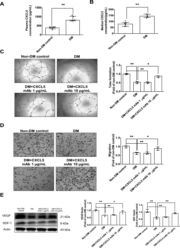
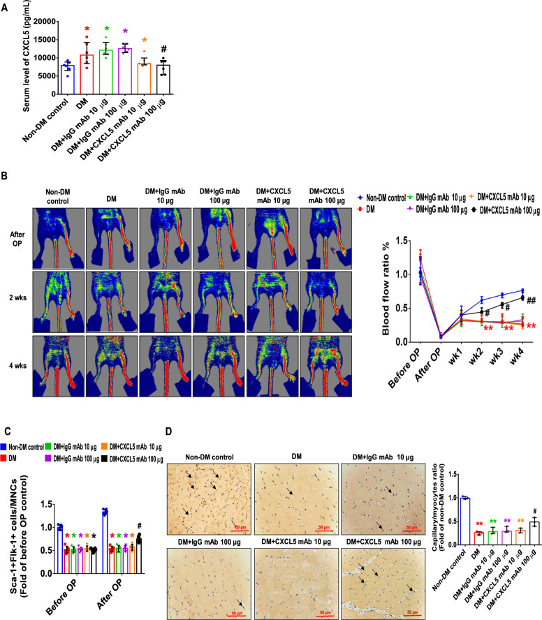
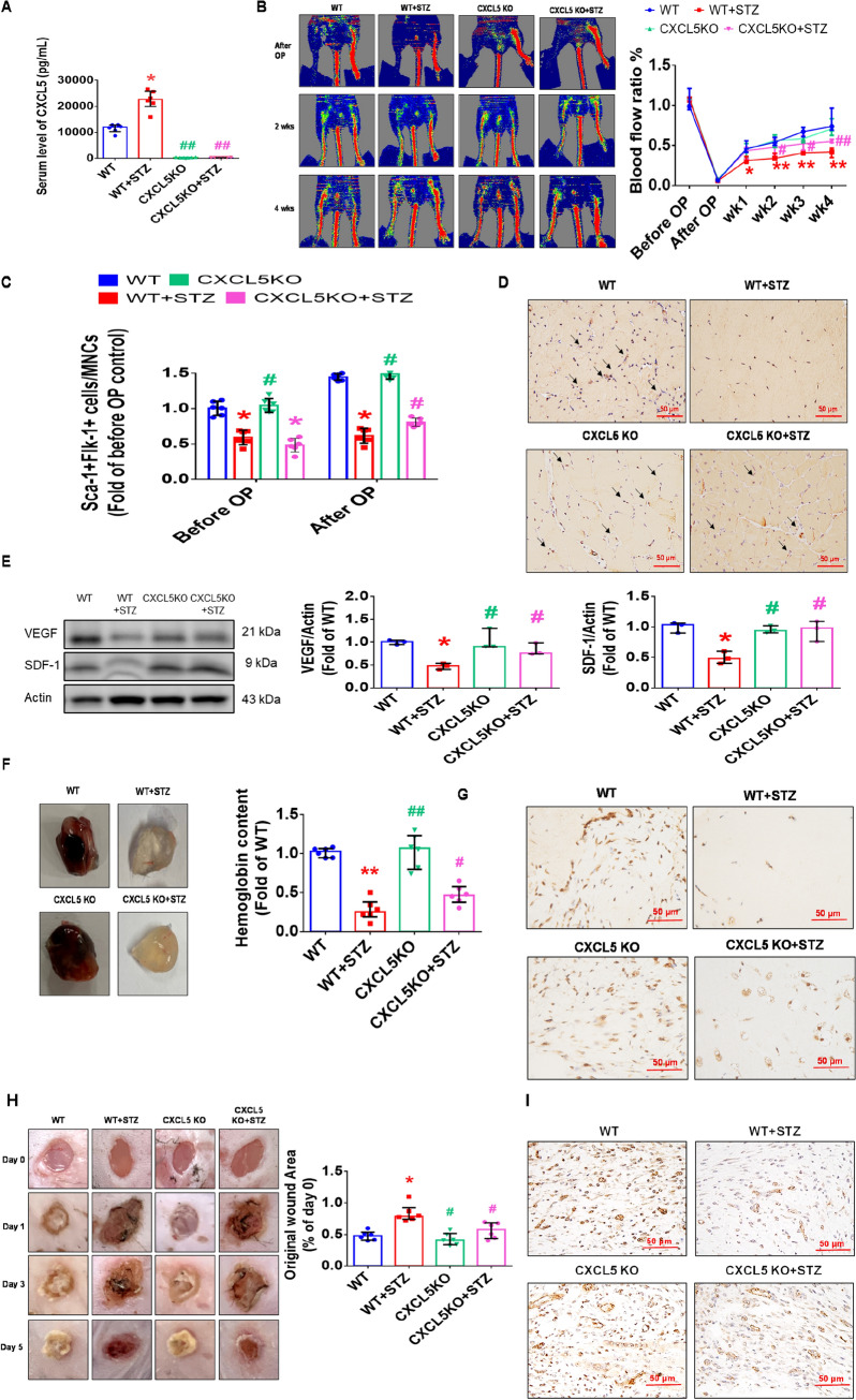
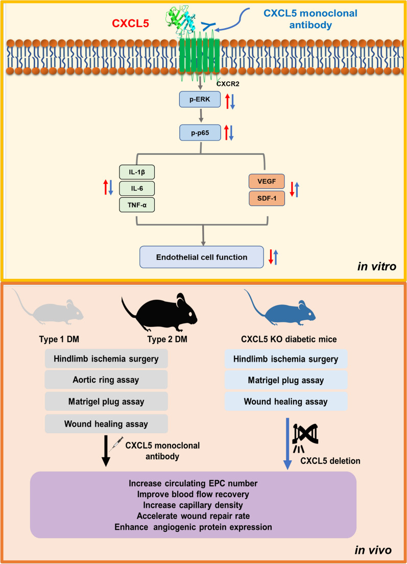
Background: Higher chemokine C-X-C motif ligand 5 (CXCL5) level was observed in type 2 diabetes mellitus (DM) patients; however, its role in diabetic vasculopathy was not clarified. This study aimed to explore the impacts and mechanistic insights of CXCL5 in neovasculogenesis and wound healing in DM.

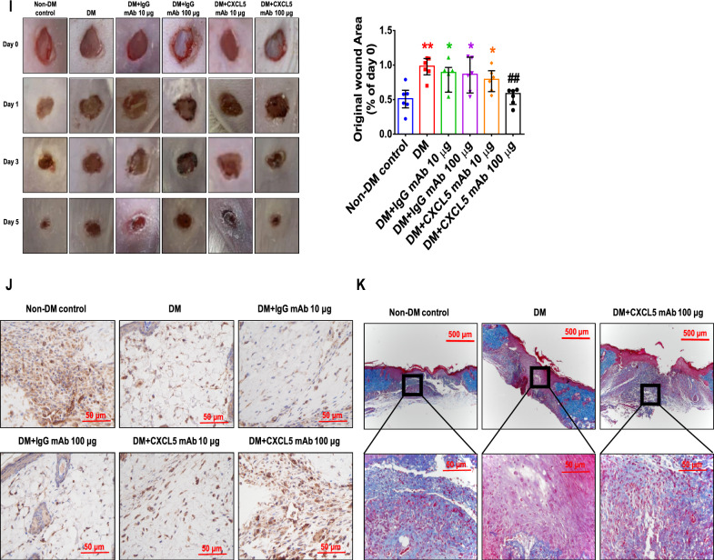
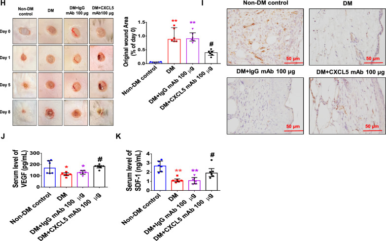
Methods: Endothelial progenitor cells (EPCs) and human aortic endothelial cells (HAECs) were used in vitro. Streptozotocin-induced diabetic mice and Leprdb/JNarl mice were used as type 1 and type 2 DM models. Moreover, CXCL5 knockout mice were used to generate diabetic mice. Hindlimb ischemia surgery, aortic ring assays, matrigel plug assay, and wound healing assay were conducted.

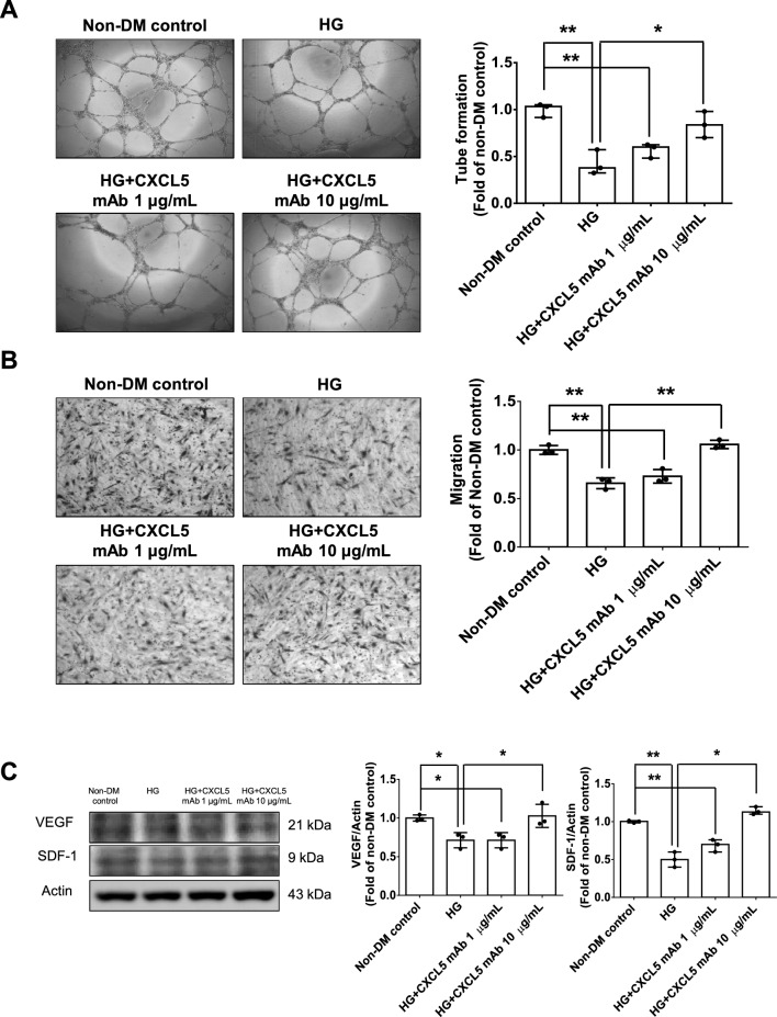
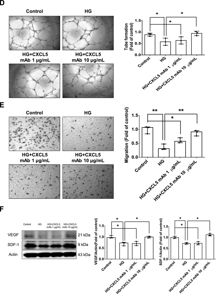
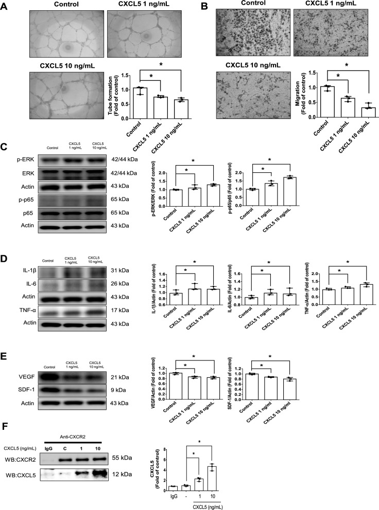
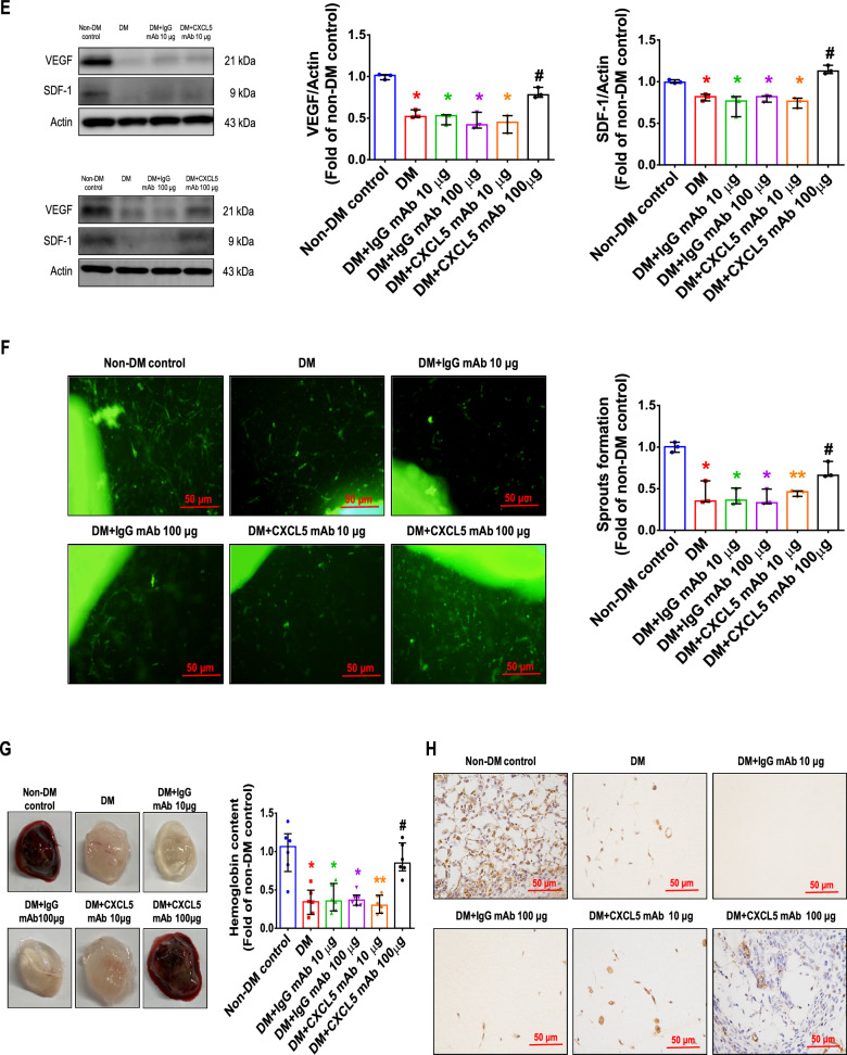
Results: CXCL5 concentrations were increased in plasma and EPCs culture medium from type 2 DM patients. CXCL5 neutralizing antibody upregulated vascular endothelial growth factor (VEGF)/stromal cell-derived factor-1 (SDF-1) and promoted cell function in EPCs from type 2 DM patients and high glucose-treated EPCs from non-DM subjects as well as HAECs. CXCL5 directly up-regulated interleukin (IL)-1β/IL-6/tumor necrosis factor-α and down-regulated VEGF/SDF-1 via ERK/p65 activation through chemokine C-X-C motif receptor 2 (CXCR2). CXCL5 neutralizing antibody recovered the blood flow after hindlimb ischemia, increased circulating EPC number, and enhanced VEGF and SDF-1 expression in ischemic muscle. CXCL5 suppression promoted neovascularization and wound healing in different diabetic animal models. The above observation could also be seen in streptozotocin-induced CXCL5 knockout diabetic mice.

Conclusions: CXCL5 suppression could improve neovascularization and wound healing through CXCR2 in DM. CXCL5 may be regarded as a potential therapeutic target for vascular complications of DM.

Keywords: CXCL5; CXCR2; Diabetes mellitus; Neovascularization; Wound healing.


Conflict of interest statement

The authors declare that they have no competing interests.

Figures

Fig. 1
Fig. 1
Treatment with CXCL5 neutralizing antibody upregulated VEGF/SDF-1 expression and promoted angiogenesis in late-EPCs from type 2 DM patients. Plasma levels of CXCL5 in type 2 DM patients and non-DM subjects (n = 6; A). EPCs medium levels of CXCL5 in type 2 DM patients and non-DM subjects (n = 6; B). The network formation and migration abilities were improved after the administration of CXCL5 mAb in EPCs from type 2 DM patients (n = 3; C, D). Western blotting and statistical analyses of VEGF and SDF-1 in EPCs from type 2 DM patients (n = 3; E). CXCL5 Chemokine C-X-C motif ligand 5, DM diabetes mellitus, EPC endothelial progenitor cell, mAb monoclonal antibody, SDF-1 stromal cell-derived factor 1, VEGF vascular endothelial growth factor. N represents cells cultured from n different individuals, and cells cultured from each individual were experimented for three independent experiments. The Mann–Whitney U test was used to determine statistically significant differences. *p < 0.05, **p < 0.01
Fig. 2
Fig. 2
Treatment with CXCL5 neutralizing antibody upregulated VEGF/SDF-1 expression and promoted angiogenesis in late-EPCs from non-DM subjects and HAECs under the HG conditions. The network formation and migration abilities were improved after the administration of CXCL5 mAb in EPCs from non-DM subjects (n = 3; A, B). Western blotting and statistical analyses of VEGF and SDF-1 in EPCs from non-DM subjects (n = 3; C). The network formation and migration abilities were improved after the administration of CXCL5 mAb in HAECs (n = 3; D, E). Western blotting and statistical analyses of VEGF and SDF-1 in HAECs (n = 3; F). CXCL5 C-X-C motif chemokine ligand 5, EPC endothelial progenitor cell, HG high glucose, HAEC human aortic endothelial cell, mAb,monoclonal antibody, SDF-1 stromal cell-derived factor 1, VEGF vascular endothelial growth factor. N represents the number of independent experiments on different days and in different experimental runs. The Mann–Whitney U test was used to determine statistically significant differences. *p < 0.05, **p < 0.01
Fig. 2
Fig. 2
Treatment with CXCL5 neutralizing antibody upregulated VEGF/SDF-1 expression and promoted angiogenesis in late-EPCs from non-DM subjects and HAECs under the HG conditions. The network formation and migration abilities were improved after the administration of CXCL5 mAb in EPCs from non-DM subjects (n = 3; A, B). Western blotting and statistical analyses of VEGF and SDF-1 in EPCs from non-DM subjects (n = 3; C). The network formation and migration abilities were improved after the administration of CXCL5 mAb in HAECs (n = 3; D, E). Western blotting and statistical analyses of VEGF and SDF-1 in HAECs (n = 3; F). CXCL5 C-X-C motif chemokine ligand 5, EPC endothelial progenitor cell, HG high glucose, HAEC human aortic endothelial cell, mAb,monoclonal antibody, SDF-1 stromal cell-derived factor 1, VEGF vascular endothelial growth factor. N represents the number of independent experiments on different days and in different experimental runs. The Mann–Whitney U test was used to determine statistically significant differences. *p < 0.05, **p < 0.01
Fig. 3
Fig. 3
CXCL5 impaired vascular endothelial function via the ERK/p65 signaling pathway in HAECs. The network formation and migration abilities were impaired after administration of CXCL5 for 2 days (n = 3; A, B). Western blotting and statistical analyses of p-ERK, p-p65, IL-1β, IL-6, and TNF-α after administration of CXCL5 for 2 days (n = 3; C, D). Western blotting and statistical analyses of VEGF and SDF-1 after administration of CXCL5 for 2 days (n = 3; E). Western blotting of CXCR2 and CXCL5 after anti-goat IgG and CXCR2 immunoprecipitation (n = 3; F). CXCL5 Chemokine C-X-C motif ligand 5, CXCR2 Chemokine C-X-C motif receptor 2, ERK extracellular signal-regulated kinase, HAEC human aortic endothelial cell, IL interleukin, SDF-1 stromal cell-derived factor 1, TNF-α tumor necrosis factor-α, VEGF vascular endothelial growth factor. N represents the number of independent experiments on different days and in different experimental runs. The Mann–Whitney U test was used to determine statistically significant differences. *p < 0.05, **p < 0.01
Fig. 4
Fig. 4
CXCL5 neutralizing antibody repaired neovascularization and wound healing in type 1 DM mice. Serum CXCL5 levels in the diabetic mice were higher than those in the non-DM control. Treatment with CXCL5 mAb reduced CXCL5 levels (n = 6; A). Representative evaluation of the ischemic (right) and nonischemic (left) hindlimbs before, immediately after 2 weeks and 4 weeks after the hindlimb ischemia surgery in STZ induced type 1 diabetic mice (n = 6; B). The number of circulating EPCs was determined by flow cytometry in STZ induced type 1 diabetic mice. Treatment with CXCL5 mAb 100 μg increased the number of circulating EPCs after ischemia surgery compared with DM (n = 6; C). Anti-CD31 immunostaining showed that CXCL5 mAb 100 μg treatment significantly increased the number of capillaries. Scale bar, 50 µm (n = 6; D) Western blotting and statistical analyses of VEGF and SDF-1 in the ischemia leg (n = 3; E). Angiogenesis in aortic ring cultures from CXCL5 mAb 100 μg mice was significantly increased the number of vessels sprouting compared with DM mice. Scale bar, 50 µm (n = 3; F). Representative matrigel plug images and analysis of hemoglobin content (n = 6; G). Representative matrigel plug images with immunostaining of CD31. CD31 positive areas were enhanced in the CXCL5 mAb 100 μg treatment mice. Scale bar, 50 µm (H). CXCL5 mAb 100 μg treatment improved wound repair ability in STZ induced type 1 diabetic mice. Representative wound areas and the closure rates of 3-mm punch biopsies were measured (n = 6; I). Representative wound area images with immunostaining of CD31. CD31 positive areas were enhanced in the CXCL5 100 μg mAb treatment mice. Scale bar, 50 µm (J). Representative skin images with masson trichrome staining. Collagen depositions were enhanced in the CXCL5 mAb 100 μg treatment mice. Scale bar, 50 and 500 µm (K). CXCL5 Chemokine C-X-C motif ligand 5, DM diabetes mellitus, EPC endothelial progenitor cell, mAb monoclonal antibody, STZ Streptozotocin, SDF-1 stromal cell-derived factor 1, VEGF vascular endothelial growth factor. The Mann–Whitney U test was used to determine statistically significant differences. *p < 0.05, **p < 0.01 compared with the non-DM control. #p < 0.05, ##p < 0.01 compared with the untreated DM group
Fig. 4
Fig. 4
CXCL5 neutralizing antibody repaired neovascularization and wound healing in type 1 DM mice. Serum CXCL5 levels in the diabetic mice were higher than those in the non-DM control. Treatment with CXCL5 mAb reduced CXCL5 levels (n = 6; A). Representative evaluation of the ischemic (right) and nonischemic (left) hindlimbs before, immediately after 2 weeks and 4 weeks after the hindlimb ischemia surgery in STZ induced type 1 diabetic mice (n = 6; B). The number of circulating EPCs was determined by flow cytometry in STZ induced type 1 diabetic mice. Treatment with CXCL5 mAb 100 μg increased the number of circulating EPCs after ischemia surgery compared with DM (n = 6; C). Anti-CD31 immunostaining showed that CXCL5 mAb 100 μg treatment significantly increased the number of capillaries. Scale bar, 50 µm (n = 6; D) Western blotting and statistical analyses of VEGF and SDF-1 in the ischemia leg (n = 3; E). Angiogenesis in aortic ring cultures from CXCL5 mAb 100 μg mice was significantly increased the number of vessels sprouting compared with DM mice. Scale bar, 50 µm (n = 3; F). Representative matrigel plug images and analysis of hemoglobin content (n = 6; G). Representative matrigel plug images with immunostaining of CD31. CD31 positive areas were enhanced in the CXCL5 mAb 100 μg treatment mice. Scale bar, 50 µm (H). CXCL5 mAb 100 μg treatment improved wound repair ability in STZ induced type 1 diabetic mice. Representative wound areas and the closure rates of 3-mm punch biopsies were measured (n = 6; I). Representative wound area images with immunostaining of CD31. CD31 positive areas were enhanced in the CXCL5 100 μg mAb treatment mice. Scale bar, 50 µm (J). Representative skin images with masson trichrome staining. Collagen depositions were enhanced in the CXCL5 mAb 100 μg treatment mice. Scale bar, 50 and 500 µm (K). CXCL5 Chemokine C-X-C motif ligand 5, DM diabetes mellitus, EPC endothelial progenitor cell, mAb monoclonal antibody, STZ Streptozotocin, SDF-1 stromal cell-derived factor 1, VEGF vascular endothelial growth factor. The Mann–Whitney U test was used to determine statistically significant differences. *p < 0.05, **p < 0.01 compared with the non-DM control. #p < 0.05, ##p < 0.01 compared with the untreated DM group
Fig. 5
Fig. 5
The neovascularization and wound healing were improved by neutralization of CXCL5 antibodies in type 2 DM mice. Treatment with CXCL5 mAb reduced CXCL5 levels in diabetic mice (n = 6; A). Representative evaluation of the ischemic (right) and nonischemic (left) hindlimbs before, immediately after 2 weeks and 4 weeks after the hindlimb ischemia surgery in db/db mice (n = 6; B). The number of circulating EPCs was determined by flow cytometry in db/db mice. Treatment with CXCL5 mAb 100 μg increased the number of circulating EPCs after ischemia surgery compared with DM (n = 6; C). Anti-CD31 immunostaining showed that CXCL5 mAb 100 μg treatment significantly increased the number of capillaries. Scale bar, 50 µm (n = 6; D). Western blotting and statistical analyses of VEGF and SDF-1 in the ischemia leg (n = 3; E). Representative matrigel plug images and analysis of hemoglobin content (n = 6; F). Representative matrigel plug images with immunostaining of CD31. CD31 positive areas were enhanced in the CXCL5 mAb 100 μg treatment mice. Scale bar, 50 µm (G). CXCL5 mAb 100 μg treatment improved wound repair ability in db/db mice. Representative wound areas and the closure rates of 3-mm punch biopsies were measured (n = 6; H). Representative wound area images with immunostaining of CD31. CD31 positive areas were enhanced in the CXCL5 mAb 100 μg treatment mice. Scale bar, 50 µm (I). Serum concentration of VEGF and SDF-1 were higher in the CXCL5 mAb 100 μg treatment mice (n = 6; J, K). CXCL5 Chemokine C-X-C motif ligand 5, DM diabetes mellitus, EPC endothelial progenitor cell, mAb monoclonal antibody, SDF-1 stromal cell-derived factor 1, VEGF vascular endothelial growth factor. The Mann–Whitney U test was used to determine statistically significant differences. *p < 0.05, **p < 0.01 compared with the non-DM control. #p < 0.05, ##p < 0.01 compared with the untreated DM group
Fig. 5
Fig. 5
The neovascularization and wound healing were improved by neutralization of CXCL5 antibodies in type 2 DM mice. Treatment with CXCL5 mAb reduced CXCL5 levels in diabetic mice (n = 6; A). Representative evaluation of the ischemic (right) and nonischemic (left) hindlimbs before, immediately after 2 weeks and 4 weeks after the hindlimb ischemia surgery in db/db mice (n = 6; B). The number of circulating EPCs was determined by flow cytometry in db/db mice. Treatment with CXCL5 mAb 100 μg increased the number of circulating EPCs after ischemia surgery compared with DM (n = 6; C). Anti-CD31 immunostaining showed that CXCL5 mAb 100 μg treatment significantly increased the number of capillaries. Scale bar, 50 µm (n = 6; D). Western blotting and statistical analyses of VEGF and SDF-1 in the ischemia leg (n = 3; E). Representative matrigel plug images and analysis of hemoglobin content (n = 6; F). Representative matrigel plug images with immunostaining of CD31. CD31 positive areas were enhanced in the CXCL5 mAb 100 μg treatment mice. Scale bar, 50 µm (G). CXCL5 mAb 100 μg treatment improved wound repair ability in db/db mice. Representative wound areas and the closure rates of 3-mm punch biopsies were measured (n = 6; H). Representative wound area images with immunostaining of CD31. CD31 positive areas were enhanced in the CXCL5 mAb 100 μg treatment mice. Scale bar, 50 µm (I). Serum concentration of VEGF and SDF-1 were higher in the CXCL5 mAb 100 μg treatment mice (n = 6; J, K). CXCL5 Chemokine C-X-C motif ligand 5, DM diabetes mellitus, EPC endothelial progenitor cell, mAb monoclonal antibody, SDF-1 stromal cell-derived factor 1, VEGF vascular endothelial growth factor. The Mann–Whitney U test was used to determine statistically significant differences. *p < 0.05, **p < 0.01 compared with the non-DM control. #p < 0.05, ##p < 0.01 compared with the untreated DM group
Fig. 6
Fig. 6
The neovascularization and wound healing were improved in STZ-induced CXCL5KO diabetic mice. CXCL5 knockout mice had rarely circulating CXCL5 (n = 6; A). Representative evaluation of the ischemic (right) and nonischemic (left) hindlimbs before, immediately after 2 weeks and 4 weeks after the hindlimb ischemia surgery in STZ induced type 1 diabetic mice (n = 6; B). The number of circulating EPCs was determined by flow cytometry in STZ induced type 1 diabetic mice. Deletion of CXCL5 expression increased the number of circulating EPCs after ischemia surgery compared with DM (n = 6; C). Anti-CD31 immunostaining showed that inhibition of CXCL5 expression significantly increased the number of capillaries. Scale bar, 50 µm (n = 6; D). Western blotting and statistical analyses of VEGF and SDF-1 in the ischemia leg (n = 3; E). Representative matrigel plug and analysis of hemoglobin content (n = 6; F). Representative matrigel plug images with immunostaining of CD31. CD31 positive areas were enhanced in the CXCL5 knockout diabetic mice. Scale bar, 50 µm (G). CXCL5 inhibition by knockout improved wound repair ability in STZ induced type 1 diabetic mice. Representative wound areas and the closure rates of 3-mm punch biopsies were measured (n = 6; H). Representative wound area images with immunostaining of CD31. CD31 positive areas were enhanced in the CXCL5 knockout mice. Scale bar, 50 µm (I). CXCL5, Chemokine C-X-C motif ligand 5; CXCL5KO, CXCL5-knockout mice; CXCL5KO+STZ, CXCL5 knockout diabetic mice. DM diabetes mellitus, EPC endothelial progenitor cell, STZ streptozotocin, WT wild-type mice, WT+STZ wild-type diabetic mice. The Mann–Whitney U test was used to determine statistically significant differences. *p < 0.05, **p < 0.01 compared with the WT group. #p < 0.05, ##p < 0.01 compared with the WT+STZ group
Fig. 7
Fig. 7
Summary of beneficial effects of CXCL5 suppression in diabetic vasculopathy. CXCL5 Chemokine C-X-C motif ligand 5, CXCR2 Chemokine C-X-C motif receptor 2, EPC endothelial progenitor cell, ERK extracellular signal-regulated kinase, DM diabetes mellitus, IL interleukin, SDF-1 stromal cell-derived factor 1, TNF-α tumor necrosis factor-α, VEGF vascular endothelial growth factor

References

    1. Galicia-Garcia U, Benito-Vicente A, Jebari S, Larrea-Sebal A, Siddiqi H, Uribe KB, Ostolaza H, Martin C. Pathophysiology of type 2 diabetes mellitus. Int J Mol Sci. 2020;21(17):6275. doi: 10.3390/ijms21176275. - DOI - PMC - PubMed
    1. Paschou SA, Papadopoulou-Marketou N, Chrousos GP, Kanaka-Gantenbein C. On type 1 diabetes mellitus pathogenesis. Endocr Connect. 2018;7(1):R38–R46. doi: 10.1530/EC-17-0347. - DOI - PMC - PubMed
    1. Zajec A, TrebusakPodkrajsek K, Tesovnik T, Sket R, Cugalj Kern B, JenkoBizjan B, Smigoc Schweiger D, Battelino T, Kovac J. Pathogenesis of type 1 diabetes: established facts and new insights. Genes. 2022;13(4):706. doi: 10.3390/genes13040706. - DOI - PMC - PubMed
    1. Thiruvoipati T, Kielhorn CE, Armstrong EJ. Peripheral artery disease in patients with diabetes: epidemiology, mechanisms, and outcomes. World J Diabetes. 2015;6(7):961–969. doi: 10.4239/wjd.v6.i7.961. - DOI - PMC - PubMed
    1. Yang SL, Zhu LY, Han R, Sun LL, Li JX, Dou JT. Pathophysiology of peripheral arterial disease in diabetes mellitus. J Diabetes. 2017;9(2):133–140. doi: 10.1111/1753-0407.12474. - DOI - PubMed

Publication types

MeSH terms