. 2023 Jun 14;23(12):5570.
doi: 10.3390/s23125570.

Development of Optical Label-Free Biosensor Method in Detection of Listeria monocytogenes from Food

Affiliations

Development of Optical Label-Free Biosensor Method in Detection of Listeria monocytogenes from Food

Ana Fernández Blanco et al. Sensors (Basel). .

Abstract

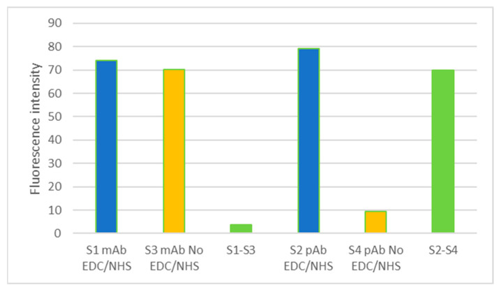
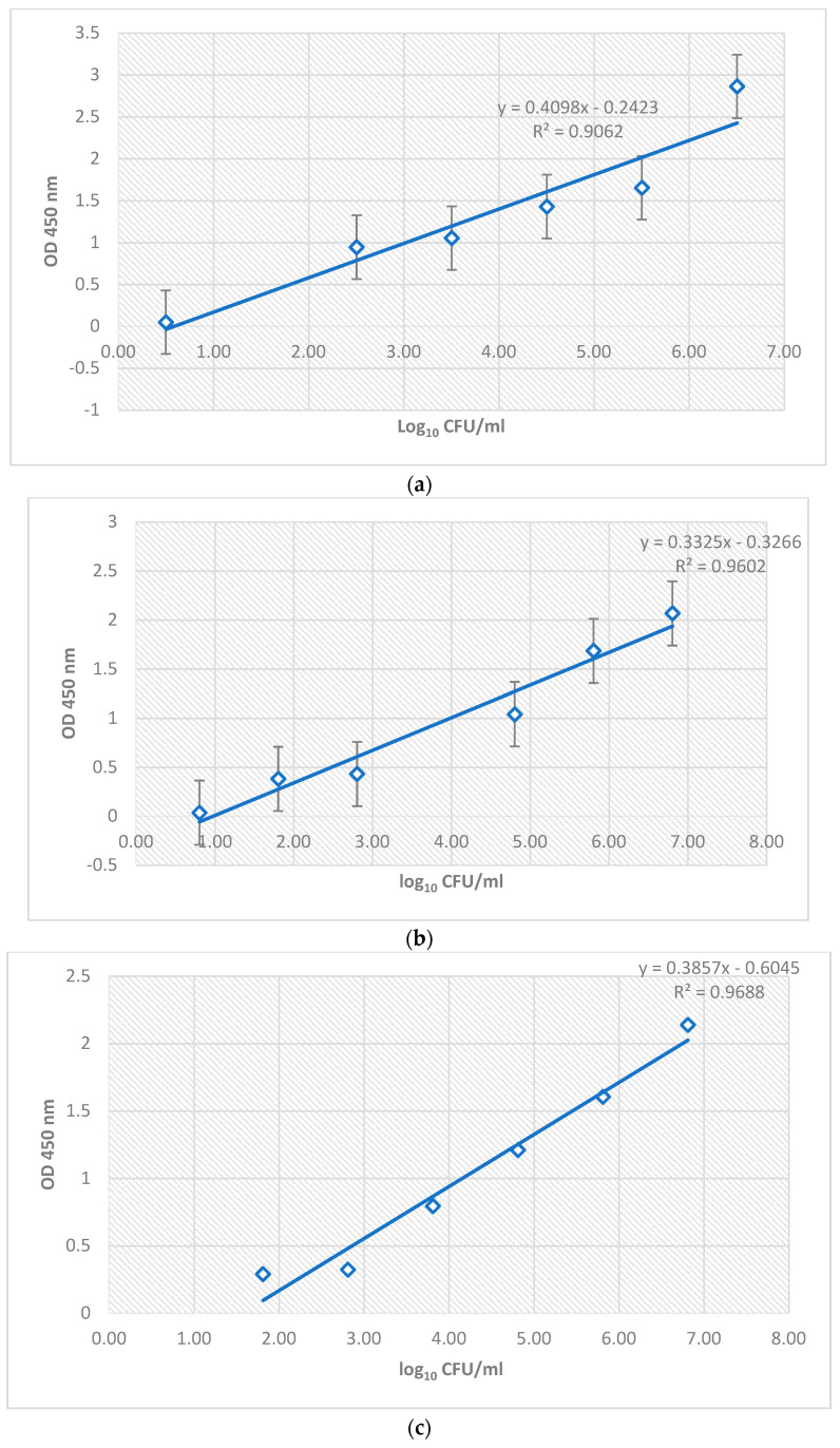
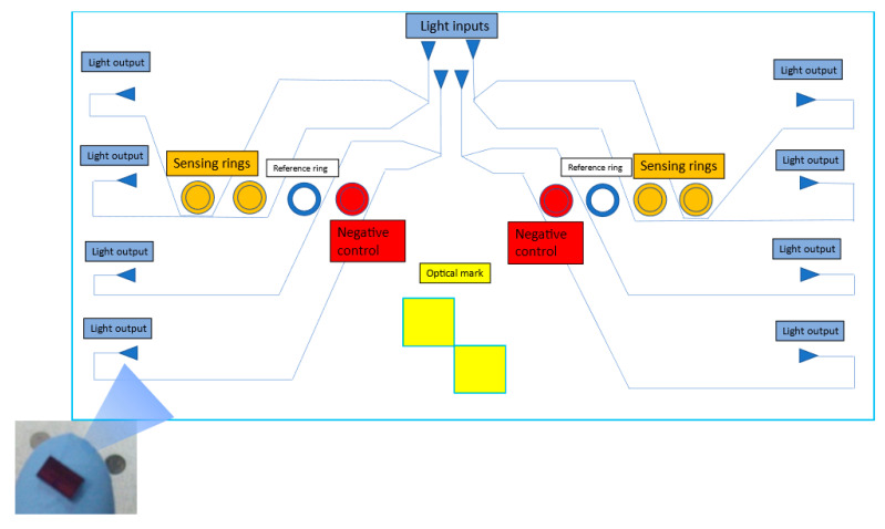
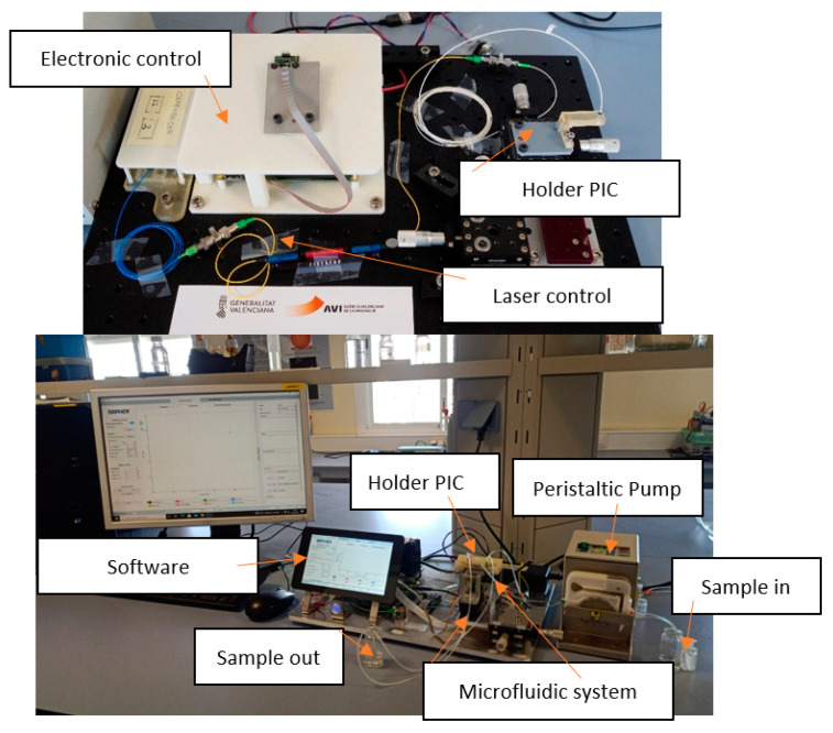
The present work describes an alternative method for detecting and identifying Listeria monocytogenes in food samples by developing a nanophotonic biosensor containing bioreceptors and optical transducers. The development of photonic sensors for the detection of pathogens in the food industry involves the implementation of procedures for selecting probes against the antigens of interest and the functionalization of the sensor surfaces on which the said bioreceptors are located. As a previous step to functionalizing the biosensor, an immobilization control of these antibodies on silicon nitride surfaces was carried out to check the effectiveness of in plane immobilization. On the one hand, it was observed that a Listeria monocytogenes-specific polyclonal antibody has a greater binding capacity to the antigen at a wide range of concentrations. A Listeria monocytogenes monoclonal antibody is more specific and has a greater binding capacity only at low concentrations. An assay for evaluating selected antibodies against particular antigens of Listeria monocytogenes bacteria was designed to determine the binding specificity of each probe using the indirect ELISA detection technique. In addition, a validation method was established against the reference method for many replicates belonging to different batches of meat-detectable samples, with a medium and pre-enrichment time that allowed optimal recovery of the target microorganism. Moreover, no cross-reactivity with other nontarget bacteria was observed. Thus, this system is a simple, highly sensitive, and accurate platform for L. monocytogenes detection.

Keywords: Listeria monocytogenes; bioreceptor; biosensor; food safety; nanophotonics; optical transducers.


Conflict of interest statement

The authors declare no conflict of interest.

Figures

Figure 1
Figure 1
Graphic representation of the array layout. (a) S1 and S2 activated surfaces with primary antibody immobilization (lines 1,2), along with a negative (line 3) and a positive (line 4) control test. (b) S1 activated surface with primary and secondary antibody anti-mouse immobilization. (c) S2 activated surface with primary and secondary antibody anti-rabbit immobilization. (d) S3 and S4 nonactivated surface with primary antibody immobilization (lines 1,2) and negative control test (line 3). (e) S3 nonactivated surface with primary and secondary antibody anti-mouse immobilization. (f) S4 nonactivated surface with primary and secondary antibody anti-rabbit immobilization.
Figure 2
Figure 2
Schematics of the overall steps of the manufacturing process of Si3N4 PICs.
Figure 3
Figure 3
Graphic representation of functionalization PIC process.
Figure 4
Figure 4
Images of silicon nitride surfaces after immobilization obtained by microarray fluorescence reader GenePix 4000B Axon Instruments. (a) The mAb activated surface S1 reacted with secondary anti-mouse antibody (line 1) and a serial dilution of an ELISA-positive control of L. monocytogenes (line 4). (b) The mAb nonactivated surface (S3) reacted insignificantly with the secondary anti-mouse antibody (line 1). (c) The pAb-activated surface S2 reacted with a secondary anti-rabbit antibody (line 2). (d) The pAb nonactivated surface (S4) reacted insignificantly with secondary anti-rabbit antibody (line 2).
Figure 5
Figure 5
Fluorescence intensity results obtained for each surface activated and not activated by EDC/NHS, on which the pAb and mAb antibodies against L. monocytogenes were immobilized.
Figure 6
Figure 6
Calibration curves were obtained after relating the absorbance values (OD 450 nm) versus bacterial concentration in log CFU/mL obtained for iELISA anti-Listeria polyclonal antibody, along with its equation and R2 value. (a) Calibration curve obtained for five increasing dilutions of wild strain M4 of L. monocytogenes against the anti-Listeria polyclonal antibody. (b) Calibration curve for five increasing dilutions of commercial strain L. monocytogenes CECT936 against the anti-Listeria polyclonal antibody. (c) Calibration curve for five increasing dilutions of the hamburger contaminated by L. monocytogenes against the anti-L. monocytogenes polyclonal antibody.
Figure 7
Figure 7
General schematic view of a PIC device. Detail of resonating structures (ring resonators with radius ~100 μm) used in the detection. In each circuit, eight rings are shown: four rings spread over two sensing areas that allow the detection of L. monocytogenes (sensing rings), two more rings (one per sensing area) to compensate for derivatives, and two additional rings as a negative control. These negative control rings bind to an analyte different from the objective, which will not be found in the matrix to be analyzed. In this case, the antibody deposited was an anti-fish antibody. The molecular receptors are represented in different colors to illustrate the multiple target capabilities of the technology. The input and respective light outputs in each channel are also indicated on the scheme, divided into four per detection channel or sensing area. The optical mark recognized by the developed hardware and software allows locating the PIC’s center, which helps the collimator focus the light on the input grating couplers. In addition, the optical mark allows presetting of the reading system. When the detection system recognizes it, it aligns the photodetector outputs with the laser so that the reading system captures the light from the output grating couplers.
Figure 8
Figure 8
Photonic setup reader and measurement equipment for PICs or photonic sensors.
Figure 9
Figure 9
Schematic representation of sensogram and acquisition of data obtained by functionalized photonic integrated circuit (PIC) surface measurement. The average flow of a contaminated sample from its initial washing is shown (to establish a baseline), along with the flow of the contaminated sample in which the binding of Listeria monocytogenes antigen to antibody fixed on the surface of the PIC (Detection) occurs, and the final washing process of the PIC surface to remove unwanted residues. The correlation between resonance in pm and time in s in a flow of a sample contaminated by Listeria monocytogenes is shown. On the right, the translation of the optical signal performed by the system is shown in the form of an internal calibration curve in which the resonance in pm is correlated with the concentration of bacteria obtained by subtracting the net difference obtained between the resonance obtained by the reference rings and the functionalized rings (rr1 func, rr2 func, rings functionalized with the antibody against the target bacteria, rr3 ref negative control reference, rr4 ref blank control reference).
Figure 10
Figure 10
Calibration curves against commercial/wild strains of L. monocytogenes and meat matrix naturally contaminated by L. monocytogenes. (A) Calibration curve of biosensor against L. monocytogenes CECT936. (B) Calibration curve of biosensor against L. monocytogenes M4. (C) Biosensor calibration curve against L. monocytogenes isolated from naturally contaminated deep-frozen hamburgers. Perform at least six determinations (preferably 10) of samples at the calculated breakpoint concentration to estimate the baseline or threshold spread s0. The corresponding CFU/mL of L. monocytogenes in the enrichment cultures was derived from Table 1. The resonance in pm is the unit of measurement obtained by the laboratory setup reader used after inserting and flowing the contaminated samples shown in Table 1. LoD (limit of detection) calculated as LoD = 3.3 s0. LoQ (limit of quantification) calculated as LoQ = 10 s0. ULOQ, upper limit of quantification.
Figure 10
Figure 10
Calibration curves against commercial/wild strains of L. monocytogenes and meat matrix naturally contaminated by L. monocytogenes. (A) Calibration curve of biosensor against L. monocytogenes CECT936. (B) Calibration curve of biosensor against L. monocytogenes M4. (C) Biosensor calibration curve against L. monocytogenes isolated from naturally contaminated deep-frozen hamburgers. Perform at least six determinations (preferably 10) of samples at the calculated breakpoint concentration to estimate the baseline or threshold spread s0. The corresponding CFU/mL of L. monocytogenes in the enrichment cultures was derived from Table 1. The resonance in pm is the unit of measurement obtained by the laboratory setup reader used after inserting and flowing the contaminated samples shown in Table 1. LoD (limit of detection) calculated as LoD = 3.3 s0. LoQ (limit of quantification) calculated as LoQ = 10 s0. ULOQ, upper limit of quantification.

Similar articles

Cited by

References

    1. Schuchat A., Swaminathan B., Broome C.V. Epidemiology of human listeriosis. J. Clin. Microbiol. 1991;4:169–183. doi: 10.1128/CMR.4.2.169. - DOI - PMC - PubMed
    1. Berrada H., Soriano J., Picó Y., Mañes J. Quantification of Listeria monocytogenes in salads by real time quantitative PCR. Int. J. Food Microbiol. 2006;107:202–206. doi: 10.1016/j.ijfoodmicro.2005.07.006. - DOI - PubMed
    1. Radhakrishnan R., Hashem A., Abd_Allah E.F. Bacillus: A Biological Tool for Crop Improvement through Bio-Molecular Changes in Adverse Environments. Front. Physiol. 2017;8:667. doi: 10.3389/fphys.2017.00667. - DOI - PMC - PubMed
    1. Gallagher L., Ebel E.D., Kause J.R. Draft FSIS Risk Assessment for Listeria, Ready-to-eat Meat and Poultry Products. Food Safety and Inspection Service; Washington, DC, USA: 2003.
    1. Commission Regulation (EC) No 2073/2005 of 15 November 2005 on microbiological criteria for foodstuffs. Off. J. Eur. Union. 2005;L338:1–26.