

MRI sequence optimisation methods to identify cranial nerve course for radiotherapy planning
- PMID: 37421243
- PMCID: PMC10715361
- DOI: 10.1002/jmrs.700
MRI sequence optimisation methods to identify cranial nerve course for radiotherapy planning
Abstract
Introduction: Magnetic resonance imaging (MRI) is being increasingly used to improve radiation therapy planning by allowing visualisation of organs at risk that cannot be well-defined on computed tomography (CT). Diagnostic sequences are increasingly being adapted for radiation therapy planning, such as the use of heavily T2-weighted 3D SPACE (Sampling Perfection with Application optimised Contrasts using different flip angle Evolution) sequence for cranial nerve identification in head and neck tumour treatment planning.
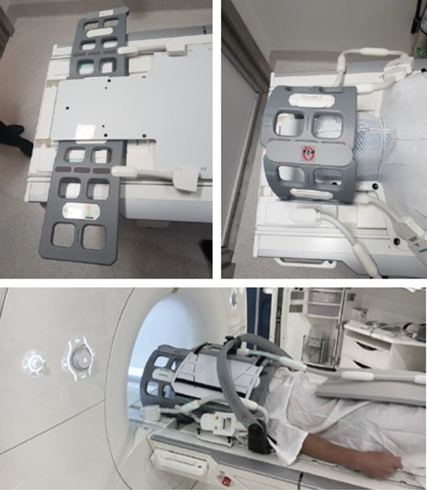
Methods: A 3D isotropic T2 SPACE sequence used for cranial nerve identification was adapted for radiation therapy purposes. Distortion was minimised using a spin-echo-based sequence, 3D distortion correction, isocentre scanning and an increased readout bandwidth. Radiation therapy positioning was accounted for by utilising two small flex, 4-channel coils. The protocol was validated for cranial nerve identification in clinical applications and distortion minimisation using an MRI QA phantom.
Results: Normal anatomy of the cranial nerves CI-CIX, were presented, along with a selection of clinical applications and abnormal anatomy. The usefulness of cranial nerve identification is discussed for several case studies, particularly in proximity to tumours extending into the base of skull region. In-house testing validated that higher bandwidths of 600 Hz resulted in minimal displacement well below 1 mm.
Conclusion: The use of MRI for radiation therapy planning allows for greater individualisation and prediction of patient outcomes. Dose reduction to cranial nerves can decrease late side effects such as cranial neuropathy. In addition to current applications, future directions include further applications of this technology for radiation therapy treatments.
Keywords: Magnetic resonance imaging; radiotherapy; radiotherapy planning, computer-assisted; radiotherapy, image-guided.
© 2023 The Authors. Journal of Medical Radiation Sciences published by John Wiley & Sons Australia, Ltd on behalf of Australian Society of Medical Imaging and Radiation Therapy and New Zealand Institute of Medical Radiation Technology.
Conflict of interest statement
The authors declare that the research was conducted in the absence of any commercial or financial relationships that could be construed as a potential conflict of interest.
Figures


Similar articles
-
ANATOMICAL STUDY OF CRANIAL NERVE EMERGENCE AND SKULL FORAMINA IN THE HORSE USING MAGNETIC RESONANCE IMAGING AND COMPUTED TOMOGRAPHY.Vet Radiol Ultrasound. 2015 Jul-Aug;56(4):391-7. doi: 10.1111/vru.12256. Epub 2015 Apr 2. Vet Radiol Ultrasound. 2015. PMID: 25832323
-
Quality comparison between three-dimensional T2-weighted SPACE and two-dimensional T2-weighted turbo spin echo magnetic resonance images for the brachytherapy planning evaluation of prostate and periprostatic anatomy.Brachytherapy. 2020 Jul-Aug;19(4):484-490. doi: 10.1016/j.brachy.2020.04.001. Epub 2020 May 10. Brachytherapy. 2020. PMID: 32402544 Free PMC article.
-
MRI distortion: considerations for MRI based radiotherapy treatment planning.Australas Phys Eng Sci Med. 2014 Mar;37(1):103-13. doi: 10.1007/s13246-014-0252-2. Epub 2014 Feb 12. Australas Phys Eng Sci Med. 2014. PMID: 24519001
-
[Anatomy of the skull base and the cranial nerves in slice imaging].Radiologe. 2009 Jul;49(7):584-97. doi: 10.1007/s00117-008-1800-0. Radiologe. 2009. PMID: 19506829 Review. German.
-
Minimizing magnetic resonance image geometric distortion at 7 Tesla for frameless presurgical planning using skin-adhered fiducials.Med Phys. 2023 Feb;50(2):694-701. doi: 10.1002/mp.16035. Epub 2022 Nov 12. Med Phys. 2023. PMID: 36301228 Review.
Cited by
-
Defining failure patterns and dynamics in locally advanced pharyngeal and laryngeal SCC following radiotherapy: Real-World Insights in the modern Era!Clin Transl Radiat Oncol. 2025 Aug 7;55:101026. doi: 10.1016/j.ctro.2025.101026. eCollection 2025 Nov. Clin Transl Radiat Oncol. 2025. PMID: 40821397 Free PMC article.
References
-
- Yousry I, Camelio S, Schmid UD, et al. Visualization of cranial nerves I‐XII: value of 3D CISS and T2‐weighted FSE sequences. Eur Radiol 2000; 10: 1061–7. - PubMed
-
- Fischbach F, Muller M, Bruhn H. High‐resolution depiction of the cranial nerves in the posterior fossa (N III‐N XII) with 2D fast spin echo and 3D gradient echo sequences at 3.0 T. Clin Imaging 2009; 33: 169–74. - PubMed
-
- Ors S, Inci E, Turkay R, Kokurcan A, Hocaoglu E. Retrospective comparison of three‐dimensional imaging sequences in the visualization of posterior fossa cranial nerves. Eur J Radiol 2017; 97: 65–70. - PubMed
MeSH terms
LinkOut - more resources
Full Text Sources
Medical
