. 2023 Jul 18;12(14):e028565.
doi: 10.1161/JAHA.122.028565. Epub 2023 Jul 8.

Individual Assessment of Perioperative Brain Growth Trajectories in Infants With Congenital Heart Disease: Correlation With Clinical and Surgical Risk Factors

Affiliations

Individual Assessment of Perioperative Brain Growth Trajectories in Infants With Congenital Heart Disease: Correlation With Clinical and Surgical Risk Factors

Daniel Cromb et al. J Am Heart Assoc. .

Abstract

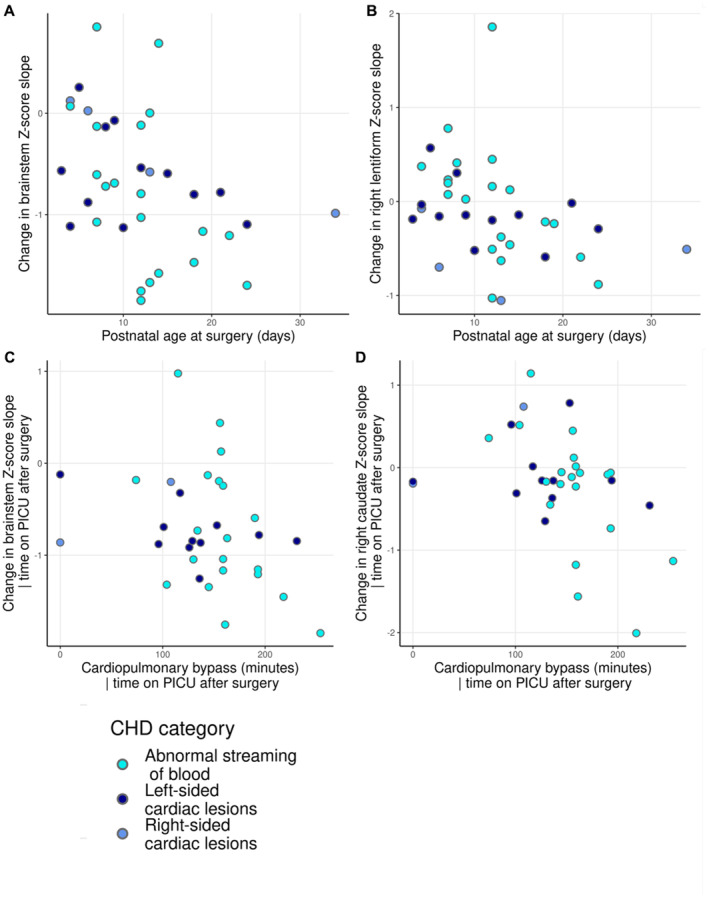
Background Infants with congenital heart disease (CHD) are at risk of neurodevelopmental impairments, which may be associated with impaired brain growth. We characterized how perioperative brain growth in infants with CHD deviates from typical trajectories and assessed the relationship between individualized perioperative brain growth and clinical risk factors. Methods and Results A total of 36 infants with CHD underwent preoperative and postoperative brain magnetic resonance imaging. Regional brain volumes were extracted. Normative volumetric development curves were generated using data from 219 healthy infants. Z-scores, representing the degree of positive or negative deviation from the normative mean for age and sex, were calculated for regional brain volumes from each infant with CHD before and after surgery. The degree of Z-score change was correlated with clinical risk factors. Perioperative growth was impaired across the brain, and it was associated with longer postoperative intensive care stay (false discovery rate P<0.05). Higher preoperative creatinine levels were associated with impaired brainstem, caudate nuclei, and right thalamus growth (all false discovery rate P=0.033). Older postnatal age at surgery was associated with impaired brainstem and right lentiform growth (both false discovery rate P=0.042). Longer cardiopulmonary bypass duration was associated with impaired brainstem and right caudate growth (false discovery rate P<0.027). Conclusions Infants with CHD can have impaired brain growth in the immediate postoperative period, the degree of which associates with postoperative intensive care duration. Brainstem growth appears particularly vulnerable to perioperative clinical course, whereas impaired deep gray matter growth was associated with multiple clinical risk factors, possibly reflecting vulnerability of these regions to short- and long-term hypoxic injury.

Keywords: brain volumes; congenital heart disease; surgery.


Figures

Figure 1
Figure 1. Example 3D reconstruction of an acquired brain MRI image and regional segmentations.
A 3D reconstruction (top) of the MRI‐derived regional brain segmentations with CSF (semitransparent), cortex, brainstem, and cerebellum visible. Axial (bottom left), coronal (bottom middle), and sagittal (bottom right) views showing MRI‐derived regional brain segmentations, including CSF (green), cortical gray matter (yellow), white matter (red), ventricles (blue), and deep gray matter structures (caudate nuclei, thalami, subthalamic nuclei, and lentiform nuclei). 3D indicates 3‐dimensional; CSF, cerebrospinal fluid; and MRI, magnetic resonance imaging.
Figure 2
Figure 2. Preoperative and postoperative volumetric brain development in neonates with CHD.
Plots showing preoperative and postoperative volumetric brain development in neonates with CHD. Shaded areas represent 1, 2, and 3 SDs from the normative model mean. Results are shown separately for female and male infants. CHD indicates congenital heart disease; CSF, cerebrospinal fluid.
Figure 3
Figure 3. Association between age at surgery and change in regional brain volume slopes preoperatively to postoperatively.
Plots to show the association between age at surgery and change in regional volume slope preoperatively to postoperatively in brainstem (A) and right caudate nucleus (B); and the relationship between duration of cardiopulmonary bypass and change in slope in brainstem (C) and right caudate nucleus (D), correcting for time on PICU postoperatively. CHD indicates congenital heart disease; PICU, pediatric intensive care unit.
Figure 4
Figure 4. Relationship between time on PICU postoperatively and change in regional brain volume slopes preoperatively to postoperatively.
Plots to show the relationship between time on PICU postoperatively and change in regional volume slope preoperatively to postoperatively for total tissue volume (A), cortical gray matter (B), brainstem (C), cerebellum (D), and left thalamus (E). CHD indicates congenital heart disease; and PICU, pediatric intensive care unit.
Figure 5
Figure 5. Relationship between postoperative renal replacement therapy and Z‐score slope change in cortical gray matter, cerebellum, and left thalamus volumes.
Plots to show the Z‐score slope change preoperatively to postoperatively in brainstem (A), cerebellum (B), cortical gray matter (C), left thalamus (D), and total brain tissue volume (E) for infants grouped according to the need for postoperative renal replacement therapy. CHD indicates congenital heart disease.
Figure 6
Figure 6. Relationship between preoperative serum creatinine levels and change in regional brain volume slopes preoperatively to postoperatively.
Plots to show the relationship between preoperative serum creatinine and change in regional volume slope preoperatively to postoperatively for brainstem (A), right thalamus (B), left caudate (C), and right caudate (D). CHD indicates congenital heart disease.

Similar articles

Cited by

References

    1. EUROCAT . European Commission. Prevalence Tables–Full Member Registries from 2012–2020. Accessed May 22, 2023. https://eu‐rd‐platform.jrc.ec.europa.eu/eurocat/eurocat‐data/prevalence_en.
    1. Marelli A, Miller SP, Marino BS, Jefferson AL, Newburger JW. The brain in congenital heart disease across the lifespan: the cumulative burden of injury. Circulation. 2016;133:1951–1962. doi: 10.1161/CIRCULATIONAHA.115.019881 - DOI - PMC - PubMed
    1. Latal B. Neurodevelopmental outcomes of the child with congenital heart disease. Clin Perinatol. 2016;43:173–185. doi: 10.1016/j.clp.2015.11.012 - DOI - PubMed
    1. Peyvandi S, Latal B, Miller SP, McQuillen PS. The neonatal brain in critical congenital heart disease: insights and future directions. NeuroImage. 2019;185:776–782. doi: 10.1016/j.neuroimage.2018.05.045 - DOI - PubMed
    1. Bonthrone AF, Dimitrova R, Chew A, Kelly CJ, Cordero‐Grande L, Carney O, Egloff A, Hughes E, Vecchiato K, Simpson J, et al. Individualized brain development and cognitive outcome in infants with congenital heart disease. Brain Commun. 2021;3:3. doi: 10.1093/braincomms/fcab046 - DOI - PMC - PubMed

Publication types

MeSH terms