. 2023 Aug 3;186(16):3350-3367.e19.
doi: 10.1016/j.cell.2023.06.004. Epub 2023 Jul 7.

Development of an α-synuclein positron emission tomography tracer for imaging synucleinopathies

Affiliations

Development of an α-synuclein positron emission tomography tracer for imaging synucleinopathies

Jie Xiang et al. Cell. .

Abstract

Synucleinopathies are characterized by the accumulation of α-synuclein (α-Syn) aggregates in the brain. Positron emission tomography (PET) imaging of synucleinopathies requires radiopharmaceuticals that selectively bind α-Syn deposits. We report the identification of a brain permeable and rapid washout PET tracer [18F]-F0502B, which shows high binding affinity for α-Syn, but not for Aβ or Tau fibrils, and preferential binding to α-Syn aggregates in the brain sections. Employing several cycles of counter screenings with in vitro fibrils, intraneuronal aggregates, and neurodegenerative disease brain sections from several mice models and human subjects, [18F]-F0502B images α-Syn deposits in the brains of mouse and non-human primate PD models. We further determined the atomic structure of the α-Syn fibril-F0502B complex by cryo-EM and revealed parallel diagonal stacking of F0502B on the fibril surface through an intense noncovalent bonding network via inter-ligand interactions. Therefore, [18F]-F0502B is a promising lead compound for imaging aggregated α-Syn in synucleinopathies.

Keywords: Lewy body; PET tracer; cryoelectron microscopy; protein aggregation; synucleinopathies.


Conflict of interest statement

Declaration of interests K.Y. is a co-founder of Shanghai Braegen Pharmaceuticals, Inc. that licensed the PET tracer patent from Emory University. No data are sponsored by the company.

Figures
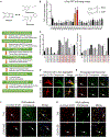
Figure 1.
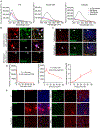
Figure 1.. Drug design strategy and screening of potential imaging agents for synucleinopathies
For a Figure360 author presentation of Figure 1, see https://doi.org/10.1016/j.cell.2023.06.004 (A) The screening strategy of α-Syn PET tracers. (B) Screening candidate chemicals using spectrophotometric assay with α-Syn PFFs. The emission wavelength was determined based on the excitation wavelength of each chemical. The PFF/monomer binding ratio was standardized by the ThT binding fold change. Error bars represent the mean ± SEM. Data are representative of 7 independent experiments. (C) The second round of screening of candidate chemicals with the α-Syn PFF versus Aβ/Tau PFF binding ratios. Error bars represent the mean ± SEM. Data are representative of 5 independent experiments. (D) Confocal fluorescence images of neurons containing α-Syn aggregates. The neurons were infected with AAV-α-Syn at days in vitro (DIV) 7 and transduced with α-Syn PFFs at DIV 12. The samples were immunostained with anti-p-α-Syn S129 (pseudocolors were converted to red) together with ThT or EU05-02B (0.1 μM). Scale bars, 10 μm. (E) Double staining of α-Syn aggregates in AAV-α-Syn A53T-injected mouse SN with pS129 antibody and EU05-02B or ThS. EU05-02B sensitively captured α-Syn aggregates in the SN (arrows). Scale bars, 20 μm. (F) Double staining of α-Syn aggregates in the cortex of DLB patients with pS129 antibody and EU05-02B or ThS. Scale bars, 20 μm. (G) Double staining of α-Syn aggregates in the SN of MSA patients with pS129 antibody and EU05-02B or ThS. Scale bars, 20 μm. See also Figures S1 and S2.
Figure 2.
Figure 2.. F0502B selectively binds to α-Syn aggregates in vitro and ex vivo
(A) The binding of ThT, EU05-02B, and F0502B to α-Syn, Aβ, and Tau PFFs. ThT binding with the same PFF concentration showed a binding peak at 485 nm. EU05-02B and F0502B showed stronger binding affinity with α-Syn PFFs than with Aβ and Tau PFFs. (B and C) Characterization of EU05-02B selectivity for aggregates in cultured neurons, mouse SN region expressing α-Syn A53T, and brain slices from DLB (cortex) and MSA (SN) patients (arrows show co-localization signals). Scale bars, 20 μm. (D) The levels of F0502B in the plasma and brain homogenates of male ICR mice that received i.v. injection of 5 mg/kg F0502B. The B/P ratio was determined at different time points. Error bars represent the mean ± SD. Data are representative of 3 mice at every time point. (E) Staining of inclusions in WT, 5xFAD, Tau P301S, 3xTg (cortex), and AAV-α-Syn A53T-injected mice (SN) intravenously administered F0502B (2 mg/kg). The tissues were prepared at 2 h after tracer administration. Scale bars, 20 μm. See also Figures S3 and S4.
Figure 3.
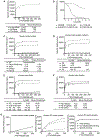
Figure 3.. [18F]-F0502B binding affinity and selectivity to different protein aggregates in vitro
(A) Quantification of binding affinities. The binding affinities of [18F]-F0502B to α-Syn, Aβ, or Tau PFFs were determined in saturation binding studies. Data points represent the mean ± SD. Similar results were obtained in more than three independent experiments. (B) [18F]-F0502B competitive binding assays with α-Syn PFFs. Data points represent the mean ± SD. (C) F0502B saturation binding assays with unfractionated AD/PD/control mouse brain homogenates. The α-Syn aggregates in SNCA transgenic mice treated with rotenone or AAV-α-Syn A53T-injected mice displayed strong binding activities with a Kd of 5.75 nM. Data points represent the mean ± SD. (D and E) F0502B saturation binding assays with unfractionated human PD/DLB/AD/control brain homogenates and insoluble protein fractions from human PD/DLB/AD/brain samples. Both PD and DLB brain homogenates and insoluble fractions exhibited robust binding activities with Kd values of 4.26/9.79 and 3.68/6.23 nM, respectively. Data points represent the mean ± SD. (F) F0502B saturation binding assays of unfractionated human PD brain homogenates from different brain areas. Bmax (fmol/nmol) = CPM/(2.22 * 1012 dpm/Ci * 0.75 cpm/dpm *2,000 Ci/mmol *10−12 mmol/fmol *0.5 nmol). All of the brain regions, including the midbrain, amygdala, and cortex, displayed prominent binding activities. Midbrain homogenates show the strongest binding. Error bars represent the mean ± SD. Data are representative of 3 independent experiments. (G) Total, specific, and non-displaceable binding (NDB) from saturation binding studies with [18F]-F0502B in the frontal cortex of healthy control, AD, and PD patients. NDB was defined by self-block at 100 × Kd concentration. Error bars represent the mean ± SD. Data are representative of 3 independent experiments. See also Figure S3F.
Figure 4.
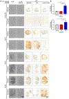
Figure 4.. [18F]-F0502B binding affinity and selectivity to different human brain slides
(A) Autoradiographic labeling of adjacent brain sections from patients with [18F]-F0502B (4 nM). Total binding of [18F]-F0502B was markedly abolished by the addition of non-radioactive F0502B (400 nM), except for the nonspecific (NS) labeling of white matter (left 2 lanes). Scale bars, 5 mm. The serial sections of each group were immunostained with antibodies against Aβ, p-Tau, and pS129 α-Syn (right 3 lanes). PD patient brains displayed specific [18F]-F0502B signals coupled with demonstrable pS129 staining. Scale bars, 5 mm. (B) Quantification of [18F]-F0502B binding in human brain slices. The total mean signal intensity for specific brain regions was normalized to the nonspecific signal intensity of the same region. Error bars represent the mean ± SEM. Statistical significance was determined using a two-way ANOVA followed by post hoc Bonferroni test for multiple group comparison. Data represent three independent experiments using 3 controls, 3 AD patients, and 3 PD patients in the midbrain and amygdala. *p < 0.05, **p < 0.01.
Figure 5.
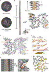
Figure 5.. Cryo-EM structure of the α-Syn fibril-F0502B complex
(A) Central slices of the 3D maps of the apo-α-Syn fibril (top) and α-Syn fibril-F0502B complex (bottom). The additional densities are indicated by arrows. (B) Cryo-EM 3D reconstruction density map of the α-Syn fibril-F0502B complex. Fibril parameters including the length of half pitch (180° helical turn), twist angle, and helical rise are indicated. Extra densities are colored in orange. (C) Cross-section view of the structural model of the α-Syn fibril-F0502B complex fitted in the density map. α-Syn is colored in gray. Ligand F0502B is colored in orange. (D) Enlarged view of the F0502B structural model superimposed with the ligand density (mesh). The dihedral angle between the phenol and benzothiazole-ethenyl planes is ~10°. The angle between the fluoro tail and the benzothiazole-ethenyl plane is ~50°. (E) Views of F0502B molecules stacking in the ligand-binding tunnel along the fibril axis (side view, left; top view, right). The surface of α-Syn fibril is shown and colored in gray. F0502B is shown in sticks and colored in orange. The angle between the F0502B body plane and fibril axis is ~50°. (F) Enlarged top view of the F0502B binding cavity with the surrounding residues shown in sticks and highlighted in yellow. See also Figure S5.
Figure 6.
Figure 6.. Interactions between F0502B and α-Syn fibril
(A) π-π stacking among F0502B molecules. The distance between neighboring aromatic rings of F0502B is ~3.7 Å. Detailed interaction distances are labeled. (B) Summary of the molecular interactions between F0502B (orange) and α-Syn fibril (black). Polar interactions (blue dashed lines) and non-dipolar interactions (red spiked arcs) are depicted with interaction distances. (C) T-shaped π-π interactions between F0502B and Tyr39 residues in three consecutive α-Syn molecules (i, i+1, i+2) along the fibril. Detailed interaction distances and the dihedral angles formed by π planes are labeled. (D) Cation-anion-π-π network between the phenol head of F0502B and α-Syn residues Lys80, Glu46, and Tyr39. Detailed interaction distances are labeled. (E) Hydrogen bonding and halogen bonding interactions between the phenol head of F0502B and α-Syn. The phenolic hydroxyl group of F0502B forms hydrogen bonds with the backbone amide of Gly41. The bromine of F0502B interacts with the sidechain hydroxyl group of Thr44 via halogen bonding. Detailed interaction distances are labeled. (F) Hydrophobic and halogen bonding interactions between the tail of F0502B and α-Syn. The methyl group of F0502B forms hydrophobic interactions with Val82. The fluorine of F0502B forms halogen bonding interactions with Glu83. Detailed interaction distances are labeled. See also Figure S6.
Figure 7.
Figure 7.. PET imaging of non-human primate PD models with [18F]-F0502B
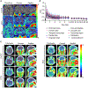
(A) [18F]-F0502B detects signals in the brains of macaques injected with α-Syn PFFs and AAV-α-Syn A53T, but not in the brains of control macaques. PET images in each row represent the standardized uptake value (percentage of injected dose per cubic centimeter [%ID/cc] × body weight) averaged over a 30 min period at 60 min post injection, co-registered to an MRI imaging template, from left to right showing axial, coronal and sagittal views. (B) Time course of the standardized uptake value (SUV) in control macaque brain regions after injection of [18F]-F0502B. Each point represents the average of the three subjects. Error bars represent the mean ± SEM. Data are representative of 3 independent experiments. (C) PET-CT images of α-Syn aggregates in the brains of control, α-Syn PFFs-, and AAV-α-Syn A53T-injected macaques. See also Figure S7 and Video S1.

Comment in

Similar articles

Cited by

References

    1. Dev KK, Hofele K, Barbieri S, Buchman VL, and van der Putten H (2003). Part II: alpha-synuclein and its molecular pathophysiological role in neurodegenerative disease. Neuropharmacology 45, 14–44. - PubMed
    1. Theillet FX, Binolfi A, Bekei B, Martorana A, Rose HM, Stuiver M, Verzini S, Lorenz D, van Rossum M, Goldfarb D, and Selenko P (2016). Structural disorder of monomeric alpha-synuclein persists in mammalian cells. Nature 530, 45–50. 10.1038/nature16531. - DOI - PubMed
    1. Olanow CW, and Brundin P (2013). Parkinson’s disease and alpha synuclein: is Parkinson’s disease a prion-like disorder? Mov. Disord 28, 31–40. 10.1002/mds.25373. - DOI - PubMed
    1. Jucker M, and Walker LC (2013). Self-propagation of pathogenic protein aggregates in neurodegenerative diseases. Nature 501, 45–51. 10.1038/nature12481. - DOI - PMC - PubMed
    1. Shahnawaz M, Mukherjee A, Pritzkow S, Mendez N, Rabadia P, Liu X, Hu B, Schmeichel A, Singer W, Wu G, et al. (2020). Discriminating alpha-synuclein strains in Parkinson’s disease and multiple system atrophy. Nature 578, 273–277. 10.1038/s41586-020-1984-7. - DOI - PMC - PubMed

Publication types

Substances

LinkOut - more resources