. 2023 Oct;193(10):1528-1547.
doi: 10.1016/j.ajpath.2023.06.009. Epub 2023 Jul 6.

Aberrations in Energetic Metabolism and Stress-Related Pathways Contribute to Pathophysiology in the Neb Conditional Knockout Mouse Model of Nemaline Myopathy

Affiliations

Aberrations in Energetic Metabolism and Stress-Related Pathways Contribute to Pathophysiology in the Neb Conditional Knockout Mouse Model of Nemaline Myopathy

Rebecca A Slick et al. Am J Pathol. 2023 Oct.

Abstract

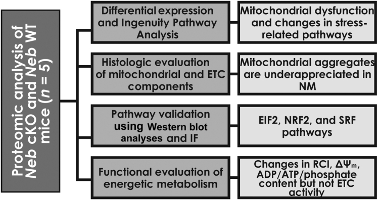
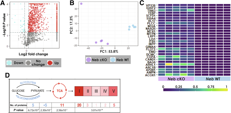
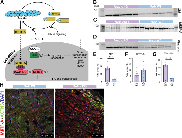
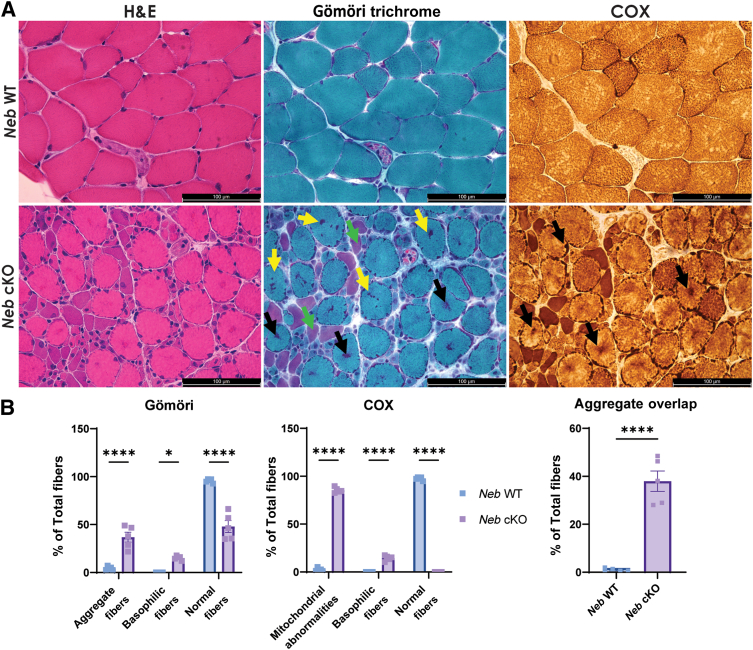
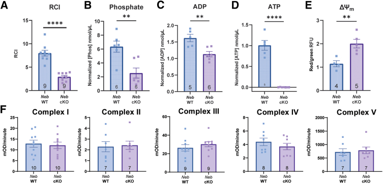
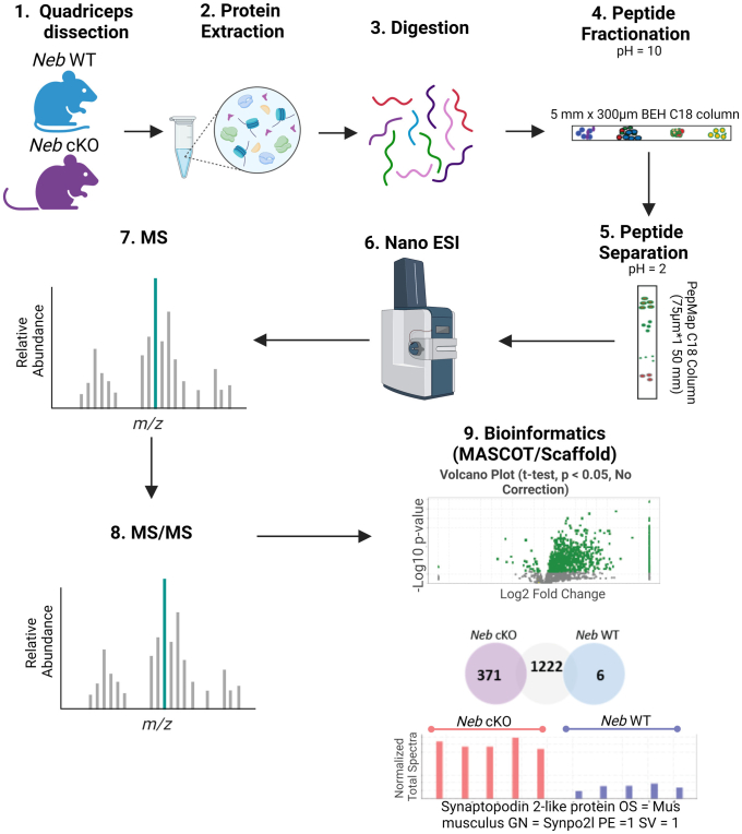
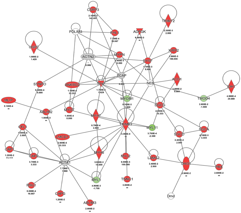
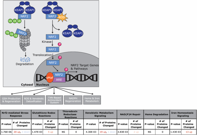
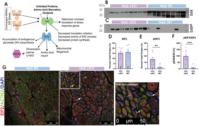
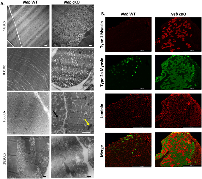
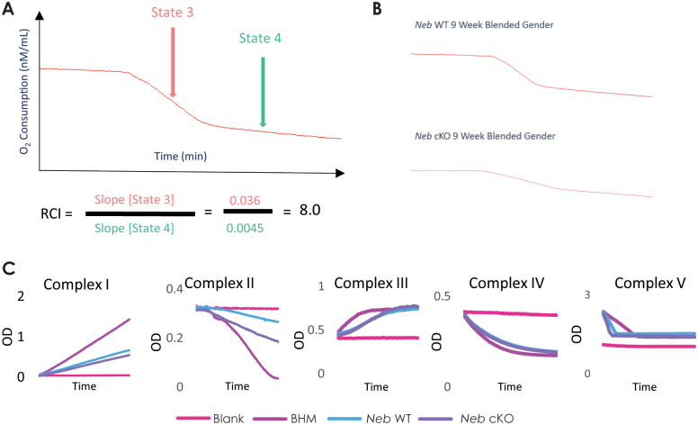
Nemaline myopathy (NM) is a genetically and clinically heterogeneous disease that is diagnosed on the basis of the presence of nemaline rods on skeletal muscle biopsy. Although NM has typically been classified by causative genes, disease severity or prognosis cannot be predicted. The common pathologic end point of nemaline rods (despite diverse genetic causes) and an unexplained range of muscle weakness suggest that shared secondary processes contribute to the pathogenesis of NM. We speculated that these processes could be identified through a proteome-wide interrogation using a mouse model of severe NM in combination with pathway validation and structural/functional analyses. A proteomic analysis was performed using skeletal muscle tissue from the Neb conditional knockout mouse model compared with its wild-type counterpart to identify pathophysiologically relevant biological processes that might impact disease severity or provide new treatment targets. A differential expression analysis and Ingenuity Pathway Core Analysis predicted perturbations in several cellular processes, including mitochondrial dysfunction and changes in energetic metabolism and stress-related pathways. Subsequent structural and functional studies demonstrated abnormal mitochondrial distribution, decreased mitochondrial respiratory function, an increase in mitochondrial transmembrane potential, and extremely low ATP content in Neb conditional knockout muscles relative to wild type. Overall, the findings of these studies support a role for severe mitochondrial dysfunction as a novel contributor to muscle weakness in NM.


Figures

Figure 1
Figure 1
Schematic of experimental design. Quadriceps muscles from Neb conditional knockout (cKO) and wild-type (WT) mice were isolated for proteomic and differential expression analyses. Ingenuity Pathway Analysis was used to determine perturbed pathways and determined changes in metabolic and stress-related pathways. These studies were followed up by pathway validation and structural and functional assays to determine their relevance to Neb nemaline myopathy (NM) pathophysiology. n = 5 Neb cKO and WT mice. ΔΨm, mitochondrial transmembrane potential; EIF2, eukaryotic translation initiation factor 2; ETC, electron transport chain; IF, immunofluorescence; NRF2, nuclear factor erythroid 2–related factor 2; RCI, respiratory control index; SRF, serum response factor.
Figure 2
Figure 2
Pathway analysis indicates metabolism and stress-related processes are perturbed in Neb nemaline myopathy. A: Volcano plot representing differentially expressed proteins in the Neb conditional knockout (cKO) quadriceps versus their wild-type (WT) counterpart. Dashed lines represent boundaries for differentially expressed proteins, defined by the parameters of a fold change of <0.5 or >1.5 with a P ≤ 0.05. B: Principal component (PC) analysis of Neb cKO versus WT differential expression data sets, where PC1 accounts for 53.8% of variability within the data set. C: Heat map representing the top 28 proteins detected solely in Neb cKO samples that contribute to data set variability. D: Protein expression in pathways related to energetic metabolism are altered in the Neb cKO mice. Increases in protein expression are depicted in shades of red, and decreases in protein expression are depicted in shades of blue. TCA, tricarboxylic acid.
Figure 3
Figure 3
Protein expression and localization related to the nuclear factor erythroid 2–related factor 2 (NRF2) pathway are altered in Neb nemaline myopathy. AF: NRF2 expression is statistically different than in wild-type (WT) mice. Kelch-like ECH-associated protein 1 (KEAP1) (P = 0.0018) and NAD(P)H dehydrogenase [quinone] 1 (NQO1) (P = 0.0030) expression levels are significantly increased in the Neb conditional knockout (cKO) animals versus their WT counterparts via Western blot analyses. All blots were quantified using total protein. G: Immunofluorescence shows NRF2, KEAP1, and NQO1 localization are altered in the Neb cKO mice. All three proteins appear to form their own aggregates as well as colocalize to nemaline rods shown using alpha-actinin 3 (ACTN3). (Blue arrows indicate individual aggregates; yellow arrows, colocalization with nemaline rods; and white arrow, nuclear localization.) ∗P < 0.05, ∗∗P < 0.01, and ∗∗∗P < 0.001. Scale bars = 50 μm (G).
Figure 4
Figure 4
Proteins related to the serum response factor (SRF) pathway are altered in expression or localization in the Neb conditional knockout (cKO) mice. A: In skeletal muscle, RhoA signaling in combination with increased expression of striated muscle activator of rho signaling (STARS) are known to activate SRF target gene transcription by enhancing actin polymerization. STARS is enriched in skeletal muscle and prominently localized to the z-disk. STARS has been postulated to be a skeletal muscle mechanosensor that activates downstream SRF signaling. In addition, eukaryotic translation initiation factor 2 (EIF2) is known to activate cyclic AMP-dependent transcription factor ATF-4 (ATF4), which promotes mitochondrial biogenesis via peroxisome proliferator-activated receptor gamma coactivator 1-alpha (PGC-1α). PGC-1α binding to the steroid hormone receptor ERR (ERR) promoter can also activate STARS transcription and, therefore, SRF signaling. B and E: SRF protein expression is significantly increased in the Neb cKO animals. C and F: Myocardin-related transcription factor A (MRTF-A) expression is not statistically different between Neb cKO and wild-type (WT) mice. D and G: Vinculin expression is significantly increased in the Neb cKO animals. H: MRTF-A is mislocalized in Neb cKO mice, forming its own aggregates in some areas and colocalizing to nemaline rods based on alpha-actinin 3 (ACTN3) expression in other areas. ∗∗∗P < 0.001, ∗∗∗∗P < 0.0001. Scale bars = 50 μm (H). MEF2, myocyte-specific enhancer 2; MyoD, myoblast determination protein 1.
Figure 5
Figure 5
Histologic changes in the Neb conditional knockout (cKO) mouse. A:Neb cKO mice have a decrease in fiber size and show significant nemaline pathology on Gömöri trichrome staining compared with wild-type (WT) animals. Neb cKO animals also show mitochondrial aggregation and changes in mitochondrial distribution on the cytochrome oxidase (COX) stain. B:Neb cKO animals show significant increases in the percentage of fibers with aggregates and percentage of basophilic fibers and a decrease in the percentage of normal fibers on Gömöri trichrome stain. The same pattern is seen on the COX stain. The Neb cKO animal shows a significant overlap in the number of aggregates seen in the Gömöri trichrome and COX stains. Examples of nemaline rods (yellow arrows), mitochondrial aggregates (black arrows), and basophilic fibers (green arrows) are highlighted. ∗P < 0.05, ∗∗∗∗P < 0.0001. Scale bars = 100 μm (A). H&E, hematoxylin and eosin.
Figure 6
Figure 6
Mitochondrial phenotype in the Neb conditional knockout (cKO) mouse. A:Neb cKO animals have a significant decrease in respiratory control index (RCI) values compared with Neb wild-type (WT) animals. BE: The Neb cKO animals have significant changes in the amount of phosphate (B), ADP (C), and ATP (D) as well as an increase of the red/green ratio of JC-1 (5,5,6,6′-tetrachloro-1,1′,3,3′ tetraethylbenzimi-dazoylcarbocyanine iodide), indicating a significant increase in transmembrane potential (E) compared with WT animals. F: The Neb cKO mouse shows no changes in any electron transport chain enzyme activity compared with WT animals. ∗∗P < 0.01, ∗∗∗∗P < 0.0001. ΔΨm, mitochondrial transmembrane potential; mOD, milli-optical density.
Supplemental Figure S1
Workflow of proteomics analysis. Proteins extracted from Neb wild-type (WT) and Neb conditional knockout (cKO) mice were digested with trypsin. Peptides were prefractionation on an online C18 fractionation column using high pH mobile phase, and each of the fractions was then separated on a C18 separation column and analyzed by tandem mass spectrometry (MS/MS) on a Thermo Fusion instrument. The data were searched on MASCOT against the most recent mouse database and then compiled in Scaffold for comparison and statistical analysis. Figure was generated with BioRender.com (Toronto, ON, Canada). ESI, electrospray ionization; GN, gene; OS, species; PE, protein existence; SV, sequence version.
Supplemental Figure S2
Proteins known to aggregate in nemaline rods are to be overexpressed in Neb conditional knockout muscle tissue The network shown here was generated using Qiagen Ingenuity Pathway Analysis. The network, built using common proteins found in nemaline rods, shows most proteins connected by protein-protein interactions have an increase in expression. Red represents a significant increase in protein expression, whereas green represents a significant decrease in protein expression.
Supplemental Figure S3
Nuclear factor erythroid 2–related factor 2 (NRF2) is likely activated in Neb conditional knockout (cKO) mice. Under normal conditions, KEAP1 inhibits NRF2, marking it for proteasomal degradation. However, when oxidative stress increases, KEAP1 is modified, allowing NRF2 to translocate to the nucleus and bind to the antioxidant response element (ARE), which is present on >500 genes. NRF2 activation results in transcriptional activation of antioxidant and metabolic genes to aid in relieving increased oxidative stress (pathway). NRF2 activation causes a variety of downstream effects, including GSH production, reactive oxygen species (ROS) detoxification, TXN antioxidation, and NADPH regeneration. These pathways are predicted to be activated to varying levels based on changes in protein expression in the Neb cKO mouse model versus its wild-type counterpart (Table). Red represents an increase in expression, whereas blue represents a decrease in expression, with the relative amount of change indicated by the shade of the colors. GSH, glutathione; KEAP1, kelch-like ECH-associated protein 1; Redox, oxidation-reduction; sMAF, small transcription factor Maf; TXN, thioredoxin.
Supplemental Figure S4
Neb nemaline myopathy results in perturbations in eukaryotic translation initiation factor 2 (EIF2) expression and localization. A: EIF2 is known to be an important component of the integrated stress response. When there are unfolded proteins, amino acid starvation, or excessive oxidants, EIF2 can be activated by being phosphorylated by PERK or GCN2, shown as EIF2AK4 and EIF2AK3. Activated EIF2 decreases global translation initiation and protein synthesis while selectively increasing translation of stress response genes. It also acts upstream of ATF4, which is important for amino acid import, mitochondrial biogenesis, and nuclear factor erythroid 2–related factor 2(NRF2) activation in response to oxidative stress. BE: Total EIF2 expression is not changed, whereas phosphorylated EIF2 (pEIF2) expression is increased in the Neb conditional knockout (cKO) animals via Western blot analysis. F: pEIF2 compared with total EIF2 expression is significantly greater in the Neb cKO animals. G: Immunofluorescence shows EIF2 is mislocalized and forms aggregates in Neb cKO muscle tissue, some of which colocalize with nemaline rods represented by ACTN3. (Blue arrows indicate individual aggregates; yellow arrows, co-localization with nemaline rods; and white arrows, nuclear localization.) ∗∗P < 0.01, ∗∗∗P < 0.001. Scale bars = 50 μm (G). ACTN3, alpha-actinin 3; ATF4, AMP-dependent transcription factor ATF-4; ER, endoplasmic reticulum; GCN2, EIF2-alpha kinase; GPx, glutathione peroxidase; PERK, proline-rich receptor-like protein kinase; WT, wild type.
Supplemental Figure S5
Representative image of total protein blot for Neb conditional knockout (cKO) and Neb wild-type (WT) quadriceps tissue. In most cases, there was more total protein in Neb WT lanes than Neb cKO lanes, although protein concentration was quantified and normalized before loading.
Supplemental Figure S6
Neb conditional knockout (cKO) animals show significant nemaline pathology on electron microscopy (EM) and an increase in oxidative fibers by fiber typing of triceps tissue. A:Neb cKO animals show large aggregations of nemaline rods (yellow arrow) and mitochondrial aggregates (black arrow) on EM). B: Laminin was used to stain the sarcolemma of individual muscle cells. Staining shows an increase in type 1 and type 2a fiber types in the Neb cKO mouse. Scale bars: 2 μm (A, white bars); 500 μm (A, black bars, and B). WT, wild type.
Supplemental Figure S7
Mitochondrial respirometry in Neb conditional knockout (cKO) mouse. A and B: Example of oxygraph output and respiratory control index (RCI) calculation (A) with example tracing from an Neb wild-type (WT) and cKO animal (B). C: Sample tracings of each electron transport chain enzyme are shown. A blank well and a well containing bovine heart mitochondria (BHM) were run as a positive control for every assay to ensure the assay is working properly. The complex I assay shows an increase in absorbance over time as the dye is reduced in the presence of NAD+. Complex II pairs the reduction of ubiquinol by DCPIP to monitor the change from a blue solution to a colorless one. Complexes III and IV work by oxidizing cytochrome c to cause a change in absorbance over time. Complex V causes a change in absorbance by oxidizing NADH over time. DCPIP, 2,6-dichlorophenolindophenol.

Similar articles

Cited by

References

    1. North K.N., Laing N.G., Wallgren-Pettersson C. Nemaline myopathy: current concepts: the ENMC International Consortium and Nemaline Myopathy. J Med Genet. 1997;34:705–713. - PMC - PubMed
    1. Sewry C.A., Laitila J.M., Wallgren-Pettersson C. Nemaline myopathies: a current view. J Muscle Res Cell Motil. 2019;40:111–126. - PMC - PubMed
    1. Kondo E., Nishimura T., Kosho T., Inaba Y., Mitsuhashi S., Ishida T., Baba A., Koike K., Nishino I., Nonaka I., Furukawa T., Saito K. Recessive RYR1 mutations in a patient with severe congenital nemaline myopathy with ophthalomoplegia identified through massively parallel sequencing. Am J Med Genet A. 2012;158a:772–778. - PubMed
    1. Pelin K., Donner K., Holmberg M., Jungbluth H., Muntoni F., Wallgren-Pettersson C. Nebulin mutations in autosomal recessive nemaline myopathy: an update. Neuromuscul Disord. 2002;12:680–686. - PubMed
    1. Pelin K., Hilpela P., Donner K., Sewry C., Akkari P.A., Wilton S.D., Wattanasirichaigoon D., Bang M.L., Centner T., Hanefeld F., Odent S., Fardeau M., Urtizberea J.A., Muntoni F., Dubowitz V., Beggs A.H., Laing N.G., Labeit S., de la Chapelle A., Wallgren-Pettersson C. Mutations in the nebulin gene associated with autosomal recessive nemaline myopathy. Proc Natl Acad Sci U S A. 1999;96:2305–2310. - PMC - PubMed

Publication types

LinkOut - more resources