

Diagnostic yield of computed tomography after non-traumatic out-of-hospital cardiac arrest
- PMID: 37422167
- PMCID: PMC11527794
- DOI: 10.1016/j.resuscitation.2023.109898
Diagnostic yield of computed tomography after non-traumatic out-of-hospital cardiac arrest
Abstract
Aim: Determine the frequency with which computed tomography (CT) after out-of-hospital cardiac arrest (OHCA) identifies clinically important findings.
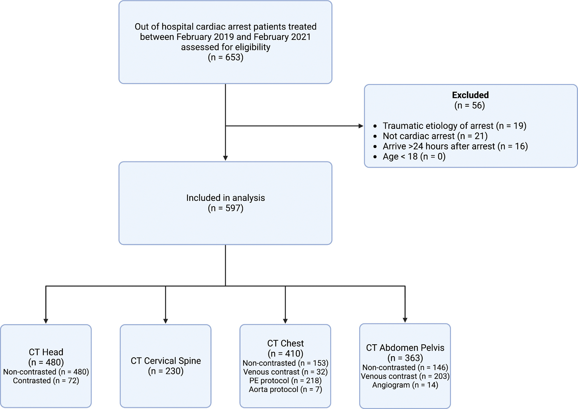
Methods: We included non-traumatic OHCA patients treated at a single center from February 2019 to February 2021. Clinical practice was to obtain CT head in comatose patients. Additionally, CT of the cervical spine, chest, abdomen, and pelvis were obtained if clinically indicated. We identified CT imaging obtained within 24 hours of emergency department (ED) arrival and summarized radiology findings. We used descriptive statistics to summarize population characteristics and imaging results, report their frequencies and, post hoc, compared time from ED arrival to catheterization between patients who did and did not undergo CT.
Results: We included 597 subjects, of which 491 (82.2%) had a CT obtained. Time to CT was 4.1 hours [2.8-5.7]. Most (n = 480, 80.4%) underwent CT head, of which 36 (7.5%) had intracranial hemorrhage and 161 (33.5%) had cerebral edema. Fewer subjects (230, 38.5%) underwent a cervical spine CT, and 4 (1.7%) had acute vertebral fractures. Most subjects (410, 68.7%) underwent a chest CT, and abdomen and pelvis CT (363, 60.8%). Chest CT abnormalities included rib or sternal fractures (227, 55.4%), pneumothorax (27, 6.6%), aspiration or pneumonia (309, 75.4%), mediastinal hematoma (18, 4.4%) and pulmonary embolism (6, 3.7%). Significant abdomen and pelvis findings were bowel ischemia (24, 6.6%) and solid organ laceration (7, 1.9%). Most subjects that had CT imaging deferred were awake and had shorter time to catheterization.
Conclusions: CT identifies clinically important pathology after OHCA.
Keywords: Cardiac arrest; Computed tomography; Heart arrest; Injuries.
Copyright © 2023 Elsevier B.V. All rights reserved.
Conflict of interest statement
Declaration of Competing Interest The authors declare that they have no known competing financial interests or personal relationships that could have appeared to influence the work reported in this paper.
References
-
- Tsao CW, Aday AW, Almarzooq ZI, et al. Heart Disease and Stroke Statistics-2023 Update: A Report From the American Heart Association. Circulation 2023. - PubMed
-
- Lee KY, So WZ, Ho JSY, et al. Prevalence of intracranial hemorrhage amongst patients presenting with out-of-hospital cardiac arrest: a systematic review and meta-analysis. Resuscitation 2022;176:136–49. - PubMed
-
- Paul M, Bougouin W, Legriel S, et al. Frequency, risk factors, and outcomes of non-occlusive mesenteric ischaemia after cardiac arrest. Resuscitation 2020;157:211–8. - PubMed
MeSH terms
Grants and funding
LinkOut - more resources
Full Text Sources
Medical
