

Pharmacokinetics, biodistribution and toxicology of novel cell-penetrating peptides
- PMID: 37422520
- PMCID: PMC10329699
- DOI: 10.1038/s41598-023-37280-0
Pharmacokinetics, biodistribution and toxicology of novel cell-penetrating peptides
Abstract
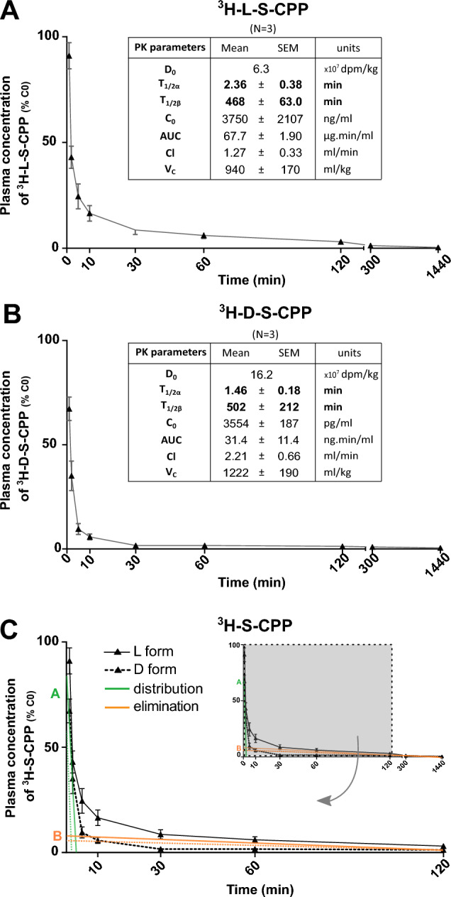
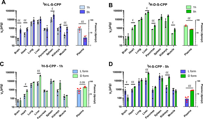
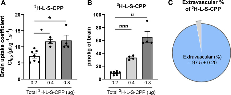
Cell-penetrating peptides (CPPs) have been used in basic and preclinical research in the past 30 years to facilitate drug delivery into target cells. However, translation toward the clinic has not been successful so far. Here, we studied the pharmacokinetic (PK) and biodistribution profiles of Shuttle cell-penetrating peptides (S-CPP) in rodents, combined or not with an immunoglobulin G (IgG) cargo. We compared two enantiomers of S-CPP that contain both a protein transduction domain and an endosomal escape domain, with previously shown capacity for cytoplasmic delivery. The plasma concentration versus time curve of both radiolabelled S-CPPs required a two-compartment PK analytical model, which showed a fast distribution phase (t1/2α ranging from 1.25 to 3 min) followed by a slower elimination phase (t1/2β ranging from 5 to 15 h) after intravenous injection. Cargo IgG combined to S-CPPs displayed longer elimination half-life, of up to 25 h. The fast decrease in plasma concentration of S-CPPs was associated with an accumulation in target organs assessed at 1 and 5 h post-injection, particularly in the liver. In addition, in situ cerebral perfusion (ISCP) of L-S-CPP yielded a brain uptake coefficient of 7.2 ± 1.1 µl g-1 s-1, consistent with penetration across the blood-brain barrier (BBB), without damaging its integrity in vivo. No sign of peripheral toxicity was detected either by examining hematologic and biochemical blood parameters, or by measuring cytokine levels in plasma. In conclusion, S-CPPs are promising non-toxic transport vectors for improved tissue distribution of drug cargos in vivo.
© 2023. The Author(s).
Conflict of interest statement
The authors declare no competing interests.
Figures


Similar articles
-
A real-time assay for cell-penetrating peptide-mediated delivery of molecular cargos.PLoS One. 2021 Sep 2;16(9):e0254468. doi: 10.1371/journal.pone.0254468. eCollection 2021. PLoS One. 2021. PMID: 34473728 Free PMC article.
-
Cell penetration: scope and limitations by the application of cell-penetrating peptides.J Pept Sci. 2014 Oct;20(10):760-84. doi: 10.1002/psc.2672. Epub 2014 Aug 11. J Pept Sci. 2014. PMID: 25112216 Review.
-
Plasma membrane depolarization reveals endosomal escape incapacity of cell-penetrating peptides.Eur J Pharm Biopharm. 2023 Mar;184:116-124. doi: 10.1016/j.ejpb.2023.01.019. Epub 2023 Jan 26. Eur J Pharm Biopharm. 2023. PMID: 36709921
-
Applications and Challenges for Use of Cell-Penetrating Peptides as Delivery Vectors for Peptide and Protein Cargos.Int J Mol Sci. 2016 Jan 30;17(2):185. doi: 10.3390/ijms17020185. Int J Mol Sci. 2016. PMID: 26840305 Free PMC article. Review.
-
New generation of cell-penetrating peptides: Functionality and potential clinical application.J Pept Sci. 2021 May;27(5):e3300. doi: 10.1002/psc.3300. Epub 2021 Feb 21. J Pept Sci. 2021. PMID: 33615648 Review.
Cited by
-
Antihypertensive, Anti-Inflammatory, and Antiangiogenic In Silico Activity of Lactoferrin-Derived Peptides of Equine Milk Hydrolysate.Biomedicines. 2024 Nov 27;12(12):2715. doi: 10.3390/biomedicines12122715. Biomedicines. 2024. PMID: 39767622 Free PMC article.
-
Antisense oligonucleotides and their applications in rare neurological diseases.Front Neurosci. 2024 Sep 23;18:1414658. doi: 10.3389/fnins.2024.1414658. eCollection 2024. Front Neurosci. 2024. PMID: 39376536 Free PMC article. Review.
-
cAmbly: A non-toxic cell-penetrating peptide derived from Amblyomin-X with targeted delivery to mitochondrial and cytoplasmic proteins.PLoS One. 2025 Mar 12;20(3):e0318119. doi: 10.1371/journal.pone.0318119. eCollection 2025. PLoS One. 2025. PMID: 40072951 Free PMC article.
-
Cell-penetrating peptides as facilitators of cargo-specific nanocarrier-based drug delivery.Nanoscale. 2025 Aug 26. doi: 10.1039/d5nr00617a. Online ahead of print. Nanoscale. 2025. PMID: 40856125 Free PMC article. Review.
-
Toxicity Studies of Cardiac-Targeting Peptide Reveal a Robust Safety Profile.Pharmaceutics. 2024 Jan 4;16(1):73. doi: 10.3390/pharmaceutics16010073. Pharmaceutics. 2024. PMID: 38258084 Free PMC article.
References
Publication types
MeSH terms
Substances
LinkOut - more resources
Full Text Sources
Miscellaneous
