

Is automatic cephalometric software using artificial intelligence better than orthodontist experts in landmark identification?
- PMID: 37422630
- PMCID: PMC10329795
- DOI: 10.1186/s12903-023-03188-4
Is automatic cephalometric software using artificial intelligence better than orthodontist experts in landmark identification?
Abstract
Background: To evaluate the techniques used for the automatic digitization of cephalograms using artificial intelligence algorithms, highlighting the strengths and weaknesses of each one and reviewing the percentage of success in localizing each cephalometric point.
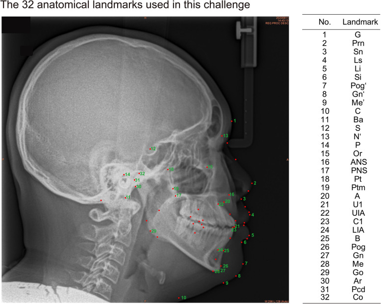
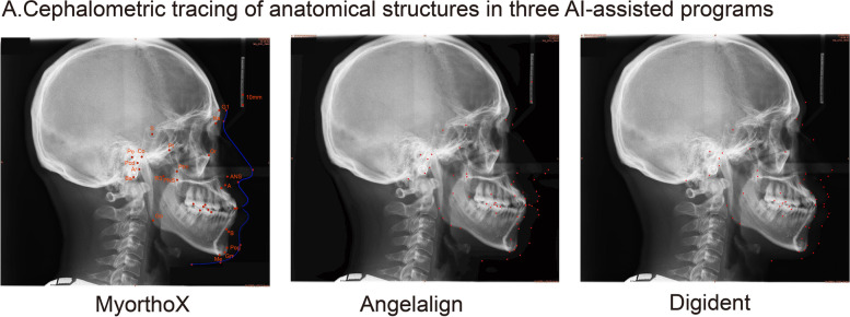
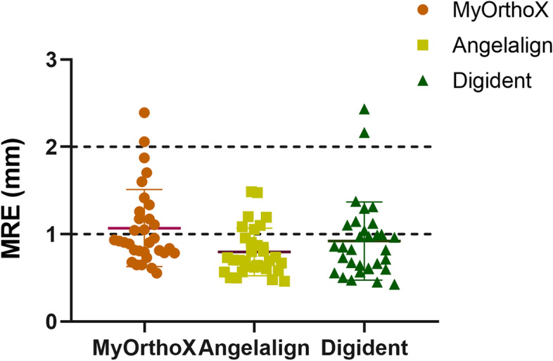
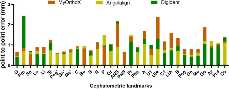
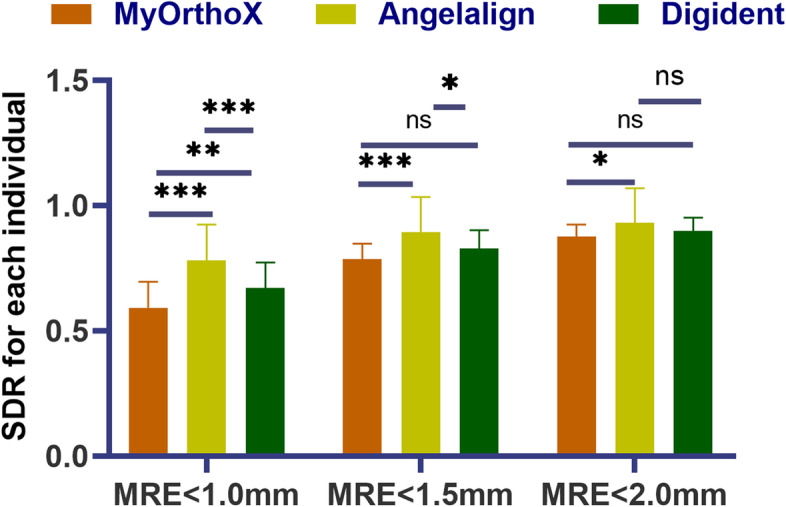
Methods: Lateral cephalograms were digitized and traced by three calibrated senior orthodontic residents with or without artificial intelligence (AI) assistance. The same radiographs of 43 patients were uploaded to AI-based machine learning programs MyOrthoX, Angelalign, and Digident. Image J was used to extract x- and y-coordinates for 32 cephalometric points: 11 soft tissue landmarks and 21 hard tissue landmarks. The mean radical errors (MRE) were assessed radical to the threshold of 1.0 mm,1.5 mm, and 2 mm to compare the successful detection rate (SDR). One-way ANOVA analysis at a significance level of P < .05 was used to compare MRE and SDR. The SPSS (IBM-vs. 27.0) and PRISM (GraphPad-vs.8.0.2) software were used for the data analysis.
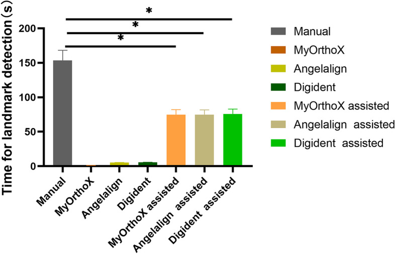
Results: Experimental results showed that three methods were able to achieve detection rates greater than 85% using the 2 mm precision threshold, which is the acceptable range in clinical practice. The Angelalign group even achieved a detection rate greater than 78.08% using the 1.0 mm threshold. A marked difference in time was found between the AI-assisted group and the manual group due to heterogeneity in the performance of techniques to detect the same landmark.
Conclusions: AI assistance may increase efficiency without compromising accuracy with cephalometric tracings in routine clinical practice and research settings.
Keywords: Artificial intelligence; Automatic digitization; Cephalometric tracings.
© 2023. The Author(s).
Conflict of interest statement
The authors declare no competing interests.
Figures
References
-
- Bilgir E, Bayrakdar IS, Celik O, Orhan K, Akkoca F, Saglam H, Odabas A, Aslan AF, Ozcetin C, Killi M, Rozylo-Kalinowska I. An artificial intelligence approach to automatic tooth detection and numbering in panoramic radiographs. BMC Med Imaging. 2021;21:124. doi: 10.1186/s12880-021-00656-7. - DOI - PMC - PubMed
Publication types
MeSH terms
LinkOut - more resources
Full Text Sources
Miscellaneous
