

Meta-analysis and open-source database for in vivo brain Magnetic Resonance spectroscopy in health and disease
- PMID: 37423487
- PMCID: PMC10561665
- DOI: 10.1016/j.ab.2023.115227
Meta-analysis and open-source database for in vivo brain Magnetic Resonance spectroscopy in health and disease
Abstract
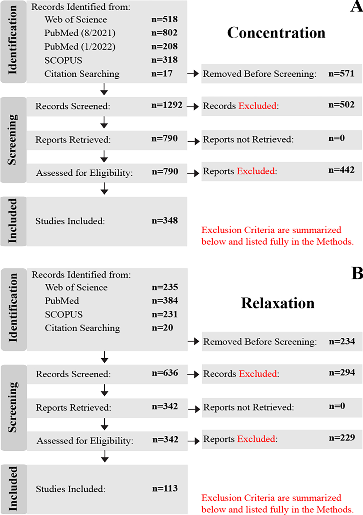
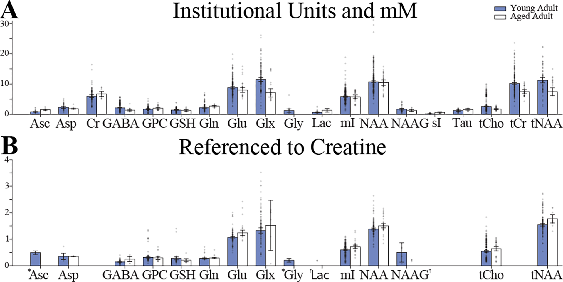
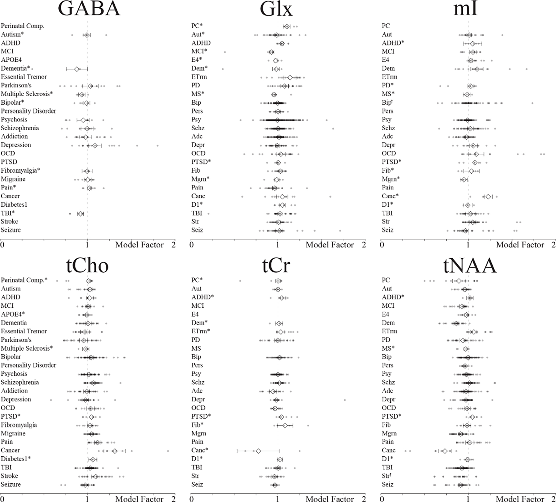
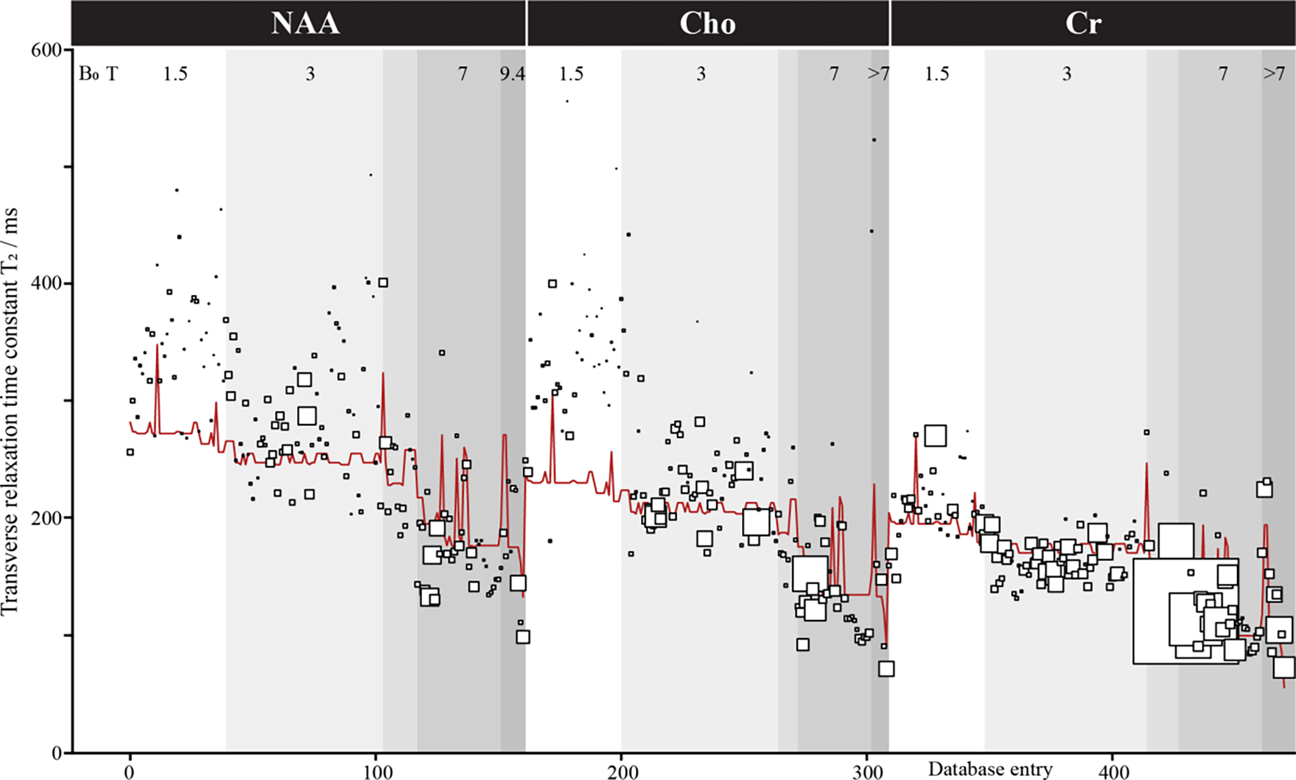
Proton (1H) Magnetic Resonance Spectroscopy (MRS) is a non-invasive tool capable of quantifying brain metabolite concentrations in vivo. Prioritization of standardization and accessibility in the field has led to the development of universal pulse sequences, methodological consensus recommendations, and the development of open-source analysis software packages. One on-going challenge is methodological validation with ground-truth data. As ground-truths are rarely available for in vivo measurements, data simulations have become an important tool. The diverse literature of metabolite measurements has made it challenging to define ranges to be used within simulations. Especially for the development of deep learning and machine learning algorithms, simulations must be able to produce accurate spectra capturing all the nuances of in vivo data. Therefore, we sought to determine the physiological ranges and relaxation rates of brain metabolites which can be used both in data simulations and as reference estimates. Using the Preferred Reporting Items for Systematic reviews and Meta-Analyses (PRISMA) guidelines, we've identified relevant MRS research articles and created an open-source database containing methods, results, and other article information as a resource. Using this database, expectation values and ranges for metabolite concentrations and T2 relaxation times are established based upon a meta-analyses of healthy and diseased brains.
Keywords: Database; Human brain; In vivo; Meta-analysis; Proton MRS; Simulation1.
Copyright © 2023 The Authors. Published by Elsevier Inc. All rights reserved.
Conflict of interest statement
Declaration of competing interest The authors declare that they have no known competing financial interests or personal relationships that could have appeared to influence the work reported in this paper
Figures
Update of
-
Meta-analysis and Open-source Database for In Vivo Brain Magnetic Resonance Spectroscopy in Health and Disease.bioRxiv [Preprint]. 2023 Jun 15:2023.02.10.528046. doi: 10.1101/2023.02.10.528046. bioRxiv. 2023. Update in: Anal Biochem. 2023 Sep 1;676:115227. doi: 10.1016/j.ab.2023.115227. PMID: 37205343 Free PMC article. Updated. Preprint.
Similar articles
-
Meta-analysis and Open-source Database for In Vivo Brain Magnetic Resonance Spectroscopy in Health and Disease.bioRxiv [Preprint]. 2023 Jun 15:2023.02.10.528046. doi: 10.1101/2023.02.10.528046. bioRxiv. 2023. Update in: Anal Biochem. 2023 Sep 1;676:115227. doi: 10.1016/j.ab.2023.115227. PMID: 37205343 Free PMC article. Updated. Preprint.
-
In vivo T(2) relaxation time measurement with echo-time averaging.NMR Biomed. 2014 Aug;27(8):863-9. doi: 10.1002/nbm.3115. Epub 2014 May 28. NMR Biomed. 2014. PMID: 24865447 Free PMC article.
-
In vivo proton magnetic resonance spectroscopic signal processing for the absolute quantitation of brain metabolites.Eur J Radiol. 2012 Apr;81(4):e653-64. doi: 10.1016/j.ejrad.2011.03.076. Epub 2011 Apr 22. Eur J Radiol. 2012. PMID: 21515011
-
In vivo quantitation of metabolite concentrations in the brain by means of proton MRS.NMR Biomed. 1995 Jun;8(4):139-48. doi: 10.1002/nbm.1940080402. NMR Biomed. 1995. PMID: 8771088 Review.
-
The use of proton magnetic resonance spectroscopy in PTSD research--meta-analyses of findings and methodological review.Neurosci Biobehav Rev. 2010 Jan;34(1):7-22. doi: 10.1016/j.neubiorev.2009.06.008. Epub 2009 Jun 24. Neurosci Biobehav Rev. 2010. PMID: 19559046 Review.
Cited by
-
WAND: Wavelet Analysis-Based Neural Decomposition of MRS Signals for Artifact Removal.NMR Biomed. 2025 Jun;38(6):e70038. doi: 10.1002/nbm.70038. NMR Biomed. 2025. PMID: 40289522 Free PMC article.
-
Metabolite T2 relaxation times decrease across the adult lifespan in a large multi-site cohort.Magn Reson Med. 2025 Mar;93(3):916-929. doi: 10.1002/mrm.30340. Epub 2024 Oct 24. Magn Reson Med. 2025. PMID: 39444343
-
The periaqueductal gray in chronic low back pain: dysregulated neurotransmitters and function.Pain. 2025 May 15;166(7):1690-1705. doi: 10.1097/j.pain.0000000000003617. Pain. 2025. PMID: 40372313 Free PMC article.
-
High-resolution 1H-MRSI at 9.4 T by integrating relaxation enhancement and subspace imaging.NMR Biomed. 2024 Oct;37(10):e5161. doi: 10.1002/nbm.5161. Epub 2024 May 8. NMR Biomed. 2024. PMID: 38715469 Free PMC article.
-
Microstructural Changes in the Corpus Callosum in Neurodegenerative Diseases.Cureus. 2024 Aug 21;16(8):e67378. doi: 10.7759/cureus.67378. eCollection 2024 Aug. Cureus. 2024. PMID: 39310519 Free PMC article. Review.
References
Publication types
MeSH terms
Substances
Grants and funding
LinkOut - more resources
Full Text Sources
Research Materials
