. 2023 Aug;40(8):1865-1879.
doi: 10.1007/s10815-023-02874-0. Epub 2023 Jul 10.

Loss of PMFBP1 Disturbs Mouse Spermatogenesis by Downregulating HDAC3 Expression

Affiliations

Loss of PMFBP1 Disturbs Mouse Spermatogenesis by Downregulating HDAC3 Expression

Weilong Xu et al. J Assist Reprod Genet. 2023 Aug.

Abstract

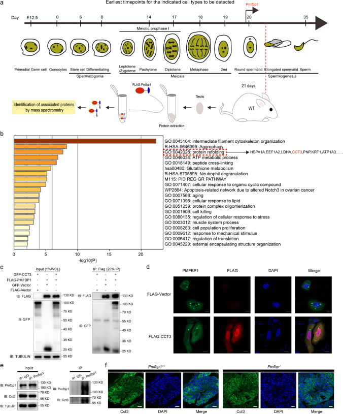
Purpose: Polyamine modulating factor 1 binding protein (PMFBP1) acts as a scaffold protein for the maintenance of sperm structure. The aim of this study was further to identify the new role and molecular mechanism of PMFBP1 during mouse spermatogenesis.

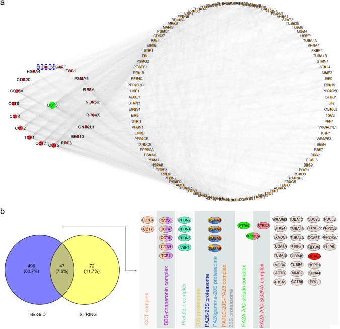
Methods and results: We identified a profile of proteins interacting with PMFBP1 by immunoprecipitation combined with mass spectrometry and demonstrated that class I histone deacetylases, particularly HDAC3 and chaperonin-containing TCP1 subunit 3 (CCT3), were potential interaction partners of PMFBP1 based on network analysis of protein-protein interactions and co-immunoprecipitation. Immunoblotting and immunochemistry assays showed that loss of Pmfbp1 would result in a decline in HDACs and change the proteomic profile of mouse testis, in which differently expressed proteins are associated with spermatogenesis and assembly of flagella, which was proved by proteomic analysis of testis tissue obtained from Pmfbp1-/- mice. After integrating with transcriptome data for Hdac3-/- and Sox30-/- round sperm obtained from a public database, RT-qPCR confirmed ring finger protein 151 (Rnf151) and ring finger protein 133 (Rnf133) were key downstream response factors of the Pmfbp1-Hdac axis affecting mouse spermatogenesis.

Conclusion: Taken together, this study indicates a previously unidentified molecular mechanism of PMFBP1 in spermatogenesis whereby PMFBP1 interacts with CCT3, affecting the expression of HDAC3, followed by the downregulation of RNF151 and RNF133, resulting in an abnormal phenotype of sperm beyond the headless sperm tails. These findings not only advance our understanding of the function of Pmfbp1 in mouse spermatogenesis but also provide a typical case for multi-omics analysis used in the functional annotation of specific genes.

Keywords: CCT3; HDAC3; PMFBP1; Protein-protein interaction; Proteome; Spermatogenesis.


Conflict of interest statement

The authors declare no competing interests.

Figures

Fig. 1
Fig. 1
CCT3 interacts with PMFBP1. a Label-free mass spectrometry analysis of proteins that interact with Pmfbp1 in the testis of a 21-day-old mouse. Graphic representation of Pmfbp1 expression during spermatogenesis. The recombinant plasmid of the mouse Pmfbp1 gene was transfected into HEK293T cells. After 24 h, the total cell proteins were extracted for incubation with Flag-probe antibody agarose beads; these bead-bound proteins were incubated as bait with whole protein extracts from the testis of a 21-day-old mouse. The proteins that interacted with the Pmfbp1 protein were obtained for label-free mass spectrometry analysis. b Bar graph showing pathway enrichment of 74 Pmfbp1 interacting proteins identified by the affinity purification mass spectrometry approach via the Metascape database. Only the significant Gene Ontology terms (P < 0.05) are shown in rows. Terms with the prefix “GO” are from the Gene Ontology Consortium; the prefix “R-HSA” from the Reactome; the prefix “WP” from the WikiPathways, and the prefix “hsa” from the KEGG database. c The immunoprecipitation experiment was carried out using FLAG-M2 beads, and the lysates of HEK293T cells were transfected with FLAG-PMFBP1 and GFP-CCT3. The isolated proteins were then analyzed by western blotting with anti-FLAG and anti-GFP antibodies, respectively. This experiment was repeated three times, and representative blots are presented. d Immunofluorescent staining indicates the subcellular localization of PMFBP1 and CCT3. The upper panel of HeLa cells was transfected with the pEGFP-N2-PMFBP1 plasmid, and the lower panel was transfected with pEGFP-N2-PMFBP1 and pCDNA3.1-CMV-3×flag-CCT3 plasmids. Subcellular localization of CCT3 (red) was probed with an anti-FLAG antibody. Cell nuclei were counterstained with DAPI. Scale bar: 10 μm. e A co-immunoprecipitation assay confirmed the interaction between endogenous mouse Cct3 and Pmfbp1 in the mouse testis. The anti-IgG or anti-Pmfbp1 antibody and protein A/G beads were used for immunoprecipitation, and anti-Cct3 and anti-Pmfbp1 antibodies were used for western blot analysis. f Immunofluorescent staining of Cct3 (green) and DAPI (nucleus, blue) on frozen sections from P21 Pmfbp1−/− and wild-type mouse testes. Scale bar, 20 μm. CCT3, chaperonin-containing TCP1 subunit 3; GFP, green fluorescent protein; KEGG, Kyoto Encyclopedia of Genes and Genomes; P21, postnatal day 21; PMFBP1, polyamine modulating factor 1 binding protein
Fig. 2
Fig. 2
Protein-protein interactions in the CCT3 network. a Protein-protein interactions in the CCT3 network (green, CCT3; red, top 19 proteins interacting with CCT3; yellow, other potentially common targets of CCT3) were generated by Cytoscape (version 3.8) (http://cytoscape.org/). b Venn diagram showing overlap of the genes identified by the BioGriD (blue) and STRING (yellow) databases. The g:Profiler based on information from the CORUM database was used to determine the specific cellular protein complexes (right panel) involved by the overlap of 47 proteins. Half of the prey belongs to different protein complexes that are highlighted by color coding. CCT3, chaperonin-containing TCP1 subunit 3; PMFBP1, polyamine modulating factor 1 binding protein
Fig. 3
Fig. 3
PMFBP1 interacts with HDAC3 and affects its expression level. a A co-immunoprecipitation assay confirmed the interaction of exogenous human GFP-PMFBP1 and FLAG-HDAC1/2/3/4/5 in HEK293T cells. FLAG-M2 beads were used for immunoprecipitation and anti-GFP and anti-FLAG antibodies for western blot analysis. The co-immunoprecipitation assay confirmed the interaction between endogenous mouse Pmfbp1 and Hdac3 in the mouse testis (b, c). Anti-Pmfbp1 and anti-Hdac3 antibodies were used for western blot analysis. (df) Hdac1, Hdac2, and Hdac3 expression in Pmfbp1−/− and wild-type mouse testes at P21. Hdac1, Hdac2, and Hdac3 expressions are shown in green and the nuclei in blue. Scale bar, 20 μm. g Western blot analysis of the Pmfbp1 and Hdac1/2/3 in Pmfbp1−/− and wild-type mouse testes at P21. GAPDH is shown as the loading control. The bands in the left panel were quantified by ImageJ software and show that the abundance of Hdac1, Hdac2, and Hdac3 proteins in the Pmfbp1−/− mouse was significantly lower than that in the wild-type animal. The expression level was set as 1 in the wild-type samples. *P < 0.05, **P < 0.01, and ***P < 0.001 (Student’s t-test). GFP, green fluorescent protein; HDAC3, histone deacetylase 3; PMFBP1, polyamine modulating factor 1 binding protein; P21, postnatal day 21
Fig. 4
Fig. 4
Pmfbp1 may cooperate with Hdac3 to affect gene expression profiling in the P21 mouse testis. a Heat map representing differentially regulated proteins (fold-change >1.5, P-value < 0.05, two-tailed Student’s t-test, false discovery rate < 0.05) in P21 mouse testis tissue after knockout of Pmfbp1. The color gradient represents the changes (log2 scale) from the most downregulated (dark blue) to the most upregulated (red) genes. Each row is a gene, and each column represents replicates (average of n = 3 replicates). b Histogram showing the Gene Ontology enrichment analysis results for 138 DEPs between the control group and the Pmfbp1−/− mouse. The tassel size corresponds to the −log10 P-value of the enrichment while the color to the genes belonging to each category. (c) Venn diagram of DEGs and DEPs. In total, 5126 DEGs and 138 DEPs were respectively identified in Hdac−/− and Pmfbp1−/− mouse testes at P21. Seventy overlapping target genes were found between the two sets. d Quantitative polymerase chain reaction analysis of key gene expression in P21 wild-type and Pmfbp1−/− testes (n = 3 each). Data are shown as the mean ± standard deviation. *P < 0.05, **P < 0.01, and ***P < 0.001 (Student’s t-test). e Western blot showing that the Efhc1, Spag6, Tekt4, Dnajb13, Nme5, and Odf1 proteins involved in mouse spermatogenesis were downregulated in Pmfbp1−/− mice. The P21 testis lysate was subjected to sodium dodecyl sulfate-polyacrylamide gel electrophoresis. α-Tubulin was used as the loading control. The right panel shows the quantified western blot bands for Pmfbp1−/− and Pmfbp1+/+ mice by ImageJ software (right). The expression level of the wild-type samples was set at 1. f The indicated proteins in sperm samples from a patient with PMFBP1 deficiency were immunodetected using specific antibodies. α-Tubulin was used for the loading control. Bands for the patient and a normal subject were quantified by ImageJ software (right). The expression level of the wild-type samples was set at 1. BP, biological process; CC, cellular component; MF, molecular function; DEGs, differentially expressed genes; DEPs, differentially expressed proteins; HDAC3, histone deacetylase 3; PMFBP1, polyamine modulating factor 1 binding protein; P21, postnatal day 21
Fig. 5
Fig. 5
Rnf151 and Rnf133 may be the response factor downstream of the Pmfbp1-Hdac3 axis in mouse spermatogenesis. a Venn diagram showing the overlapping downregulated genes among P21 Hdac3−/− round spermatids, P23 Sox30−/− round spermatids, and P21 Sox30−/− round spermatids compared with wild-type round spermatids. The 193 overlapping genes in these three groups were subjected to gene functional analysis using the Metascape database. The most significantly enriched pathway of these genes is “GO:0007283 spermatogenesis,” which harbors Rnf133 and Rnf151. b The Hdac3 ChIP-seq and Sox30 ChIP-seq reads in wild-type and Sox30 knockout testes at P20–P21 are aligned to the genomic sequence of Rnf151 (left) and Rnf133 (right) in the mouse reference genome. (C) Pmfbp1, Rnf151, and Rnf133 mRNA levels in testis tissue from Pmfbp1−/− (n = 3) and wild-type (n = 3) mice at P21. Values were determined by quantitative real-time polymerase chain reaction and are expressed as the mean ± standard error of triplicate experiments after normalization to BACT mRNA levels. The expression level of the wild-type samples was set at 1. The data are shown as the mean ± standard deviation. *P < 0.05, **P < 0.01, and ***P < 0.001 (Student’s t-test). GO, Gene Ontology; HDAC3, histone deacetylase 3; PMFBP1, polyamine modulating factor 1 binding protein; P20, postnatal day 20; P21, postnatal day 21; P23, postnatal day 23; Rnf, ring finger protein

Similar articles

Cited by

References

    1. Ni FD, Hao SL, Yang WX. Multiple signaling pathways in Sertoli cells: recent findings in spermatogenesis. Cell Death Dis. 2019;10:541. doi: 10.1038/s41419-019-1782-z. - DOI - PMC - PubMed
    1. Sharma S, Hanukoglu A, Hanukoglu I. Localization of epithelial sodium channel (ENaC) and CFTR in the germinal epithelium of the testis, Sertoli cells, and spermatozoa. J Mol Histol. 2018;49:195–208. doi: 10.1007/s10735-018-9759-2. - DOI - PubMed
    1. Zhu F, Liu C, Wang F, Yang X, Zhang J, Wu H, Zhang Z, He X, Zhang Z, Zhou P, et al. Mutations in PMFBP1 cause acephalic spermatozoa syndrome. Am J Hum Genet. 2018;103:188–199. doi: 10.1016/j.ajhg.2018.06.010. - DOI - PMC - PubMed
    1. Mazaheri Moghaddam M, Mazaheri Moghaddam M, Hamzeiy H, Baghbanzadeh A, Pashazadeh F, Sakhinia E. Genetic basis of acephalic spermatozoa syndrome, and intracytoplasmic sperm injection outcomes in infertile men: a systematic scoping review. J Assist Reprod Genet. 2021;38:573–586. doi: 10.1007/s10815-020-02008-w. - DOI - PMC - PubMed
    1. Zhang Y, Liu C, Wu B, Li L, Li W, Yuan L. The missing linker between SUN5 and PMFBP1 in sperm head-tail coupling apparatus. Nat Commun. 2021;12:4926. doi: 10.1038/s41467-021-25227-w. - DOI - PMC - PubMed

LinkOut - more resources