

The increased anterior talofibular ligament-posterior talofibular ligament angle on MRI may help evaluate chronic ankle instability
- PMID: 37423946
- PMCID: PMC10533641
- DOI: 10.1007/s00276-023-03196-7
The increased anterior talofibular ligament-posterior talofibular ligament angle on MRI may help evaluate chronic ankle instability
Abstract
Purpose: This study intended to compare the difference between the anterior talofibular ligament (ATFL) and posterior talofibular ligament (PTFL) angle with chronic ankle instability (CAI) patients and healthy volunteers, and to confirm whether using the ATFL-PTFL angle could be a reliable assessment method for CAI, so as to improve the accuracy and specificity of clinical diagnosis.
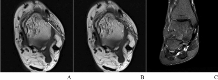
Methods: This retrospective study included 240 participants: 120 CAI patients and 120 healthy volunteers between 2015 and 2021. The ATFL-PTFL angle of the ankle region was gaged in the cross-sectional supine position on MRI between two groups. After participants undergoing a comprehensive MRI scanning, ATFL-PTFL angles were regarded as the main indicator of patients with the injured ATFLs and healthy volunteers to compare, and were measured by an experienced musculoskeletal radiologist. Moreover, other qualitative and quantitative indicators referring to anatomical and morphological characteristics of the AFTL were included in this study with MRI, such as the length, width, thickness, shape, continuity, and signal intensity of the ATFL, which can be used as secondary indicators.
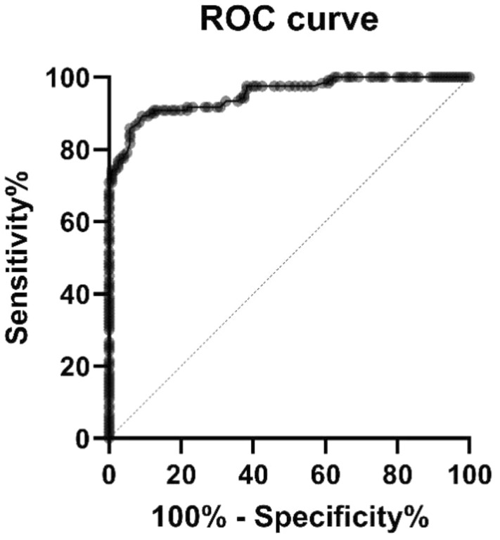
Results: In the CAI group, the ATFL-PTFL angle was 90.8° ± 5.7°, which was significantly different from the non-CAI group where the ATFL-PTFL angle for 80.0° ± 3.7° (p < 0.001). As for the ATFL-MRI characteristics, the length (p = 0.003), width (p < 0.001), and thickness (p < 0.001) in the CAI group were also significantly different from the non-CAI group. Over 90% of the cases, patients of the CAI group had injured ATFL with an irregular shape, non-continuous, and high or mixed signal intensity.
Conclusion: Compared with healthy people, the ATFL-PTFL angle of most CAI patients is larger, which can be used as a secondary index to diagnose CAI. However, the MRI characteristic changes of ATFL may not relate to the increased ATFL-PTFL angle.
Keywords: Angle; Anterior talofibular ligament; Chronic ankle instability; MRI; Posterior talofibular ligament.
© 2023. The Author(s).
Conflict of interest statement
The authors declare no competing interests.
Figures

References
-
- Alvarez C, Hattori S, Kato Y, et al. Dynamic high-resolution ultrasound in the diagnosis of calcaneofibular ligament injury in chronic lateral ankle injury: a comparison with three-dimensional magnetic resonance imaging. J Med Ultrason. 2020;47:313–317. doi: 10.1007/s10396-019-00993-9. - DOI - PubMed
MeSH terms
Grants and funding
- 82004458/the National Natural Science Foundation of China (Youth Science Foundation Project)
- 2021ZYD0078/Central Funds Guiding the Local Science and Technology Development General Program of Sichuan Provincial Science and Technology Department
- 2022-CXTD-08/Scientific Research Cultivation Project of The Affiliated Traditional Chinese Medicine Hospital of Southwest Medical University
- 2022HJXNYD04/Hejiang People's Hospital - Southwest Medical University Science and Technology Strategic Cooperation Project (major project)
- 2023MS248/General Project of Sichuan Traditional Chinese Medicine Administration Traditional Chinese Medicine Research Special Project (Fundamentals of Traditional Chinese Medicine)
LinkOut - more resources
Full Text Sources
Research Materials
