. 2023 Jul 10;21(1):250.
doi: 10.1186/s12916-023-02963-y.

Transcriptional level of inflammation markers associates with short-term brain structural changes in first-episode schizophrenia

Affiliations

Transcriptional level of inflammation markers associates with short-term brain structural changes in first-episode schizophrenia

Long-Biao Cui et al. BMC Med. .

Abstract

Background: Inflammation has been implicated in the pathology of schizophrenia and may cause neuronal cell death and dendrite loss. Neuroimaging studies have highlighted longitudinal brain structural changes in patients with schizophrenia, yet it is unclear whether this is related to inflammation. We aim to address this question, by relating brain structural changes with the transcriptional profile of inflammation markers in the early stage of schizophrenia.

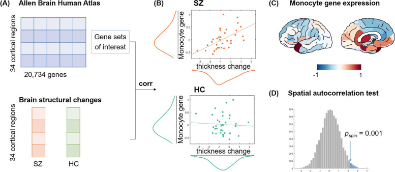
Methods: Thirty-eight patients with first-episode schizophrenia and 51 healthy controls were included. High-resolution T1-weighted magnetic resonance imaging (MRI) and clinical assessments were performed at baseline and 2 ~ 6 months follow-up for all subjects. Changes in the brain structure were analyzed using surface-based morphological analysis and correlated with the expression of immune cells-related gene sets of interest reported by previous reviews. Transcriptional data were retrieved from the Allen Human Brain Atlas. Furthermore, we examined the brain structural changes and peripheral inflammation markers in association with behavioral symptoms and cognitive functioning in patients.

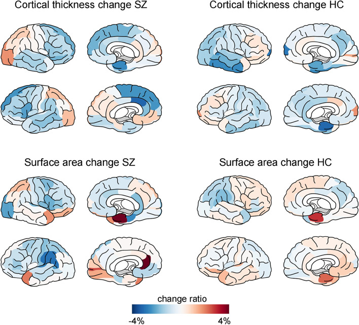
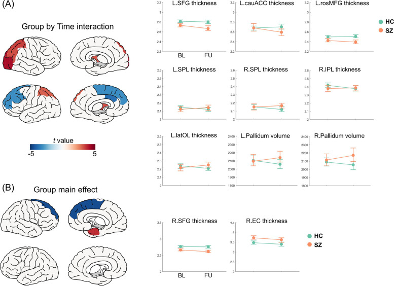
Results: Patients exhibited accelerated cortical thickness decrease in the left frontal cortices, less decrease or an increase in the superior parietal lobule and right lateral occipital lobe, and increased volume in the bilateral pallidum, compared with controls. Changes in cortical thickness correlated with the transcriptional level of monocyte across cortical regions in patients (r = 0.54, p < 0.01), but not in controls (r = - 0.05, p = 0.76). In addition, cortical thickness change in the left superior parietal lobule positively correlated with changes in digital span-backward test scores in patients.

Conclusions: Patients with schizophrenia exhibit regional-specific cortical thickness changes in the prefrontal and parietooccipital cortices, which is related to their cognitive impairment. Inflammation may be an important factor contributing to cortical thinning in first-episode schizophrenia. Our findings suggest that the immunity-brain-behavior association may play a crucial role in the pathogenesis of schizophrenia.

Keywords: Brain structure; Inflammation; Longitudinal alterations; Schizophrenia; Transcriptome.


Conflict of interest statement

The authors declare that they have no competing interests.

Figures

Fig. 1
Fig. 1
Percentage of cortical thickness (upper panel) and surface area (lower panel) changes in patients with schizophrenia (left) and healthy controls (HC). Blue color indicates a decreased value at follow-up compared to baseline measurements, whereas red color indicates an increased value
Fig. 2
Fig. 2
Significant group by time interaction effect (A) and group effect (B) on brain structural measurements. T-statistics from linear mix-effects model was plotted on the left, with blue color indicating lower values or accelerated decrease in patients, while red indicates the opposite. For significant regions, cortical thickness and volume in patients (SZ) and controls (HC) at baseline (BL) and follow-up (FU) were plotted on the right.  Abbreviations: L: left, R: right, SFG: superior frontal gyrus, cauACC: caudal anterior cingulate cortex, rosMFG, rostral middle frontal gyrus, SPL: superior parietal lobule, IPL: inferior parietal lobule, latOL: lateral occipital lobe, EC: entorhinal cortex
Fig. 3
Fig. 3
Association between brain structural changes and transcriptional profiles of inflammation and antipsychotic response. A Transcriptional data were retrieved from the Allen Human Brain Atlas (AHBA) [25] (http://human.brain-map.org/) and calculated for gene sets of interests [–31] (Additional file 1: Table S1). Brain structural changes (cortical thickness and surface area) were calculated for patients with schizophrenia (SZ) and healthy controls (HC) as illustrated in Fig. 1. B A significant correlation was found between monocyte-related gene expression and cortical thickness changes in patients with schizophrenia (r = 0.54, p < 0.01) but not in healthy controls (r =  − 0.05, p = 0.76). C The normalized monocyte gene expression profile from a systematic review on leukocyte and subtypes [29]. D Association between monocyte gene expression and cortical thickness changes in patients is significantly higher than the null model generated by correlations between 5000 surrogate maps and monocyte gene expression profile (pspin = 0.001)
Fig. 4
Fig. 4
Correlation analysis between brain structure changes and cognitive score changes in patients (A) and controls (B). A significant association was found between the left superior parietal lobule (SPL) cortical thickness change and WAIS digital span (backward) change in patients (r = 0.57, p = 3.4 × 10−4) but not in controls (r = 0.09, p = 0.54)

References

    1. James SL, Abate D, Abate KH, Abay SM, Abbafati C, Abbasi N, et al. Global, regional, and national incidence, prevalence, and years lived with disability for 354 diseases and injuries for 195 countries and territories, 1990–2017: a systematic analysis for the Global Burden of Disease Study 2017. Lancet. 2018;392(10159):1789–858. - PMC - PubMed
    1. Wu X-S, Yan T-C, Wang X-Y, Cao Y, Liu X-F, Fu Y-F, et al. Magnetic resonance imaging-guided and navigated individualized repetitive transcranial magnetic stimulation for cognitive impairment in schizophrenia. Neurosci Bull. 2021;37(9):1365–1369. doi: 10.1007/s12264-021-00727-3. - DOI - PMC - PubMed
    1. Zhao Q, Li J, Xiao Y, Cao H, Wang X, Zhang W, et al. Distinct neuroanatomic subtypes in antipsychotic-treated patients with schizophrenia classified by the predefined classification in a never-treated sample. Psychoradiology. 2021;1(4):212–224. doi: 10.1093/psyrad/kkab018. - DOI - PMC - PubMed
    1. Cheng W, Frei O, van der Meer D, Wang Y, O’Connell KS, Chu Y, et al. Genetic association between schizophrenia and cortical brain surface area and thickness. JAMA Psychiat. 2021;78(9):1020–1030. doi: 10.1001/jamapsychiatry.2021.1435. - DOI - PMC - PubMed
    1. Patel Y, Parker N, Shin J, Howard D, French L, Thomopoulos SI, et al. Virtual histology of cortical thickness and shared neurobiology in 6 psychiatric disorders. JAMA Psychiat. 2021;78(1):47–63. doi: 10.1001/jamapsychiatry.2020.2694. - DOI - PMC - PubMed

Publication types