. 2022 Jun 28;3(3):153-159.
doi: 10.36518/2689-0216.1431. eCollection 2022.

Presentations of Cutaneous Disease in Various Skin Pigmentations: Cutaneous Abscesses

Affiliations

Presentations of Cutaneous Disease in Various Skin Pigmentations: Cutaneous Abscesses

Christopher M Wong et al. HCA Healthc J Med. .

Abstract

Description Cutaneous abscesses are collections of pus resulting from skin and soft tissue bacterial infections. They clinically exhibit the four cardinal inflammatory signs of pain, warmth, swelling, and erythema. In patients with darkly pigmented skin, classically-associated erythema may be challenging to appreciate and can lead to missed or delayed diagnosis. We compare abscess presentations in different skin types. Recognition of varying presentations of cutaneous abscesses in diverse skin colors will help clinicians utilize additional clues to identify and diagnose this entity correctly.

Keywords: Fitzpatrick skin types; abscess/diagnosis; bacterial infections; carbuncle; cellulitis/diagnosis; cutaneous abscess; dermatology; erythema; furunculosis; skin of color; skin pigmentation.


Conflict of interest statement

Conflicts of Interest The authors declare they have no conflicts of interest.

Figures

Figure 1
Figure 1
The Fitzpatrick scale provides a classification system for an individual’s skin type based on the ability to burn and/or tan when exposed to ultraviolet light. It is used to approximate the degree of skin pigmentation.
Figure 2
Figure 2
Fitzpatrick type II (mostly burns, rarely tans) skin on a middle-aged male’s right posteromedial proximal thigh demonstrating a violaceous nodule with central desquamation on a background of a brightly erythematous, edematous, and indurated plaque with poorly defined borders.
Figure 3
Figure 3
Fitzpatrick type III (sometimes burns, often tans) skin on a young adult male’s left posterior mid-lower leg demonstrating a violaceous nodule with surrounding erythema. The nodule has overlying desquamation. The erythema has poorly defined borders and is less bright than the Fitzpatrick type II (mostly burns, rarely tans) patient in Figure 2.
Figure 4
Figure 4
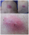
Fitzpatrick IV (rarely burns, mostly tans) skin on the right posterior thigh of a male child demonstrating a subcutaneous nodule with overlying erythema. There is a pustule at the center of the erythema. The erythema has a violaceous hue and is poorly defined peripherally.
Figure 5
Figure 5
Fitzpatrick type V (almost never burns, always tans) skin (A) in the right axilla of an adult female showing a subcutaneous nodule without surface changes. (B) When viewed from a lateral angle, the elevation of the nodule is more pronounced than when viewed at a perpendicular angle. The overlying erythema is faintly visible to extend beyond the borders of the nodule (black arrows).
Figure 6
Figure 6
Fitzpatrick type V (almost never burns, always tans) skin (A) in the right axilla of an adult male demonstrating several erythematous subcutaneous nodules. While the nodules are visibly pink, the border of the erythema is imperceptible. (B) The nodules were deroofed, revealing several abscesses.
Figure 7
Figure 7
Fitzpatrick type II (mostly burns, rarely tans) skin showing an inflamed epidermal inclusion cyst. On the mid-central back of an older-aged male, there is a brightly erythematous, edematous, indurated nodule. The erythematous borders are well-defined compared to the previous images. On closer examination, several pustules and a focus of hemorrhagic crust are visible within the nodule. The bacterial culture of the drainage from this lesion was negative.
Figure 8
Figure 8
Fitzpatrick type V (almost never burns, always tans) skin showing an inflamed epidermal inclusion cyst. A tender subcutaneous nodule is on the mid-upper back of a middle-aged male. Several puncta are visible at the nodule’s superomedial and right lateral aspects. The nodule has overlying hyperpigmentation accompanied by faintly appreciable pink erythema. The borders of the discoloration are well-defined from the surrounding uninvolved skin.

References

    1. Raff AB, Kroshinsky D. Cellulitis: a review. JAMA. 2016;316(3):325–337. doi: 10.1001/jama.2016.8825. - DOI - PubMed
    1. Ren Z, Silverberg JI. Burden, risk factors, and infectious complications of cellulitis and erysipelas in US adults and children in the emergency department setting. J Am Acad Dermatol. 2021;84(5):1496–1503. doi: 10.1016/j.jaad.2020.11.021. - DOI - PubMed
    1. Fitzpatrick TB. The validity and practicality of sun-reactive skin types I through VI. Arch Dermatol. 1988;124(6):869–871. doi: 10.1001/archderm.1988.01670060015008. - DOI - PubMed
    1. Scheufele CJ, Weis D, Weis SE. Presentations of cutaneous disease in various skin pigmentations: an introduction. HCA Healthcare Journal of Medicine. 2022;3(3):135–138. doi: 10.36518/2689-0216.1483. - DOI - PMC - PubMed
    1. Del Bino S, Duval C, Bernerd F. Clinical and biological characterization of skin pigmentation diversity and its consequences on UV impact. Int J Mol Sci. 2018;19(9):2668. doi: 10.3390/ijms19092668. Published 2018 Sep 8. - DOI - PMC - PubMed

LinkOut - more resources