. 2023 Jun 22:13:1191806.
doi: 10.3389/fcimb.2023.1191806. eCollection 2023.

Monoclonal antibodies against lipopolysaccharide protect against Pseudomonas aeruginosa challenge in mice

Affiliations

Monoclonal antibodies against lipopolysaccharide protect against Pseudomonas aeruginosa challenge in mice

Jason Kang et al. Front Cell Infect Microbiol. .

Abstract

Pseudomonas aeruginosa is a common cause of hospital-acquired infections, including central line-associated bloodstream infections and ventilator-associated pneumonia. Unfortunately, effective control of these infections can be difficult, in part due to the prevalence of multi-drug resistant strains of P. aeruginosa. There remains a need for novel therapeutic interventions against P. aeruginosa, and the use of monoclonal antibodies (mAb) is a promising alternative strategy to current standard of care treatments such as antibiotics. To develop mAbs against P. aeruginosa, we utilized ammonium metavanadate, which induces cell envelope stress responses and upregulates polysaccharide expression. Mice were immunized with P. aeruginosa grown with ammonium metavanadate and we developed two IgG2b mAbs, WVDC-0357 and WVDC-0496, directed against the O-antigen lipopolysaccharide of P. aeruginosa. Functional assays revealed that WVDC-0357 and WVDC-0496 directly reduced the viability of P. aeruginosa and mediated bacterial agglutination. In a lethal sepsis model of infection, prophylactic treatment of mice with WVDC-0357 and WVDC-0496 at doses as low as 15 mg/kg conferred 100% survival against challenge. In both sepsis and acute pneumonia models of infection, treatment with WVDC-0357 and WVDC-0496 significantly reduced bacterial burden and inflammatory cytokine production post-challenge. Furthermore, histopathological examination of the lungs revealed that WVDC-0357 and WVDC-0496 reduced inflammatory cell infiltration. Overall, our results indicate that mAbs directed against lipopolysaccharide are a promising therapy for the treatment and prevention of P. aeruginosa infections.

Keywords: O5; Pseudomonas aeruginosa; ammonium metavanadate; anti-lipopolysaccharide antibody; immunotherapeutics; monoclonal antibody (mAb); pneumonia; sepsis.


Conflict of interest statement

Authors JK, MB, and FD are inventors on a pending patent application related to the sequences of WVDC-0357 and WVDC-0496. The remaining authors declare that the research was conducted in the absence of any commercial or financial relationships that could be construed as a potential conflict of interest.

Figures

Figure 1
Figure 1
Protection of convalescent mice against lethal P. aeruginosa challenge after prior infection with P. aeruginosa PAO1 cultured on Pseudomonas isolation agar supplemented with ammonium metavanadate (PIAAMV). CD-1 mice were challenged with a sub-lethal intranasal dose (1.4x10⁷ CFU) of P. aeruginosa PAO1 cultured on PIAAMV and allowed to convalesce before being challenged with a lethal intranasal dose of P. aeruginosa PAO1 cultured on Pseudomonas isolation agar (PIA). Mice were euthanized at 16 h post-challenge. (A) Comparison of colony morphology between P. aeruginosa PAO1 cultured on PIA and P. aeruginosa PAO1 cultured on PIAAMV used for dose preparation. Growth of P. aeruginosa PAO1 on PIAAMV induced a mucoid colony morphology. (B) Serum anti-P. aeruginosa IgG titers determined by ELISA are represented by area under the curve (AUC) values. Differences in serum antibody titers were determined using a two-tailed student’s t-test. ***P<0.001 compared to naïve mice. (C) Bacterial burden in the lung and nasal wash was determined at 16 hours post-challenge. Differences in bacterial burden were determined using a two-tailed student’s t-test. **P<0.01 and ***P<0.001 compared to naïve mice. LOD, limit of detection.
Figure 2
Figure 2
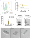
Characterization of anti-P. aeruginosa mAbs, WVDC-0357 and WVDC-0496. (A) Fluorescence-activated cell sorting (FACS) analysis of binding to P. aeruginosa was performed using WVDC-0357, WVDC-0496, and an isotype control mAb. (B) Binding kinetic analysis revealed dissociation constants (KD) of 14.96 ± 6.73 nM and 11.45 ± 4.32 nM for WVDC-0357 and WVDC-0496, respectively, using a non-linear regression model. (C) ELISA was used to determine anti-P. aeruginosa IgG titers against different serogroups, represented as area under the curve (AUC) values. (D, E) Immunoblot analysis of P. aeruginosa outer membrane fractions treated with proteinase K or sodium periodate, and probed with WVDC-0357 and WVDC-0496. (F-H) Electron microscopy imaging of P. aeruginosa treated with an isotype control mAb, WVDC-0357, or WVDC-0496, labeled with a gold-conjugated secondary antibody. PK, proteinase K; NaIO4, sodium periodate.
Figure 3
Figure 3
WVDC-0357 and WVDC-0496 promote bacterial agglutination. (A) In UV-transparent cuvettes, fresh cultures of P. aeruginosa PAO1 in lysogeny broth were incubated with an isotype control mAb, WVDC-0357, or WVDC-0496 at 100 µg/ml. Cuvettes were incubated statically at 37°C and the absorbance of growth cultures at 600 nm were measured over the course of 24 h. Differences in absorbance were analyzed using a one-way ANOVA with Dunnett’s multiple comparison test. *P<0.05, ***P<0.001, ****P<0.0001 compared to the isotype control. (B) After incubation for 24 h, cuvettes were photographed for visual inspection of turbidity. (C-E) Representative fluorescent microscopy images of P. aeruginosa PAO1 stained with BacLight Red and treated with an isotype control mAb, WVDC-0357, or WVDC-0496. (F, G) Random area images were analyzed with an aggregation cluster pipeline in CellProfiler for number of aggregates per image and aggregate diameter. Differences in aggregates were calculated using the Kruskal Wallis test with Dunn’s multiple comparison. **P<0.01, ***P<0.001, ****P<0.0001 compared to the isotype control.
Figure 4
Figure 4
Antibacterial activity of WVDC-0357 and WVDC-0496. (A) Antibacterial activity against P. aeruginosa PAO1. Mid-log phase growth cultures of P. aeruginosa PAO1 were treated with varying concentrations of an isotype control mAb, WVDC-0357 or WVDC-0496 before plating onto Pseudomonas isolation agar (PIA) plates for CFU enumeration. The percent killing was determined by normalizing the number of CFUs from each sample to the mean CFUs of the no-antibody control. Differences in CFUs were calculated using a one-way ANOVA with Dunnett’s multiple comparison test. **P < 0.01, ****P<0.0001 compared to the isotype control. (B) Antibacterial activity against O5 serogroup strains. P. aeruginosa strains PAO1, MRSN 1601, MRSN 2101, MRSN 12368, and MRSN 14981 was treated with 100 µg/mL of an isotype control mAb, WVDC-0357, and WVDC-0496, and then plated on PIA for CFU enumeration. Each data point represents the average percent killing for a specific strain. The percent killing was determined by normalizing the number of CFUs from each sample to the mean CFUs of the no-antibody control. Differences in CFUs were calculated using a one-way ANOVA with Dunnett’s multiple comparison test. *P<0.05 compared to the isotype control.
Figure 5
Figure 5
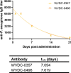
Pharmacokinetics of WVDC-0357 and WVDC-0496. CD-1 mice were intraperitoneally (IP) administered 45 mg/kg of WVDC-0357 or WVDC-0496 and submandibular bled at various time points over the course of 21 d to assess serum IgG titers against P. aeruginosa PAO1 using ELISA. The half-life (t1/2) of WVDC-0357 and WVDC-0496 was determined using a one-phase exponential decay model.
Figure 6
Figure 6
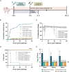
WVDC-0357 and WVDC-0496 protect mice following lethal bloodstream challenge with P. aeruginosa. (A) CD-1 mice were prophylactically administered either an isotype control mAb, P. aeruginosa whole cell vaccine (Pa WCV) serum, WVDC-0357, or WVDC-0496. After 12 h, mice were challenged with an intraperitoneal (IP) dose of P. aeruginosa PAO1 (5x10⁵ CFU) and survival was monitored for 96 h. A separate set of mice was euthanized at 6 h post-challenge to determine bacterial burden in the blood, kidney, and spleen. (B) In the survival model, clinical scores were calculated at the indicated time points. Shown is the cumulative total of average clinical scores for each group. (C) Temperature was a key indicator of morbidity in mice. Shown is the average percent temperature change of each group, with the standard deviation of the isotype control mice shown in black error bars and the standard deviation of all other groups shown as gray shaded regions. (D) The survival of mice is represented as Kaplan-Meier curves, with 5-10 mice in each group. Differences in survival were calculated using the log-rank test. ****P<0.0001 compared to isotype control. (E) At 6 h post-challenge, the blood, kidney, and spleen homogenate were serially diluted and plated to determine bacterial burden. Differences in CFUs were calculated using the Kruskal Wallis test with Dunn’s multiple comparison. **P<0.01 compared to the isotype control. Abbreviations: LOD, limit of detection.
Figure 7
Figure 7
Level of inflammatory cytokines and chemokines in serum following lethal bloodstream challenge with P. aeruginosa. CD-1 mice were prophylactically administered an isotype control mAb, P. aeruginosa whole cell vaccine (Pa WCV) serum, WVDC-0357, or WVDC-0496, and challenged 12 h later with an intraperitoneal (IP) dose of P. aeruginosa PAO1 (5x10⁵ CFU). Mice were euthanized at 6 h post-challenge, and the serum concentration of (A) CXCL-1, (B) IFN-γ, (C) TNF-α, (D) IL-1β, (E) IL-6, and (F) IL-10 was measured with the Luminex assay. Differences in cytokines and chemokines were calculated using the Kruskal Wallis test with Dunn’s multiple comparison. *P<0.05, **P<0.01, ***P<0.001 compared to the isotype control.
Figure 8
Figure 8
WVDC-0357 and WVDC-0496 enhance bacterial clearance in an acute pneumonia model. (A) CD-1 mice were prophylactically administered either an isotype control mAb, P. aeruginosa whole cell vaccine (Pa WCV) serum, WVDC-0357, or WVDC-0496, and after 12 h were challenged with an intranasal (IN) dose of P. aeruginosa PAO1 (2x10⁷ CFU). Mice were euthanized at 16 h post-challenge. (B) Bacterial burden in the lung and nasal wash of mice. Differences in CFUs were calculated using a one-way ANOVA with Dunnett’s multiple comparison test. *P<0.05, ***P<0.001, ****P<0.0001 compared to the isotype control. (C) Lung weights of mice were recorded at euthanasia. The dashed line represents the lung weight of naïve, non-challenged mice. Differences in lung weight were calculated using a one-way ANOVA with Dunnett’s multiple comparison test. **P<0.01 compared to the isotype control. (D) Rectal temperatures of mice at were recorded at euthanasia. The dashed line represents the rectal temperature of naïve, non-challenged mice. Differences in temperature were calculated using a one-way ANOVA with Dunnett’s multiple comparison test. ***P<0.001, ****P<0.0001 compared to the isotype control. Abbreviations: LOD, limit of detection.
Figure 9
Figure 9
Level of inflammatory cytokines and chemokines in the lung following acute pneumonia challenge. CD-1 mice were prophylactically administered either an isotype control mAb, Pa WCV serum, WVDC-0357, or WVDC-0496, and challenged 12 h later with an intranasal dose of P. aeruginosa PAO1 (2x10⁷ CFU). Mice were euthanized 16 h post-challenge and the concentration of (A) CXCL-1, (B) IFN-γ, (C) TNF-α, (D) IL-1β, (E) IL-6, and (F) IL-10 in the lung supernatant was measured using the Luminex assay. Differences in cytokines and chemokines were calculated using a one-way ANOVA with Dunnett’s multiple comparison test. *P<0.05, **P<0.01, ***P<0.001, ****P<0.0001 compared to the isotype control.
Figure 10
Figure 10
Representative hematoxylin and eosin (H&E) and Gram-stained sections obtained from mouse lungs following acute pneumonia challenge. (A) Lung from non-treated, non-challenged (NTNC) mouse showing (A1) unremarkable pulmonary parenchyma with no inflammation (A2) normal lung architecture with no alveolar or peribronchial acute inflammation, and (A3, A4) Gram stain revealing an absence of bacteria and inflammatory cells in the alveoli, bronchial lumen and peribronchial areas. (B) Lung from isotype control mAb-treated mouse displaying (B1) diffuse acute inflammation, (B2) acute inflammation of alveoli characterized by loose sheets of neutrophils, and (B3, B4) Gram stain revealing aggregates of bacilli in the alveoli and interstitium. (C) Lung from P. aeruginosa whole cell vaccine (Pa WCV) serum-treated mouse showing (C1) decreased net inflammation with inflammation largely restricted to peribronchial zones, (C2) acute inflammation surrounding bronchioles with relative preservation of bronchioles, and (C3, C4) Gram stain revealing an absence of bacilli in alveoli, interstitium and bronchi with surrounding peribronchial neutrophils. (D) Lung from WVDC-0357 treated mouse displaying (D1) patchy peribronchial inflammation, (D2) peribronchial acute inflammation similar to Pa WCV serum mice, and (D3, D4) Gram stain revealing an absence of bacilli in alveoli and bronchus and a layer of peribronchial neutrophils. (E) Lung from WVDC-0496 treated mouse demonstrating (E1) reduced acute inflammation restricted to rare peribronchial zones, (E2) peribronchial acute inflammation with rare, minor intraluminal infiltrate, and (E3, E4) Gram stain revealing an absence of bacilli and some neutrophils present along bronchial wall. (F) Mean lung area affected by inflammation across all groups. Differences in mean area of inflammation were calculated using a one-way ANOVA with Dunnett’s multiple comparison test. *P<0.05, **P<0.01, ****P<0.0001 compared to the isotype control. (G) Mean proportion of bronchial lumina affected by inflammation across all groups.

References

    1. Adams P. G., Lamoureux L., Swingle K. L., Mukundan H., Montaño G. A. (2014). Lipopolysaccharide-induced dynamic lipid membrane reorganization: tubules, perforations, and stacks. Biophys. J. 106 (11), 2395–2407. doi: 10.1016/j.bpj.2014.04.016 - DOI - PMC - PubMed
    1. Akiyama M., Oishi K., Tao M., Matsumoto K., Pollack M. (2000). Antibacterial properties of Pseudomonas aeruginosa immunotype 1 lipopolysaccharide-specific monoclonal antibody (MAb) in a murine thigh infection model: combined effects of MAb and ceftazidime. Microbiol. Immunol. 44 (8), 629–635. doi: 10.1111/j.1348-0421.2000.tb02543.x - DOI - PubMed
    1. Centers for Disease Control (2019). Antibiotic Resistance Threats in the United States 2019. Atlanta, GA: U.S. Department of Health and Human Services. doi: >http://dx.doi.org/10.15620/cdc:82532 - DOI
    1. Barton H. A., Johnson Z., Cox C. D., Vasil A. I., Vasil M. L. (1996). Ferric uptake regulator mutants of Pseudomonas aeruginosa with distinct alterations in the iron-dependent repression of exotoxin a and siderophores in aerobic and microaerobic environments. Mol. Microbiol. 21 (5), 1001–1017. doi: 10.1046/j.1365-2958.1996.381426.x - DOI - PubMed
    1. Blackwood C. B., Mateu-Borrás M., Sen-Kilic E., Pyles G. M., Miller S. J., Weaver K. L., et al. . (2022). Bordetella pertussis whole cell immunization protects against Pseudomonas aeruginosa infections. NPJ Vaccines 7 (1), 1–12. doi: 10.1038/s41541-022-00562-1 - DOI - PMC - PubMed

Publication types

MeSH terms

LinkOut - more resources