miR-19a mediates the mechanism by which SPHK2 regulates hypopharyngeal squamous cell carcinoma progression through the PI3K/AKT axis
- PMID: 37424828
- PMCID: PMC10326568
miR-19a mediates the mechanism by which SPHK2 regulates hypopharyngeal squamous cell carcinoma progression through the PI3K/AKT axis
Abstract
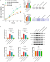
This study explored the expression of sphingosine kinase 2 (SPHK2) and microRNA miR-19a-3p (miR-19a-3p) in patients with Hypopharyngeal squamous cell carcinoma (HSCC) together with pathways affecting HSCC invasion and metastasis. Quantitative real-time polymerase chain reaction (qRT-PCR) and Western blotting (WB) were performed to assess the differential expression of SPHK2 and miR-19a-3p in patients with HSCC lymph node metastasis (LNM). Immunohistochemical (IHC) results were analyzed together with clinical information to evaluate their clinical significance. Subsequently, the functional effects of SPHK2 overexpression and knockdown on FaDu cells were evaluated in in vitro experiments. We performed in vivo experiments using nude mouse to assess the effects of SPHK2 knockdown on tumor formation, growth and LNM. Finally, we explored upstream and downstream signaling pathways associated with SPHK2 in HSCC. SPHK2 was significantly elevated in HSCC patients with LNM and survival was lower in patients with enhanced SPHK2 expression (P < 0.05). We also demonstrated that SPHK2 overexpression accelerated the proliferation, migration, and invasion. Using animal models, we further verified that SPHK2 deletion abrogated tumor growth and LNM. In terms of mechanism, we found that miR-19a-3p was significantly reduced in HSCC patients with LNM and was negatively associated with SPHK2. MiR-19a-3p and SPHK2 could regulate tumor proliferation and invasion through the PI3K/AKT axis. SPHK2 was found to contribute significantly to both LNM and HSCC patient prognosis and was shown to be an independent risk factor for LNM and staging in HSCC patients. The miR-19a-3p/SPHK2/PI3K/AKT axis was found to contribute to the development and outcome of HSCC.
Keywords: PI3P/AKT pathway; SPHK2; hypopharyngeal squamous cell carcinoma; lymph node metastasis; miRNA-19a-3p.
AJCR Copyright © 2023.
Conflict of interest statement
None.
Figures


Similar articles
-
Overexpression of microRNA-19a-3p promotes lymph node metastasis of esophageal squamous cell carcinoma via the RAC1/CDC42-PAK1 pathway.Transl Cancer Res. 2021 Jun;10(6):2694-2706. doi: 10.21037/tcr-21-254. Transl Cancer Res. 2021. PMID: 35116581 Free PMC article.
-
Overexpression of microRNA-107 suppressed proliferation, migration, invasion, and the PI3K/Akt signaling pathway and induced apoptosis by targeting Nin one binding (NOB1) protein in a hypopharyngeal squamous cell carcinoma cell line (FaDu).Bioengineered. 2022 Mar;13(3):7881-7893. doi: 10.1080/21655979.2022.2051266. Bioengineered. 2022. PMID: 35294329 Free PMC article.
-
Stathmin1 promotes lymph node metastasis in hypopharyngeal squamous cell carcinoma via regulation of HIF‑1α/VEGF‑A axis and MTA1 expression.Mol Clin Oncol. 2023 Feb 3;18(3):21. doi: 10.3892/mco.2023.2617. eCollection 2023 Mar. Mol Clin Oncol. 2023. PMID: 36844464 Free PMC article.
-
circ0005027 Acting as a ceRNA Affects the Malignant Biological Behavior of Hypopharyngeal Squamous Cell Carcinoma by Modulating miR-548c-3p/CDH1 Axis.Biochem Genet. 2024 Aug;62(4):2853-2868. doi: 10.1007/s10528-023-10570-y. Epub 2023 Nov 29. Biochem Genet. 2024. PMID: 38019338
-
Common gene signatures and key pathways in hypopharyngeal and esophageal squamous cell carcinoma: Evidence from bioinformatic analysis.Medicine (Baltimore). 2020 Oct 16;99(42):e22434. doi: 10.1097/MD.0000000000022434. Medicine (Baltimore). 2020. PMID: 33080677 Free PMC article.
Cited by
-
Decoding the interaction between miR-19a and CBX7 focusing on the implications for tumor suppression in cancer therapy.Med Oncol. 2023 Dec 19;41(1):21. doi: 10.1007/s12032-023-02251-y. Med Oncol. 2023. PMID: 38112798 Review.
-
PANDORA-Seq Unveils the Hidden Small Non-Coding RNA Landscape in Hypopharyngeal Carcinoma.Int J Mol Sci. 2025 Jun 21;26(13):5972. doi: 10.3390/ijms26135972. Int J Mol Sci. 2025. PMID: 40649749 Free PMC article.
References
-
- Li M, Lorenz RR, Khan MJ, Burkey BB, Adelstein DJ, Greskovich JF Jr, Koyfman SA, Scharpf J. Salvage laryngectomy in patients with recurrent laryngeal cancer in the setting of nonoperative treatment failure. Otolaryngol Head Neck Surg. 2013;149:245–251. - PubMed
-
- Buckley JG, MacLennan K. Cervical node metastases in laryngeal and hypopharyngeal cancer: a prospective analysis of prevalence and distribution. Head Neck. 2000;22:380–385. - PubMed
-
- Cooper JS, Porter K, Mallin K, Hoffman HT, Weber RS, Ang KK, Gay EG, Langer CJ. National cancer database report on cancer of the head and neck: 10-year update. Head Neck. 2009;31:748–758. - PubMed
-
- Roberts TJ, Colevas AD, Hara W, Holsinger FC, Oakley-Girvan I, Divi V. Number of positive nodes is superior to the lymph node ratio and American Joint Committee on Cancer N staging for the prognosis of surgically treated head and neck squamous cell carcinomas. Cancer. 2016;122:1388–1397. - PubMed
LinkOut - more resources
Full Text Sources
