. 2023 Jun 29:98:167-220.
doi: 10.3897/mycokeys.98.102816. eCollection 2023.

Culturable fungi from urban soils in China II, with the description of 18 novel species in Ascomycota (Dothideomycetes, Eurotiomycetes, Leotiomycetes and Sordariomycetes)

Affiliations

Culturable fungi from urban soils in China II, with the description of 18 novel species in Ascomycota (Dothideomycetes, Eurotiomycetes, Leotiomycetes and Sordariomycetes)

Zhi-Yuan Zhang et al. MycoKeys. .

Abstract

As China's urbanisation continues to advance, more people are choosing to live in cities. However, this trend has a significant impact on the natural ecosystem. For instance, the accumulation of keratin-rich substrates in urban habitats has led to an increase in keratinophilic microbes. Despite this, there is still a limited amount of research on the prevalence of keratinophilic fungi in urban areas. Fortunately, our group has conducted in-depth investigations into this topic since 2015. Through our research, we have discovered a significant amount of keratinophilic fungi in soil samples collected from various urban areas in China. In this study, we have identified and characterised 18 new species through the integration of morphological and phylogenetic analyses. These findings reveal the presence of numerous unexplored fungal taxa in urban habitats, emphasising the need for further taxonomic research in urban China.

Keywords: 18 new taxa; Fungal taxonomy; keratinophilic fungi; morphological characters; phylogeny.


Conflict of interest statement

The authors have declared that no competing interests exist.

Figures

Figure 1.
Figure 1.
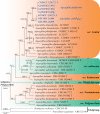
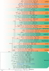
Concatenated phylogeny of the ITS, LSU, EF1A, TUB and RPB2 gene regions of species in Sympoventuriaceae. Thirty-five strains are used. The tree is rooted with Pseudoanungiteavaccinii (CPC 30523) and P.vaccinii (CBS 143164). The tree topology of the BI was similar to the ML analysis. Bayesian posterior probability (≥ 0.8) and ML bootstrap values (≥ 80%) are indicated along branches (PP/ML). Novel species are in blue and bold font and “T” indicates type derived sequences.
Figure 2.
Figure 2.
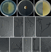
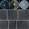
Echinocatenasinensis (from ex-holotype CGMCC 3.20775) a–c upper and reverse views of cultures on PDA, MEA and OA 14 d after inoculation d–g phialides and conidia. Scale bars: 10 µm (d–g).
Figure 3.
Figure 3.
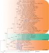
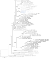
Concatenated phylogeny of the ITS, TUB, CaM, RPB2 and TSR1 gene regions of species in Aspergillus from subgenus Polypaecilum. Thirty-five strains are used. The tree is rooted in Hamigeraavellanea (CBS 295.48) and Penicilliumexpansum (CBS 325.48). The tree topology of the BI was similar to the ML analysis. Bayesian posterior probability (≥ 0.8) and ML bootstrap values (≥ 80%) are indicated along branches (PP/ML). Novel species are in blue and bold font and “T” indicates type derived sequences.
Figure 4.
Figure 4.
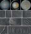
Aspergilluscylindricus (from ex-holotype CGMCC 3.20771) a–c upper and reverse views of cultures on PDA, MEA and OA 14 d after inoculation d–l phialides and conidia. Scale bars: 10 µm (d–l).
Figure 5.
Figure 5.
Aspergillusdoliiformis (from ex-holotype CGMCC 3.20772) a–c upper and reverse views of cultures on PDA, MEA and OA 14 d after inoculation d–k phialides and conidia. Scale bars: 10 µm (d–k).
Figure 6.
Figure 6.
Concatenated phylogeny of the ITS, TUB, CaM, RPB2 and TSR1 gene regions of species in Penicillium from section Citrina. Forty-eight strains are used. The tree is rooted in Aspergillusniger (CBS 554.65) and Hamigeraavellanea (CBS 295.48). The tree topology of the BI was similar to the ML analysis. Bayesian posterior probability (≥ 0.8) and ML bootstrap values (≥ 80%) are indicated along branches (PP/ML). Novel species are in blue and bold font and “T” indicates type derived sequences.
Figure 7.
Figure 7.
Penicilliumfujianense (from ex-holotype CGMCC 3.20781) a–c upper and reverse views of cultures on PDA, MEA and OA 14 d after inoculation d–k phialides and conidia. Scale bars: 10 µm (d–k).
Figure 8.
Figure 8.
Concatenated phylogeny of the ITS, TUB, CaM and RPB2 gene regions of species in Talaromyces from sections Islandici, Bacillispori and Subinflati. Fifty-six strains are used. The tree is rooted with Talaromycesbrunneosporus (FMR 16566) and T.tenuis (CBS 141840). The tree topology of the BI was similar to the ML analysis. Bayesian posterior probability (≥ 0.8) and ML bootstrap values (≥ 80%) are indicated along branches (PP/ML). Novel species are in blue and bold font and “T” indicates type derived sequences.
Figure 9.
Figure 9.
Talaromycesguiyangensis (from ex-holotype CGMCC 3.20782) a–c upper and reverse views of cultures on PDA, MEA and OA 14 d after inoculation d–g phialides and conidia h conidia chain. Scale bars: 10 µm (d–h).
Figure 10.
Figure 10.
Talaromycesjiangxiensis (from ex-holotype CGMCC 3.20783) a–c upper and reverse views of cultures on PDA, MEA and OA 14 d after inoculation d–k phialides and conidia. Scale bars: 10 µm (d–k).
Figure 11.
Figure 11.
Talaromycespaecilomycetoides (from ex-holotype CGMCC 3.20785) a–c upper and reverse views of cultures on PDA, MEA and OA 14 d after inoculation d–k phialides and conidia l conidia. Scale bars: 10 µm (d–l).
Figure 12.
Figure 12.
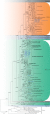
Concatenated phylogeny of the ITS, LSU, TUB, TEF3 and RP 60S L1 gene regions of species in Arthrodermataceae. Three hundred and eighteen strains are used. The tree topology of the BI was similar to the ML analysis. Bayesian posterior probability (≥ 0.7) and ML bootstrap values (≥ 70%) are indicated along branches (PP/ML). Novel species are in blue and bold font, new isolates are in blue and “T” indicates type derived sequences.
Figure 13.
Figure 13.
Nannizziasinensis (from ex-holotype CGMCC 3.20873) a–c upper and reverse views of cultures on PDA, MEA and OA 14 d after inoculation d subspherical to spherical microconidia e–h conidiophores with sessile or stalked microconidia i–k conidiophores and macroconidia l macroconidia. Scale bars: 10 µm (d–l).
Figure 14.
Figure 14.
Concatenated phylogeny of the ITS, LSU, EF1A, RPB2 and MCM7 gene regions of species in Thelebolaceae. Ninety-nine strains are used. The tree is rooted in Leuconeurosporapulcherrima (CBS 343.76) and Leuconeurospora sp. (15PA04 and 02NH04). The tree topology of the BI was similar to the ML analysis. Bayesian posterior probability (≥ 0.7) and ML bootstrap values (≥ 70%) are indicated along branches (PP/ML). Novel species are in blue and bold font, new isolates are in blue and “T” indicates type derived sequences.
Figure 15.
Figure 15.
Pseudogymnoascusbotryoides (from ex-holotype CGMCC 3.20875) a–c upper and reverse views of cultures on PDA, MEA and OA 14 d after inoculation d–h conidiophores and conidia i, j cluster conidia. Scale bars: 10 µm (d–j).
Figure 16.
Figure 16.
Pseudogymnoascuscamphorae (from ex-holotype CGMCC 3.20876) a–c upper and reverse views of cultures on PDA, MEA and OA 14 d after inoculation d–i conidiophores and conidia j conidia. Scale bars: 10 µm (d–j).
Figure 17.
Figure 17.
Pseudogymnoascuspapyriferae (from ex-holotype CGMCC 3.20877) a–c upper and reverse views of cultures on PDA, MEA and OA 14 d after inoculation d–i conidiophore and conidia. Scale bars: 10 µm (d–i).
Figure 18.
Figure 18.
Pseudogymnoascuszongqii (from ex-holotype CGMCC 3.20878) a–c upper and reverse views of cultures on PDA, MEA and OA 14 d after inoculation d–f, h, i conidia and intercalary conidia g–k conidiophore and conidia. Scale bars: 10 µm (d–k).
Figure 19.
Figure 19.
Concatenated phylogeny of the ITS and TUB gene regions of species in Clonostachys. Sixty strains are used. The tree is rooted in Fusariumacutatum (CBS 402.97) and Calonectriailicicola (CBS 190.50). The tree topology of the BI was similar to the ML analysis. Bayesian posterior probability (≥ 0.7) and ML bootstrap values (≥ 70%) are indicated along branches (PP/ML). Novel species are in blue and bold font and “T” indicates type derived sequences.
Figure 20.
Figure 20.
Clonostachysshanghaiensis (from ex-holotype CGMCC 3.20773) a–c upper and reverse views of cultures on PDA, MEA and OA 14 d after inoculation d–g conidiophore and conidia. Scale bars: 10 µm (d–g).
Figure 21.
Figure 21.
Concatenated phylogeny of the ITS, LSU, RPB2 and EF1A gene regions of species in Cyanonectria and its allied genera. Thirty-three strains are used. The tree is rooted in Ilyonectriadestructans (CBS 264.65) and I.capensis (CBS 132815). The tree topology of the BI was similar to the ML analysis. Bayesian posterior probability (≥ 0.7) and ML bootstrap values (≥ 70%) are indicated along branches (PP/ML). Novel species are in blue and bold font and “T” indicates type derived sequences.
Figure 22.
Figure 22.
Cyanonectriabispora (from ex-holotype CGMCC 3.20774) a–c upper and reverse views of cultures on PDA, MEA and OA 14 d after inoculation d–f monophialide and conidia g, h polyphialides with multiple conidiogenous loci i sporodochial conidiophore and conidiogenous cells j conidia. Scale bars: 10 µm (d–j).
Figure 23.
Figure 23.
Concatenated phylogeny of the ITS, LSU, RPB2 and EF1A gene regions of species in Fusarium. Sixty-three strains are used. The tree is rooted in Ilyonectriadestructans (CBS 264.65) and I.capensis (CBS 132815). The tree topology of the BI was similar to the ML analysis. Bayesian posterior probability (≥ 0.7) and ML bootstrap values (≥ 70%) are indicated along branches (PP/ML). Novel species are in blue and bold font and “T” indicates type derived sequences.
Figure 24.
Figure 24.
Fusariumbrachypodum (from ex-holotype CGMCC 3.20776) a–c upper and reverse views of cultures on PDA, MEA and OA 14 d after inoculation d, e conidiophores and phialides on aerial mycelium f–i sporodochia, sporodochial conidiophores and conidia j spiral hyphae k chlamydospores l conidia. Scale bars: 10 µm (d–l).
Figure 25.
Figure 25.
Concatenated phylogeny of the ITS, LSU, EF1A and ACT gene regions of species in Niesslia. Sixty strains are used. The tree is rooted in Trichodermaaggressivumf.europaeum (CBS 100526). The tree topology of the BI was similar to the ML analysis. Bayesian posterior probability (≥ 0.7) and ML bootstrap values (≥ 70%) and are indicated along branches (PP/ML). Novel species are in blue and bold font and “T” indicates type derived sequences.
Figure 26.
Figure 26.
Niessliaguizhouensis (from ex-holotype CGMCC 3.20780) a–c upper and reverse views of cultures on PDA, MEA and OA 14 d after inoculation d–j monocillium-like conidiophores and conidia k hyphal coil with conidiophores. Scale bars: 10 µm (d–k).
Figure 27.
Figure 27.
Concatenated phylogeny of the ITS, LSU, TUB and RPB2 gene regions of species in Microdochiaceae. Thirty-two strains are used. The tree is rooted in Cryptostromacorticale (CBS 218.52 and CBS 217.52). The tree topology of the BI was similar to the ML analysis. Bayesian posterior probability (≥ 0.8) and ML bootstrap values (≥ 80%) are indicated along branches (PP/ML). Novel species are in blue and bold font, and “T” indicates type derived sequences.
Figure 28.
Figure 28.
Idriellachlamydospora (from ex-holotype CGMCC 3.20778) a–c upper and reverse views of cultures on PDA, MEA and OA 14 d after inoculation d–g chlamydospores. Scale bars: 10 µm (d–g).
Figure 29.
Figure 29.
Idriellamultiformispora (from ex-holotype CGMCC 3.20779) a–c upper and reverse views of cultures on PDA, MEA and OA 14 d after inoculation d–f conidiogenous cells and conidia g hyphal coil with phialides h–k chlamydospores. Scale bars: 10 µm (d–k).

Similar articles

Cited by

References

    1. Antrop M. (2004) Landscape change and the urbanization process in Europe. Landscape and Urban Planning 67(1–4): 9–26. 10.1016/S0169-2046(03)00026-4 - DOI
    1. Auerswald B. (1869) Synopsis pyrenomycetum europaeorum. In: Gonnerman W, Rabenhorst GL (Eds) Mycologia Europaea.
    1. Baert F, Stubbe D, D’hooge E, Packeu A, Hendrickx M. (2020) Updating the taxonomy of dermatophytes of the BCCM/IHEM collection according to the new standard: A phylogenetic approach. Mycopathologia 185: 161–168. 10.1007/s11046-019-00338-7 - DOI - PubMed
    1. Barbosa RN, Bezerra JDP, Souza-Motta CM, Frisvad JC, Samson RA, Oliveira NT, Houbraken J. (2018) New Penicillium and Talaromyces species from honey, pollen and nests of stingless bees. Antonie van Leeuwenhoek 111(10): 1883–1912. 10.1007/s10482-018-1081-1 - DOI - PMC - PubMed
    1. Benjamin CR. (1955) Ascocarps of Aspergillus and Penicillium. Mycologia 47(5): 669–687. 10.1080/00275514.1955.12024485 - DOI

LinkOut - more resources