. 2020 Nov-Dec;24(11n12):1320-1360.
doi: 10.1142/s1088424620300098.

Photodynamic therapy, priming and optical imaging: Potential co-conspirators in treatment design and optimization - a Thomas Dougherty Award for Excellence in PDT paper

Affiliations

Photodynamic therapy, priming and optical imaging: Potential co-conspirators in treatment design and optimization - a Thomas Dougherty Award for Excellence in PDT paper

Pushpamali De Silva et al. J Porphyr Phthalocyanines. 2020 Nov-Dec.

Abstract

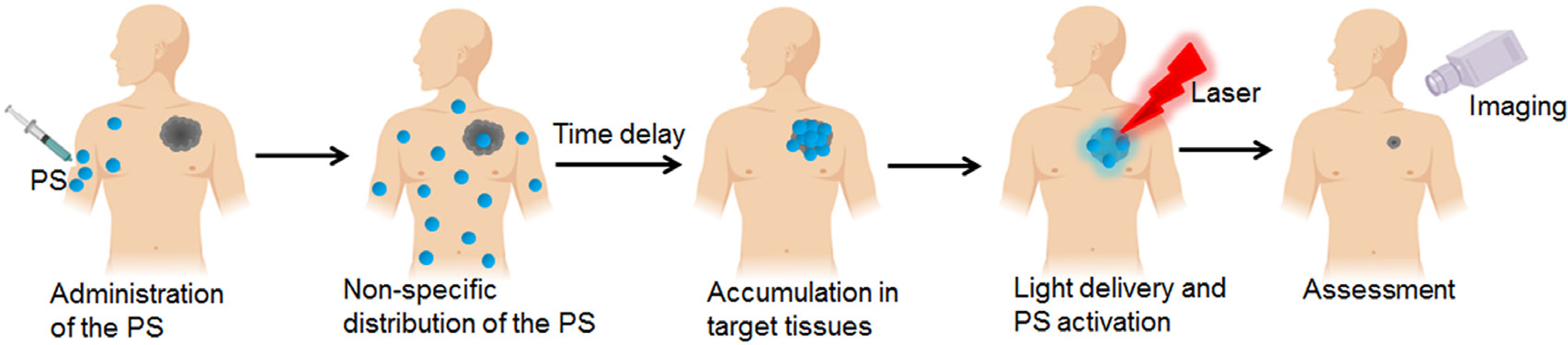
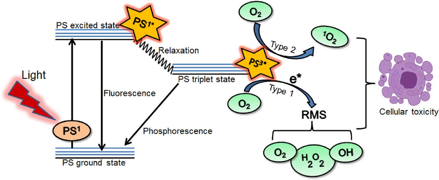
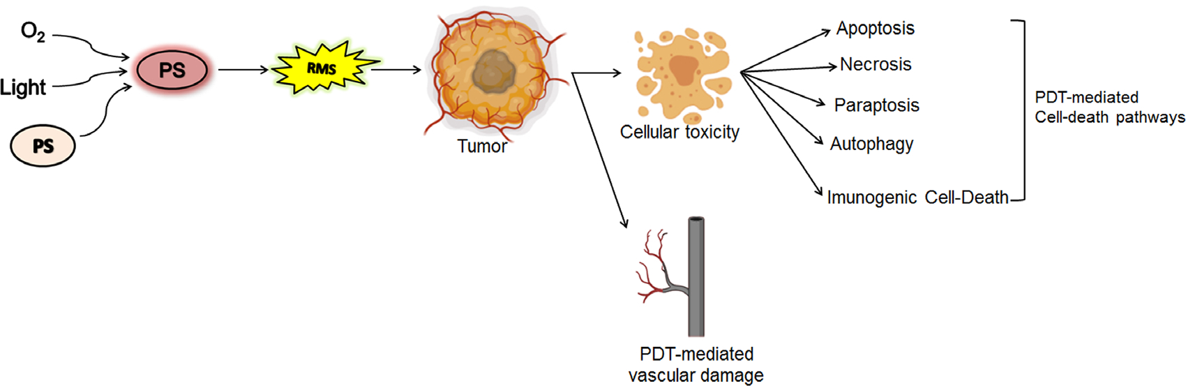
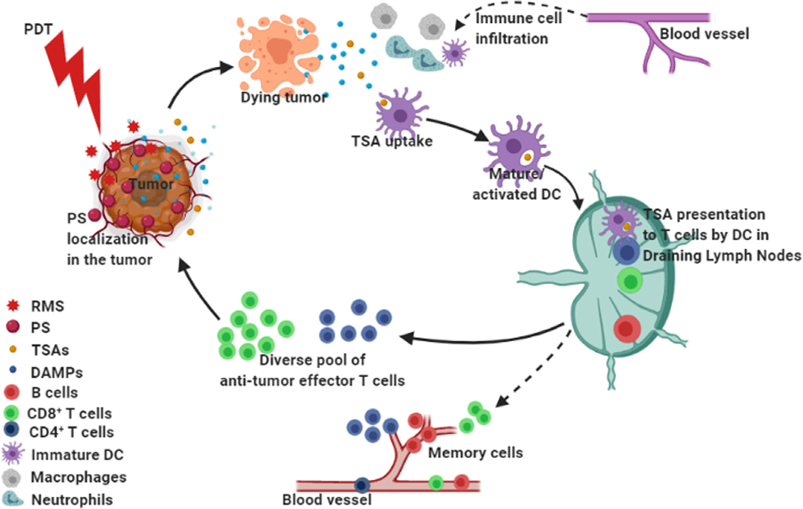
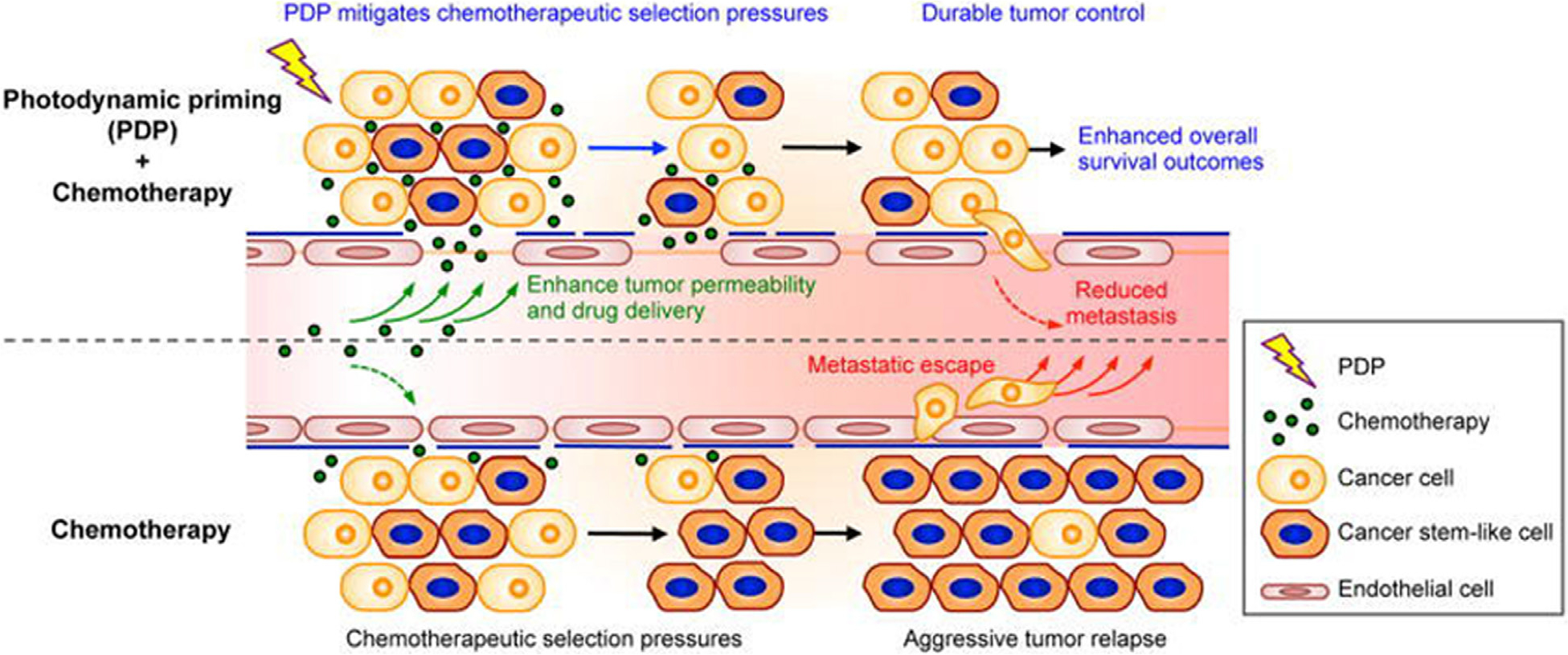
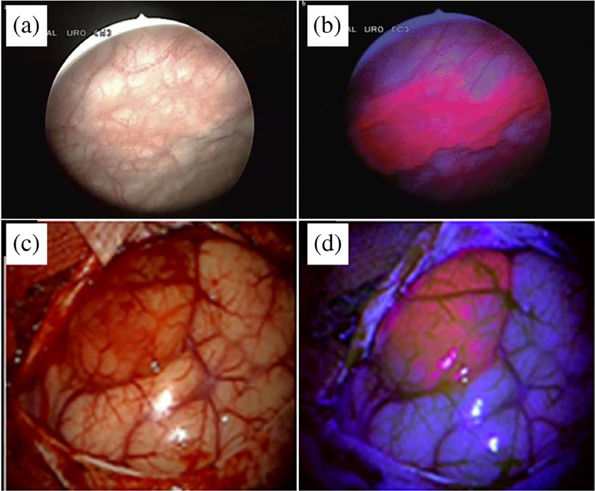
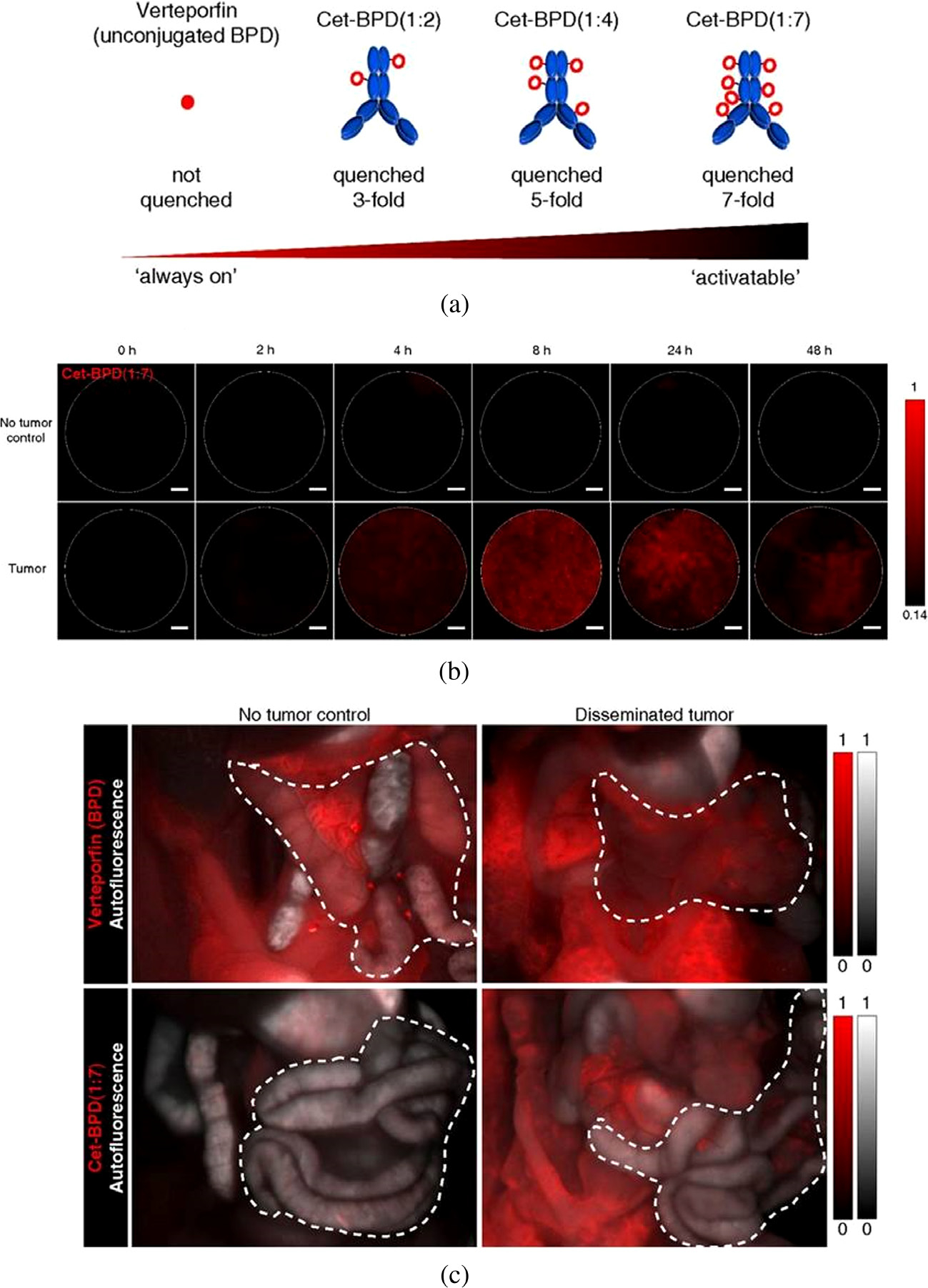
Photodynamic therapy is a photochemistry-based approach, approved for the treatment of several malignant and non-malignant pathologies. It relies on the use of a non-toxic, light activatable chemical, photosensitizer, which preferentially accumulates in tissues/cells and, upon irradiation with the appropriate wavelength of light, confers cytotoxicity by generation of reactive molecular species. The preferential accumulation however is not universal and, depending on the anatomical site, the ratio of tumor to normal tissue may be reversed in favor of normal tissue. Under such circumstances, control of the volume of light illumination provides a second handle of selectivity. Singlet oxygen is the putative favorite reactive molecular species although other entities such as nitric oxide have been credibly implicated. Typically, most photosensitizers in current clinical use have a finite quantum yield of fluorescence which is exploited for surgery guidance and can also be incorporated for monitoring and treatment design. In addition, the photodynamic process alters the cellular, stromal, and/or vascular microenvironment transiently in a process termed photodynamic priming, making it more receptive to subsequent additional therapies including chemo- and immunotherapy. Thus, photodynamic priming may be considered as an enabling technology for the more commonly used frontline treatments. Recently, there has been an increase in the exploitation of the theranostic potential of photodynamic therapy in different preclinical and clinical settings with the use of new photosensitizer formulations and combinatorial therapeutic options. The emergence of nanomedicine has further added to the repertoire of photodynamic therapy's potential and the convergence and co-evolution of these two exciting tools is expected to push the barriers of smart therapies, where such optical approaches might have a special niche. This review provides a perspective on current status of photodynamic therapy in anti-cancer and anti-microbial therapies and it suggests how evolving technologies combined with photochemically-initiated molecular processes may be exploited to become co-conspirators in optimization of treatment outcomes. We also project, at least for the short term, the direction that this modality may be taking in the near future.

Keywords: antimicrobial PDT; combination therapies; immunogenic cell death; optical imaging; photodynamic priming; photodynamic therapy; photoimmunoconjugates.


Conflict of interest statement

Conflict of interest none

Figures

Fig. 1.
Fig. 1.
Photodynamic therapy as a single modality for therapy and imaging. Photosensitizer (PS) is administered systemically, following which it preferentially localizes at the desired site. The time delay following PS administration and its subsequent irradiation is referred to as the drug-light-interval. Irradiation of the PS results in reactive molecular species generation and fluorescence emission, which could be used for inducing cytotoxicity and imaging, respectively.
Fig. 2.
Fig. 2.
Photochemical and photophysical reactions associated with photodynamic therapy. When the photosensitizer (PS; ground state) absorbs light (photon) at a particular wavelength, it is first excited to singlet state (PS1*) and then converted to a more stable triplet state (PS3*). This triplet state (PS3*) can react with molecular oxygen and other biomolecules through the so-called type 1 and type 2 reactions, creating highly reactive molecular species (RMS) and singlet oxygen (1O2), both of which can cause cellular toxicity.
Fig. 3.
Fig. 3.
Generation of reactive molecular species (RMS) upon light activation of the photosensitizer (PS) and PDT associated cell death pathways and vascular damage that could occur as a result.
Fig. 4.
Fig. 4.
Cellular changes associated with (a) necrosis, (b, d) apoptosis and (c, e) paraptosis. Panels (a, b) and (c) show phase-contrast images of cells undergoing apoptosis and paraptosis (d) Nuclear condensation and fragmentation typical of apoptosis. (e) Nuclear condensation and fragmentation are not observed during paraptosis. The fluorescent label Ho33342 was used to probe nuclear morphology in panels (d) and (e). Figure (a) adapted from Lee et al. (2018) [85]) and Figure panels (b–e) adapted from Kessel and Oleinick (2018) [80].
Fig. 5.
Fig. 5.
PDT mediated anti-tumor immune processes that may occur in the tumor microenvironment (TME). Irradiation of PS-loaded tumor cells generates RMS that leads to tumor cell death. The photodynamic process results in priming of the TME where the dying cells express or release damage associated molecular patterns (DAMPs). Photodamaged tumor cells and tissue-resident immune cells may release numerous cytokines and chemokines that may induce extravasation of innate immune cells to the tumor site causing local inflammation. Professional antigen presenting cells such as Dendritic cells (DCs) play a key role in bridging the innate immune response with adaptive immunity. Tumor specific antigens (TSAs) captured by immature DCs become activated and migrate to the draining lymph nodes and help to prime naïve T cells (CD4+ helper T or CD8+ cytotoxic T cells), which differentiate into effector or memory T cell subsets. Anti-tumor effector T cell populations, especially CD8+ cytotoxic T cells may migrate to the tumor site in search of the cognate TSAs and kill the tumor cells. We postulate that PDT may enrich a diverse pool of antitumor T cell clones in the TME to kill the tumor and possibly expand long lived memory T and B cells that get into the peripheral circulation to maintain immune surveillance.
Fig. 6.
Fig. 6.
The impact of PDP on modulating multiple compartments in the tumor microenvironment. Firstly, PDP of tumor microvasculature and parenchyma simultaneously improves therapeutic agent accessibility and overcomes chemotherapeutic selection pressures. Secondly, sublethal PDP increases tumor permeability to enhance intratumoral accumulation of chemotherapeutic agents for a prolonged period of time. Thirdly, PDP attenuates the insidious surge of stemness marker expression that is typically observed after multiple cycles of chemotherapy. PDT-mediated immune enhancement discussed above is an example of such a priming process (PDP) where the impact reaches far beyond the cells directly targeted by PDT. Figure adapted from Huang et al. (2018) [42].
Fig. 7.
Fig. 7.
Bladder (a and b) and brain (c and d) tumor detection with regular white light (a and c), and fluorescence (b and d) imaging of PpIX. Images in (a) and (b) were acquired by an equipment for photodynamic diagnosis equipped with a light source (short-arc xenon lamp with a specially designed dielectric short-pass filter (375–440 nm)) for excitation light that can be transmitted through modified cytoscopes and lenses to maximally enhance the contrast between benign tissue and fluorescence from malignancies. Images in (c) and (d) were acquired by neurosurgical microscope equipped with a fluorescent 400 nm UV light module. Figures adapted from Zaak et al. (2005) [191] and Goraynov et al. (2019) [190].
Fig. 8:
Fig. 8:
Target activatable fluorescence detection and photo-immunotherapy (taPIT). (a) PICs (Cetuximab-BPD) with different PS loading ratios, leading to varying BPD self-quenching efficiencies. (b) Longitudinal microendoscopic images of the peritoneal cavity demonstrate the efficacy of PICs to identify micrometastatic sites of ovarian cancer at 8–24 h post administration. (c) BPD fluorescence (red) and autofluorescence (gray scale) of the peritoneal cavity 2 h after free BPD or 48 h after PIC (Cetuximab-BPD) administration. Figure adapted from Spring et al. (2014) [198].
Fig. 9.
Fig. 9.
Experimental setup (a), anatomical images of pre- and post-PDT treated (b) and control (d) mice. BOLD-MRI intensity of control (c) and treated mice (e). Color coding represents ratio of signal intensity over baseline. Figure adapted from Gross et al. (2003) [251].
Fig. 10.
Fig. 10.
(a), (b) Ultrasound and photoacoustic images of oxygen saturation (StO2) and total hemoglobin (HbT) demonstrating mapping of hypoxic (blue) and oxygenated (red) regions in a murine model of glioblastoma. 1 h DLI and 3 h DLI refer to initiation of irradiation 1- or 3-h following PS-application. Green region outlines tumor region identified using ultrasound (c), (d) Mean StO2 and HbT values at pre-PDT, post-PDT, 6 h, and 2 h. Figure adapted from Mallidi et al. (2015) [16].
Fig. 11.
Fig. 11.
Schematic diagram of a targeted multi-agent nanoconstruct. (1) The surface is grafted with targeting moieties to endow molecular selectivity. (2) Light activation/illumination of the photosensitizer can be used for (3) fluorescence-based imaging (4), photochemical generation of cytotoxic reactive molecular species (RMS) for PDT (5), sequential, controlled release of synergistic therapeutic agents for combinatorial cancer therapy, and for (6) photodynamic priming of tumor microenvironment. Figure adapted from Huang and Hasan (2014) [24].
Fig. 12.
Fig. 12.
Concept of spatiotemporal-synchronized combination therapy using PMILs. (a) NIR light activation of PMILs within the tumour microvasculature for simultaneous neovascular damage, tumor cell apoptosis and necrosis and liposomal disruption to initiate sustained multikinase inhibition. (b) Schematic of a three-way interactive combination therapy with photodynamic tumour cell and microvasculature damage and inhibition of treatment escape pathways. Figure adapted from Spring et al. (2016) [41].
Fig. 13.
Fig. 13.
Schematic illustration of the formation of angiogenesis vessel-targeting NPs (AVT-NPs), generation of cytotoxic Tirapazamine (TPZ) radical under hypoxic conditions in cancer cells, and illustration of AVT-NP/TPZ based PDT that induces a local hypoxic environment and promotes angiogenesis for targeted drug delivery and synergistic chemo-phototherapy. Figure adapted from Guo et al. (2017) [342].
Fig. 14.
Fig. 14.
Mechanism of action of tumor-penetrating nanoparticles in a combined PDT and hypoxia-activated treatment strategy. Figure adapted from Wang et al. (2017), Copyright © 2017, American Chemical Society [346].
Fig. 15.
Fig. 15.
A schematic presentation of targeting ligands used to functionalize nanoconstructs to mediate the molecular selectivity of PDT damage. These include mAbs and their fragments, glycans, folate molecules targeting the folate receptor, and peptides. Figure adapted from Obaid et al. (2016), reproduced by permission of The Royal Society of Chemistry [280].
Fig. 16.
Fig. 16.
Light-controlled two-step approach for selective delivery of cell penetrating light activatable nanoconstructs for targeted photoinactivation of Ki-67 protein. Figure adapted from Wang et al. (2015), Copyright © 2015, American Chemical Society [402].
Fig. 17.
Fig. 17.
Mouse with wounds infected with E. coli, treated with conjugates (PS plus polymer) followed by illumination at 660–665 nm with different fluence rates along with phototoxicity analysis in vitro. (a) Four excisional wounds, (b) wound 1 and 2 received topical application of the conjugate, (c)–(f) wounds 1 and 2 were then illuminated with different fluence rates (40–160 J/cm2), (g), (h) the same mouse 24 h and 48 h later. Phototoxicity of the conjugates towards bacteria determined by (i) CFU counts, and (j) luminescence assays. Figure adapted from Hamblin et al. (2002) [434].
Fig. 18.
Fig. 18.
Efficacy of PDT against Leishmania in preclinical models. (a) Control ears with no treatment, (b) Topical ALA-PDT on cutaneous leishmaniasis (CL), (c) Quantification of L. major parasites. (d)–(g) Clinical outcome of patients with cutaneous leishmaniasis before (d) and (f) and after treatment (e and g) with daylight-activated photodynamic therapy (DA-PDT). Figure adapted from Akilov et al. (2007) and Enk et al. (2015) [459, 462].
Fig. 19.
Fig. 19.
Schematic representation of the β-LEAF and β-LEAP strategies.
Fig. 20.
Fig. 20.
Schematic diagram and results of ALA-PDT against malaria (a) Depiction of ALA-uptake and porphyrin biosynthesis pathways in Plasmodium-infected erythrocytes. (b) Growth of malaria parasites in the presence or absence of ALA and succinyl acetone (SA, an agent used to block PpIX synthesis), with light exposure. Figure adapted from Sigala et al. (2015) [480].

Similar articles

Cited by

References

    1. Hamblin MR and Hasan T. Photochem. Photobiol. Sci. 2004; 3: 436–450. - PMC - PubMed
    1. Baskaran R, Lee J and Yang SG. Biomater. Res. 2018; 22: 25. - PMC - PubMed
    1. Photodiagn Huang Z.. Photodyn. Ther. 2008; 5: 285–287. - PubMed
    1. Blanco Kate C, Inada Natalia M, Salvio Ana Gabriela, Vollet-Filho José Dirceu and VS Bagnato. J. Tumor 2016; 4: 386–392.
    1. Frochot C and Mordon S. J. Porphyrins Phthalocyanines 2019; 23: 347–357.

LinkOut - more resources