

Mueller-matrix imaging polarimetry elevated by wavelet decomposition and polarization-singular processing for analysis of specific cancerous tissue pathology
- PMID: 37425430
- PMCID: PMC10329407
- DOI: 10.1117/1.JBO.28.10.102903
Mueller-matrix imaging polarimetry elevated by wavelet decomposition and polarization-singular processing for analysis of specific cancerous tissue pathology
Abstract
Significance: Mueller-matrix polarimetry is a powerful method allowing for the visualization of malformations in biological tissues and quantitative evaluation of alterations associated with the progression of various diseases. This approach, in fact, is limited in observation of spatial localization and scale-selective changes in the poly-crystalline compound of tissue samples.
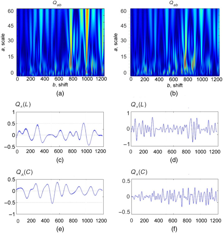
Aim: We aimed to improve the Mueller-matrix polarimetry approach by implementing the wavelet decomposition accompanied with the polarization-singular processing for express differential diagnosis of local changes in the poly-crystalline structure of tissue samples with various pathology.
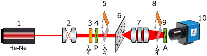
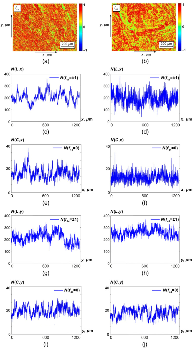
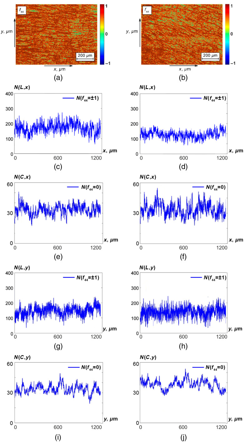
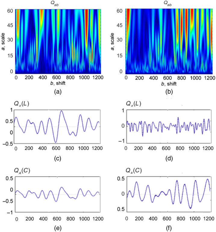
Approach: Mueller-matrix maps obtained experimentally in transmitted mode are processed utilizing a combination of a topological singular polarization approach and scale-selective wavelet analysis for quantitative assessment of the adenoma and carcinoma histological sections of the prostate tissues.
Results: A relationship between the characteristic values of the Mueller-matrix elements and singular states of linear and circular polarization is established within the framework of the phase anisotropy phenomenological model in terms of linear birefringence. A robust method for expedited (up to ) polarimetric-based differential diagnosis of local variations in the poly-crystalline structure of tissue samples containing various pathology abnormalities is introduced.
Conclusions: The benign and malignant states of the prostate tissue are identified and assessed quantitatively with a superior accuracy provided by the developed Mueller-matrix polarimetry approach.
Keywords: Mueller-matrix; birefringence; cancer; imaging polarimetry; polarization-singular; polarized light; wavelet decomposition.
© 2023 The Authors.
Figures
Similar articles
-
Differential Mueller matrix imaging of partially depolarizing optically anisotropic biological tissues.Lasers Med Sci. 2020 Jun;35(4):877-891. doi: 10.1007/s10103-019-02878-2. Epub 2019 Nov 20. Lasers Med Sci. 2020. PMID: 31749042 Free PMC article.
-
Mueller matrix-based characterization of cervical tissue sections: a quantitative comparison of polar and differential decomposition methods.J Biomed Opt. 2024 May;29(5):052916. doi: 10.1117/1.JBO.29.5.052916. Epub 2024 Feb 7. J Biomed Opt. 2024. PMID: 38328279 Free PMC article.
-
Mueller matrix imaging of prostate bulk tissues; Polarization parameters as a discriminating benchmark.Photodiagnosis Photodyn Ther. 2019 Jun;26:90-96. doi: 10.1016/j.pdpdt.2019.02.017. Epub 2019 Feb 20. Photodiagnosis Photodyn Ther. 2019. PMID: 30797118
-
Polarized light imaging in biomedicine: emerging Mueller matrix methodologies for bulk tissue assessment.J Biomed Opt. 2015 Jun;20(6):61104. doi: 10.1117/1.JBO.20.6.061104. J Biomed Opt. 2015. PMID: 25793658 Review.
-
Characterization of cervical tissue using Mueller matrix polarimetry.Lasers Med Sci. 2023 Jan 20;38(1):46. doi: 10.1007/s10103-023-03712-6. Lasers Med Sci. 2023. PMID: 36662327 Review.
Cited by
-
3D polarization-interference holographic histology for wavelet-based differentiation of the polycrystalline component of biological tissues with different necrotic states. Forensic applications.J Biomed Opt. 2024 May;29(5):052920. doi: 10.1117/1.JBO.29.5.052920. Epub 2024 Mar 15. J Biomed Opt. 2024. PMID: 38495527 Free PMC article.
-
Insights into polycrystalline microstructure of blood films with 3D Mueller matrix imaging approach.Sci Rep. 2024 Jun 13;14(1):13679. doi: 10.1038/s41598-024-63816-z. Sci Rep. 2024. PMID: 38871757 Free PMC article.
References
-
- Tuchin V. V., “Light scattering study of tissues,” Physics-Uspekhi 40(5), 495 (1997).PHUSEY10.1070/PU1997v040n05ABEH000236 - DOI
-
- Ushenko A., et al. , “Stokes-correlometry analysis of biological tissues with polycrystalline structure,” IEEE J. Sel. Top. Quantum Electron. 25(1), 7101612 (2019).IJSQEN10.1109/JSTQE.2018.2865443 - DOI
-
- Ushenko V., et al. , “3D Mueller-matrix diffusive tomography of polycrystalline blood films for cancer diagnosis,” Photonics 5(4), 54 (2018).10.3390/photonics5040054 - DOI
-
- Ushenko V. A., et al. , “Biomedical application of jones-matrix tomography to polycrystalline films of biological fluids,” J. Innov. Opt. Health Sci. 12(6), 1950017 (2019).10.1142/S1793545819500172 - DOI
Publication types
MeSH terms
LinkOut - more resources
Full Text Sources
Medical
