

Right Coronary Artery Button Pseudoaneurysm After the Modified Bentall Procedure
- PMID: 37425512
- PMCID: PMC10329497
- DOI: 10.7759/cureus.40144
Right Coronary Artery Button Pseudoaneurysm After the Modified Bentall Procedure
Abstract
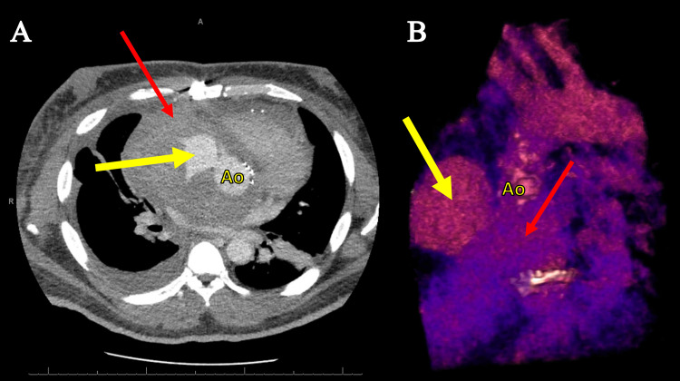
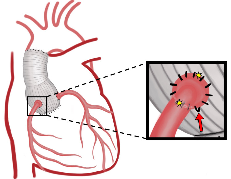
Anastomoses of the coronary buttons are the Achilles' heel of the modified Bentall procedure (MBP) for the repair of the aortic root and ascending aorta. We present a rare case of post-MBP right coronary artery button pseudoaneurysm in a 30-year-old man. The contained leak, attributed to a pseudoknot in the polypropylene suture, was visualized via computed tomography angiography and transesophageal echocardiogram and repaired under deep hypothermic circulatory arrest.
Keywords: bentall; coronary button pseudoaneurysm; coronary ostial complication; modified bentall procedure; pseudoaneurysm; pseudoknot.
Copyright © 2023, Nguyen et al.
Conflict of interest statement
The authors have declared that no competing interests exist.
Figures

Similar articles
-
[MEP-12] Rare and Catastrophic Complication After Bentall Operation: Right Coronary Artery Button Pseudoaneursym Repaired with Modified Cabrol Technique.Turk Gogus Kalp Damar Cerrahisi Derg. 2024 Dec 31;32(4 Suppl 2):108-109. doi: 10.5606/tgkdc.dergisi.2024.mep-12. eCollection 2024 Nov. Turk Gogus Kalp Damar Cerrahisi Derg. 2024. PMID: 40322167 Free PMC article.
-
Pseudoaneurysm of the left coronary ostial anastomoses as a complication of the modified Bentall procedure diagnosed by echocardiography and multislice computed tomography.Heart Surg Forum. 2007;10(3):E191-2. doi: 10.1532/HSF98.20061206. Heart Surg Forum. 2007. PMID: 17389208
-
Fate of coronary ostial anastomoses after the modified Bentall procedure.Ann Thorac Surg. 2003 Jun;75(6):1797-801; discussion 1802. doi: 10.1016/s0003-4975(03)00015-8. Ann Thorac Surg. 2003. PMID: 12822618
-
Successful Hybrid Zone 0 Landing Thoracic Endovascular Aortic Repair for Ascending Aortic Pseudoaneurysm after Bentall Procedure and Coronary Artery Bypass Grafting in Takayasu Arteritis.Ann Vasc Surg. 2019 Jan;54:335.e7-335.e10. doi: 10.1016/j.avsg.2018.06.018. Epub 2018 Aug 13. Ann Vasc Surg. 2019. PMID: 30114506
-
The modified Bentall procedure for aortic root replacement.AORN J. 2006 Jul;84(1):52-5, 58-70; quiz 71-4. doi: 10.1016/s0001-2092(06)60098-7. AORN J. 2006. PMID: 16881491 Review.
Cited by
-
Research Progress on Aortic Root Aneurysms.Med Sci Monit. 2024 Feb 9;30:e943216. doi: 10.12659/MSM.943216. Med Sci Monit. 2024. PMID: 38332569 Free PMC article. Review.
References
-
- Fate of coronary ostial anastomoses after the modified Bentall procedure. Milano AD, Pratali S, Mecozzi G, Boraschi P, Braccini G, Magagnini E, Bortolotti U. Ann Thorac Surg. 2003;75:1797–1801. - PubMed
-
- A 23-year experience with composite valve graft replacement of the aortic root. Dossche KM, Schepens MA, Morshuis WJ, de la Rivière AB, Knaepen PJ, Vermeulen FE. Ann Thorac Surg. 1999;67:1070–1077. - PubMed
-
- The modified Bentall procedure: a single-institution experience in 249 patients with a maximum follow up of 21.5 years. Celiento M, Ravenni G, Margaryan R, Ferrari G, Blasi S, Pratali S, Bortolotti U. https://pubmed.ncbi.nlm.nih.gov/28009948/ J Heart Valve Dis. 2016;25:448–455. - PubMed
-
- Post-Bentall ascending aortic pseudoaneurysm due to coronary button dehiscence. Sivakumar K, Sagar P. Ann Thorac Surg. 2021;111:0–4. - PubMed
-
- Aortic pseudoaneurysm repair after Bentall procedure using the IntraClude device. Mayr B, Alalawi Z, Ziegelmüller JA, Nöbauer C, Krane M, Lange R, Voss B. J Card Surg. 2020;35:3617–3619. - PubMed
Publication types
LinkOut - more resources
Full Text Sources
Miscellaneous