

Juvenile Ossifying Fibroma and Socioeconomic Barriers to Specialty Care: A Pediatric Case Study
- PMID: 37425522
- PMCID: PMC10325821
- DOI: 10.7759/cureus.40059
Juvenile Ossifying Fibroma and Socioeconomic Barriers to Specialty Care: A Pediatric Case Study
Abstract
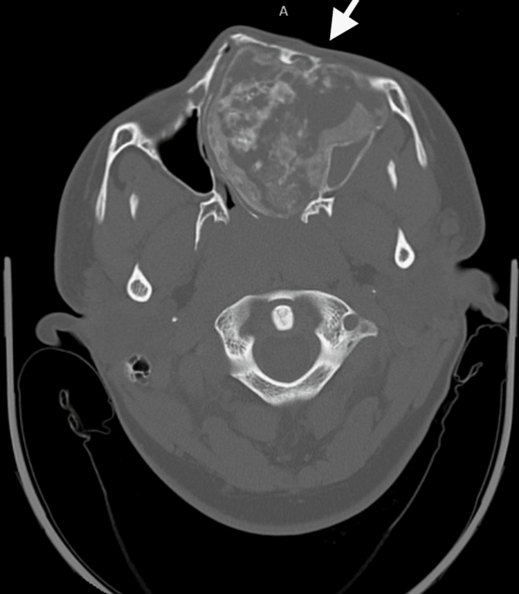
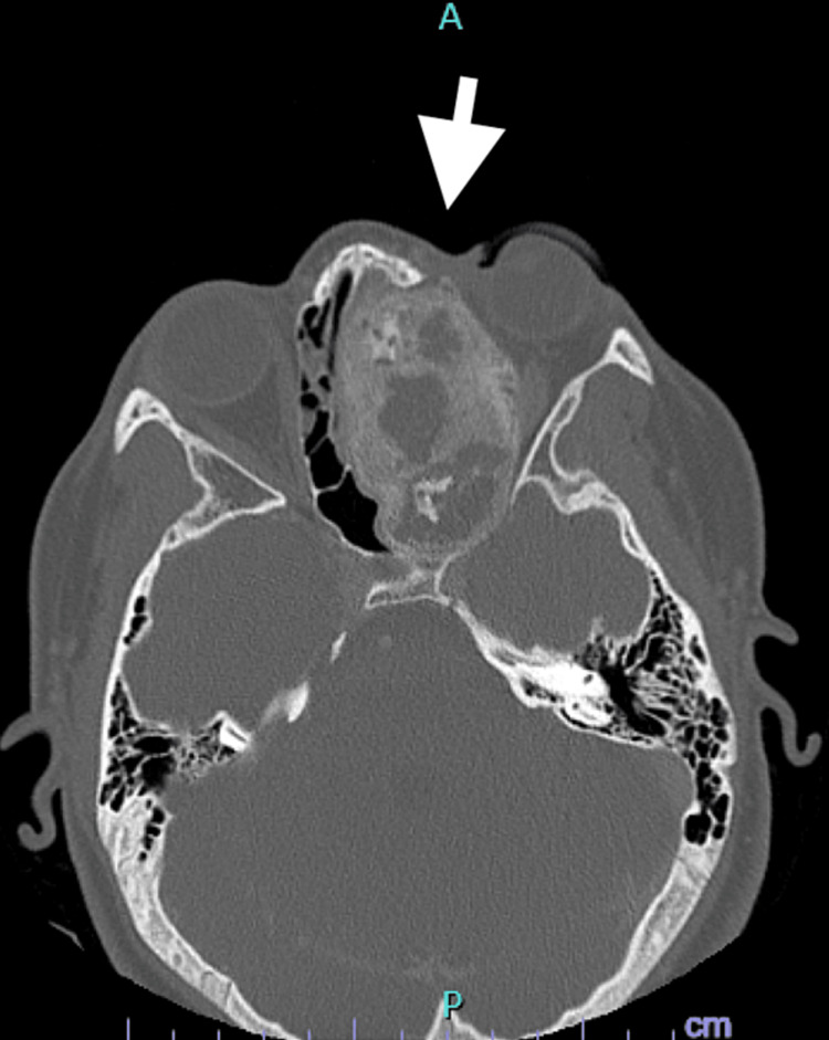
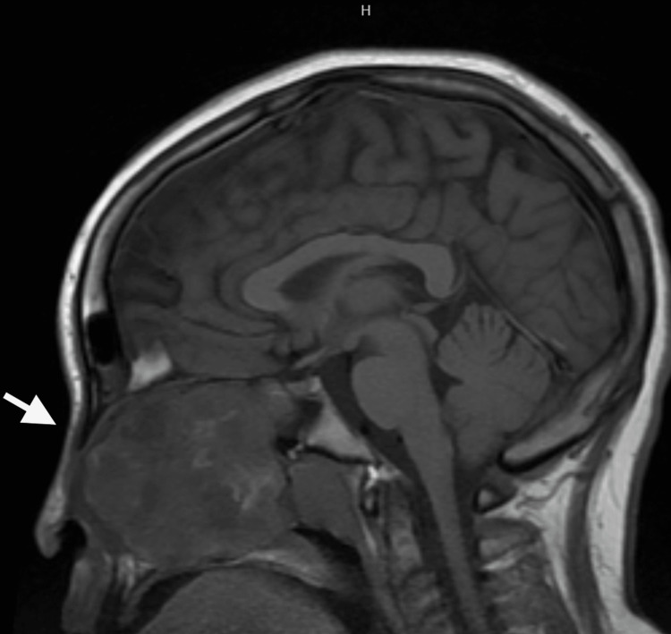
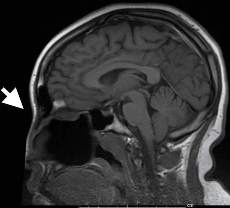
Juvenile ossifying fibroma (JOF) is a rare benign neoplastic fibro-osseous tumor commonly found in the maxilla and mandible of children usually between the ages of five and 15. Patients often present with aggressive, painless growth which is well demarcated from surrounding bone resulting in severe facial asymmetry. JOFs have high recurrence rates if not completely resected and should therefore be treated by a multidisciplinary team of physicians including a neurosurgeon to assess cranial nerve function. This case describes a child who presented to the ED after being referred by his primary care provider for facial swelling. The patient was diagnosed with JOF and had a delay in care due to a lack of access to multidisciplinary specialties to provide care due to payer difficulties which placed the patient at high risk of complications.
Keywords: delayed diagnosis; juvenile trabecular ossifying fibroma; medicaid population; multidisciplinary approach; paediatric otolaryngology; socioeconomic; socioeconomic disparities; telemedicine.
Copyright © 2023, Acosta et al.
Conflict of interest statement
The authors have declared that no competing interests exist.
Figures
Similar articles
-
Juvenile psammomatoid ossifying fibroma: An unusual case report.Contemp Clin Dent. 2013 Oct;4(4):566-8. doi: 10.4103/0976-237X.123094. Contemp Clin Dent. 2013. PMID: 24403813 Free PMC article.
-
Juvenile trabecular ossifying fibroma: A case of extensive lesion of the maxilla.Int J Surg Case Rep. 2023 Oct;111:108620. doi: 10.1016/j.ijscr.2023.108620. Epub 2023 Aug 22. Int J Surg Case Rep. 2023. PMID: 37703694 Free PMC article.
-
Simultaneous presentation of juvenile ossifying fibroma in the maxilla and mandible: a case report.Int J Surg Case Rep. 2020;71:285-289. doi: 10.1016/j.ijscr.2020.05.025. Epub 2020 May 22. Int J Surg Case Rep. 2020. PMID: 32480339 Free PMC article.
-
Rare case of a recurrent juvenile ossifying fibroma of the lumbosacral spine.J Neurosurg Spine. 2018 Jun;28(6):647-653. doi: 10.3171/2017.10.SPINE17947. Epub 2018 Mar 9. J Neurosurg Spine. 2018. PMID: 29521580 Review.
-
Juvenile psammomatoid ossifying fibroma of maxillary sinus: case report with review of literature.J Maxillofac Oral Surg. 2014 Jun;13(2):109-14. doi: 10.1007/s12663-013-0479-6. Epub 2013 Feb 7. J Maxillofac Oral Surg. 2014. PMID: 24822000 Free PMC article. Review.
References
-
- Barnes L, Everson J, Reichart P, Sidransky D. Vol. 9. Albany, NY: WHO Publications Center; 2005. Pathology and Genetics of Head and Neck Tumours ; p. 2005.
-
- Cranial juvenile psammomatoid ossifying fibroma: case report. Barrena López C, Bollar Zabala A, Úrculo Bareño E. J Neurosurg Pediatr. 2016;17:318–323. - PubMed
-
- National study of barriers to timely primary care and emergency department utilization among Medicaid beneficiaries. Cheung PT, Wiler JL, Lowe RA, Ginde AA. Ann Emerg Med. 2012;60:4–10. - PubMed
Publication types
LinkOut - more resources
Full Text Sources