

Classification and Locoregional Treatment of Rectal Neuroendocrine Tumors
- PMID: 37425523
- PMCID: PMC10329420
- DOI: 10.7759/cureus.40128
Classification and Locoregional Treatment of Rectal Neuroendocrine Tumors
Abstract
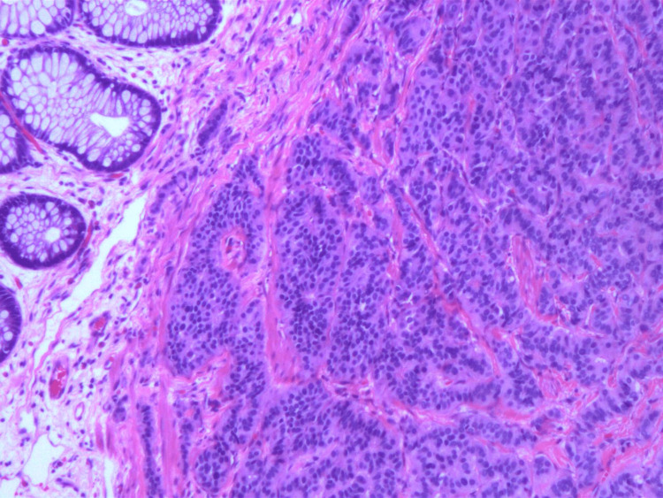
A 43-year-old male presented to his primary care physician's office with a complaint of painless rectal bleeding with a concomitant weight loss of 10-15 pounds and intermittent abdominal pain. Endoscopic evaluation was remarkable for a 5 mm rectal polyp roughly 10 cm from the anal verge. Resection was performed and the pathology was consistent with a low-grade neuroendocrine/carcinoid tumor. Immunostaining for synaptophysin, chromogranin, CD56, and CAM5.2 were positive while staining for CK20 was negative. Given the absence of metastasis on radiographic and endoscopic evaluation, the patient was managed conservatively thereafter with observation. Despite having an indolent clinical course, resection is recommended for all rectal neuroendocrine tumors. Locoregional endoscopic resection versus radical resection can be used for adequate tissue removal depending on the characteristics of the tumor and the degree of invasion.
Keywords: endoscopic mucosal resection; endoscopy; gastroenterology; malignancy; rectal neuroendocrine tumor.
Copyright © 2023, Singh et al.
Conflict of interest statement
The authors have declared that no competing interests exist.
Figures


Similar articles
-
Endoscopic submucosal resection with an endoscopic variceal ligation device for the treatment of rectal neuroendocrine tumors.Int J Colorectal Dis. 2018 Dec;33(12):1703-1708. doi: 10.1007/s00384-018-3152-1. Epub 2018 Aug 30. Int J Colorectal Dis. 2018. PMID: 30167779
-
[Application of dental floss traction-assisted endoscopic submucosa dissection to rectal neuroendocrine neoplasm].Zhonghua Wei Chang Wai Ke Za Zhi. 2019 Apr 25;22(4):377-382. doi: 10.3760/cma.j.issn.1671-0274.2019.04.011. Zhonghua Wei Chang Wai Ke Za Zhi. 2019. PMID: 31054553 Chinese.
-
Endoscopic management of a primary duodenal carcinoid tumor.Case Rep Gastroenterol. 2012 Jan;6(1):135-42. doi: 10.1159/000337870. Epub 2012 May 23. Case Rep Gastroenterol. 2012. PMID: 22679400 Free PMC article.
-
Review article: the investigation and management of rectal neuroendocrine tumours.Aliment Pharmacol Ther. 2016 Aug;44(4):332-45. doi: 10.1111/apt.13697. Epub 2016 Jun 15. Aliment Pharmacol Ther. 2016. PMID: 27302838 Review.
-
[Clinicopathologic features of primary renal neuroendocrine carcinoma].Zhonghua Bing Li Xue Za Zhi. 2018 Nov 8;47(11):851-856. doi: 10.3760/cma.j.issn.0529-5807.2018.11.007. Zhonghua Bing Li Xue Za Zhi. 2018. PMID: 30423609 Review. Chinese.
References
-
- Rectal carcinoid tumor: diagnosis and management. Viana C, Marques I, Staubus A, Martins SF. J Coloproctology. 2019;39:184–189.
Publication types
LinkOut - more resources
Full Text Sources
Research Materials