

Twenty Intracranial Skull Base Variations in the Same Specimen
- PMID: 37425550
- PMCID: PMC10328379
- DOI: 10.7759/cureus.40096
Twenty Intracranial Skull Base Variations in the Same Specimen
Abstract
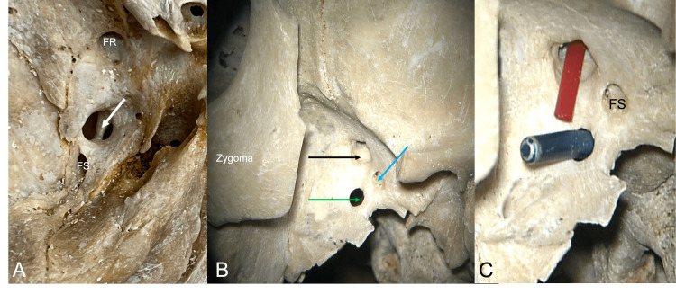
Anatomists and clinicians often encounter single bony anatomical variations in dry skulls and on imaging. However, a constellation of 20 such variants some that, to our knowledge, have not been previously described is noteworthy. Here, we describe an adult skull with multiple bony variations, and these are detailed and discussed. These included clival canals, an interclinoid bar with resultant foramen at the uppermost aspect of the clivus, middle clinoid process, posterior petroclinoid ligament, pterygoalar plate, septated hypoglossal canal, foramen through the anterior clinoid process, septated foramen ovale, shortened superior orbital fissure, and crista muscularis. Knowledge of individual differences in the structure of the skull may be of use to both anatomists and clinicians in the treatment of intracranial procedures as well as cranial imaging studies. Taken together, such a unique specimen is of archival value.
Keywords: anatomy; cadaver; cranium; intracranial; skull; variations.
Copyright © 2023, Couldwell et al.
Conflict of interest statement
The authors have declared that no competing interests exist.
Figures


Similar articles
-
Anatomical Study of Pterygospinous and Pterygoalar Bar in Human Skulls with their Phylogeny and Clinical Significance.J Clin Diagn Res. 2014 Sep;8(9):AC10-3. doi: 10.7860/JCDR/2014/9326.4888. Epub 2014 Sep 20. J Clin Diagn Res. 2014. PMID: 25386415 Free PMC article.
-
Incidence and morphometry of sellar bridges and related foramina in dry skulls: Their significance in middle cranial fossa surgery.J Craniomaxillofac Surg. 2018 Apr;46(4):635-644. doi: 10.1016/j.jcms.2018.01.008. Epub 2018 Feb 10. J Craniomaxillofac Surg. 2018. PMID: 29534911
-
The Modular Concept in Skull Base Surgery: Anatomical Basis of the Median, Paramedian and Lateral Corridors.Acta Biomed. 2021 Aug 26;92(S4):e2021411. doi: 10.23750/abm.v92iS4.12115. Acta Biomed. 2021. PMID: 34437364 Free PMC article.
-
Common surgical pitfalls in the skull.Surg Neurol. 2003 Mar;59(3):228-31; discussion 231. doi: 10.1016/s0090-3019(02)01038-8. Surg Neurol. 2003. PMID: 12681561 Review.
-
Review of the Petroclinoid Ligament.Kurume Med J. 2022 Mar 11;67(1):5-10. doi: 10.2739/kurumemedj.MS671007. Epub 2022 Jan 31. Kurume Med J. 2022. PMID: 35095019 Review.
Cited by
-
Superior orbital fissure in children: shape analysis, measurements, and surgical importance.Anat Sci Int. 2025 Mar;100(2):198-206. doi: 10.1007/s12565-024-00802-5. Epub 2024 Sep 22. Anat Sci Int. 2025. PMID: 39306830
References
-
- Clinical significance of a mysterious clival canal. Nayak SR, Saralaya VV, Prabhu LV, Pai MM, Krishnamurthy A. https://rjme.ro/RJME/resources/files/480407427429.pdf. Rom J Morphol Embryol. 2007;48:427–429. - PubMed
-
- Variations and classification of bony septations of the jugular foramen: an anatomic and histologic study with application to imaging and surgery of the skull base. Fang Y, Olewnik Ł, Iwanaga J, Loukas M, Dumont AS, Tubbs RS. World Neurosurg. 2022;163:0–70. - PubMed
-
- Moore KL, Persaud TV, Torchia MG. Philadelphia: Elsevier; 2016. The Developing Human: Clinically Oriented Embryology.
-
- The human calvaria: a review of embryology, anatomy, pathology, and molecular development. Tubbs RS, Bosmia AN, Cohen-Gadol AA. Childs Nerv Syst. 2012;28:23–31. - PubMed
Publication types
LinkOut - more resources
Full Text Sources