[Preprint]. 2023 May 2:2023.05.02.539157.
doi: 10.1101/2023.05.02.539157.

Toxic anti-phage defense proteins inhibited by intragenic antitoxin proteins

Affiliations

Toxic anti-phage defense proteins inhibited by intragenic antitoxin proteins

Aoshu Zhong et al. bioRxiv. .

Update in

Abstract

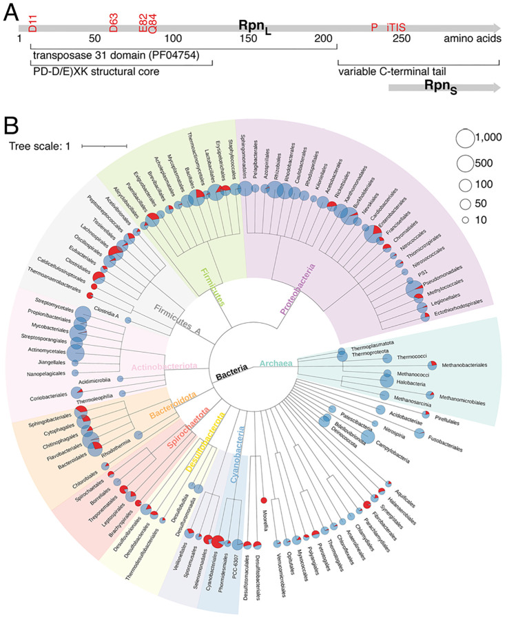
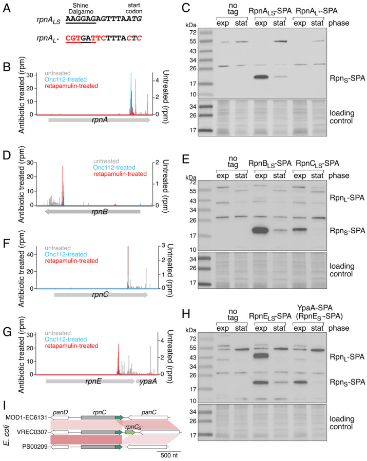
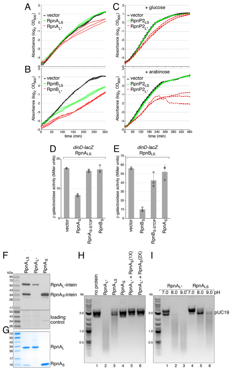
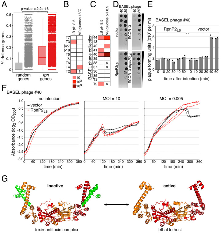
Recombination-promoting nuclease (Rpn) proteins are broadly distributed across bacterial phyla, yet their functions remain unclear. Here we report these proteins are new toxin-antitoxin systems, comprised of genes-within-genes, that combat phage infection. We show the small, highly variable Rpn C -terminal domains (Rpn S ), which are translated separately from the full-length proteins (Rpn L ), directly block the activities of the toxic full-length proteins. The crystal structure of RpnA S revealed a dimerization interface encompassing a helix that can have four amino acid repeats whose number varies widely among strains of the same species. Consistent with strong selection for the variation, we document plasmid-encoded RpnP2 L protects Escherichia coli against certain phages. We propose many more intragenic-encoded proteins that serve regulatory roles remain to be discovered in all organisms.

Significance: Here we document the function of small genes-within-genes, showing they encode antitoxin proteins that block the functions of the toxic DNA endonuclease proteins encoded by the longer rpn genes. Intriguingly, a sequence present in both long and short protein shows extensive variation in the number of four amino acid repeats. Consistent with a strong selection for the variation, we provide evidence that the Rpn proteins represent a phage defense system.


Figures

Fig. 1.
Fig. 1.
Broadly-distributed rpn genes move by horizontal gene transfer. (A) Diagram of rpn genes adapted from (7). Positions of catalytic residues for RpnA and internal promoter (P) and iTIS are indicated in red font. (B) Species distribution of rpn orthologs. Clades with more than 15 children are displayed. The pie charts represent the percentage of species with (red) and without (blue) a rpn ortholog. The size of the pie chart represents the number of species with rpn orthologs in that clade.
Fig. 2.
Fig. 2.
The rpn genes encode smaller proteins (RpnS). (A) Sequence of RpnAS ribosome binding site, and mutations to eliminate rpnAS ribosome binding and start codon. Browser images of ribosome profiling data for rpnA (B), rpnB (D), rpnC (F), rpnE-ypaA/rpnEŚ (G). Ribosome density for an untreated control (gray) and cells treated with Onc112 (blue) (4) or retapamulin (red) (3) are shown. c, e, h, Immunoblot analysis of the levels of SPA-tagged RpnAS (C) and RpnBS, and RpnCS (E) RpnES and YpaA/RpnEŚ (H). E. coli MG1655 strains were grown to exponential (exp) and stationary (stat) phase in LB. The SPA tag was detected with monoclonal anti-FLAG M2-peroxidase (HRP) antibody. Ponceau S staining of membranes served as loading controls. (I) Diagram showing an example of insertion and deletion of rpnŚ genes (green) among strains of E. coli, with red connections linking orthologous regions and color indicating identity (darker=higher, lighter=lower).
Fig. 3.
Fig. 3.
RpnS proteins function as antitoxins. RpnAL (A) and RpnBL (B) inhibit E. coli growth. E. coli ER2170 cells harboring indicated plasmids were grown in LB. Three biological replicates are shown. (C) RpnP2L inhibits E. coli growth. E. coli MG1655 cells harboring indicated plasmids were grown in LB with added glucose or arabinose. For A, B, and C, three biological replicates are shown. (D) RpnAS, but not RpnBS, blocks RpnAL induction, and (E) RpnBS, but not RpnAS, blocks RpnBL induction of the dinD-lacZ reporter of the SOS DNA damage response. RpnALS and RpnBLS are overexpressed from the rhamnose-inducible PrhaB promoter. RpnAS and RpnBS are overexpressed from the arabinose-inducible PBAD promoter. For (D) and (E), average of three independent biological repeats is given with standard deviation. For (A)-(E), the ER2170 or MG1655 strain backgrounds were WT for the rpn genes. (F) Immunoblot analysis of intein-tagged RpnALS, RpnAL* and RpnAS overexpression. Ponceau S staining of membrane served as loading control. (G) Coomassie-stained Tris-glycine SDS-PAGE gel of purified RpnALS, RpnAL* and RpnAS. (H) Purified RpnAS blocks dsDNA endonuclease activity of RpnAL*. Indicated purified proteins were incubated with pUC19. (I) RpnAL activity is pHdependent. Purified RpnAL* (lanes 1–3) or RpnALS (lanes 4–6) were incubated with pUC19 at pH 7.0, pH 8.0 or pH 9.0. Images in (H) and (I) are inverted from the original. Each of the nuclease assays was repeated at least twice.
Fig. 4.
Fig. 4.
RpnAL and RpnAS form a complex. (A) Size exclusion chromatograms of indicated proteins on a Superdex 200 column. Although the RpnALS peak migrates similarly to the 158 kDa marker peak, SEC-MALS analysis, which is less influenced by protein shape, indicates that the complex has a molecular weight of 74.1 ± 0.5 kDa. The RpnAL* protein migrates as a large molecular weight complex indicative of an aggregate, but we do not observe precipitates of this protein. (B) Sequence alignments for E. coli RpnAS and RpnCS as well as C. difficile RpnS. The sequence logo was built based on all the sequences for each group though only representative individual sequences are shown. The four amino acid repeat that varies between strains is colored in the alignment, with a bracket indicating the number of repeats. The alignments were manually curated. (C) Crystal structure of RpnAS. Pink asterisks represents approximate position where four amino acid insertions occur (H261 in RpnAL). (D) Only RpnCS with same number of four amino acid repeats blocks RpnCL induction of dinD-lacZ. RpnCLS and RpnCLS+REPEAT are expressed from the rhamnose-inducible PrhaB promoter, while RpnCS and RpnCS+REPEAT are expressed from the arabinose-inducible PBAD promoter. For the REPEAT derivatives, a four amino acid repeat (KGIE) is inserted in the same position. Average of four independent biological repeats is given with standard deviation.
Fig. 5.
Fig. 5.
Rpn proteins block phage infection. (A) Enrichment of phage defense genes in the vicinity of rpn genes compared to random genes. Box plots (left) and dot plots (right) display the percentage and distribution of defense-associated genes in proximity to these genes, with P-values indicating the significance levels determined by the Mann-Whitney U Test. (B) Efficiency of plaquing (EOP) for the indicated phages infecting cells producing RpnP2LS grown at 30°C on LB medium buffered to pH 8.5 and 18°C on M9 glucose medium pH 7.1. (C) EOP for the indicated phages infecting cells producing RpnP2LS grown in LB and M9 glucose, pH 8.5. For (B) and (C), “s” denotes smaller plaques were observed. (D) Images of BASEL phage #40 and #39 plaques for MG1655 cells carrying vector control or producing RpnP2LS (left) or ECOR13 or ECOR13 ΔrpnP2LS cells (right), from SI Appendix, Fig. S8. The back wedge denotes the 10-fold dilution series. (E) Growth of vector control or RpnP2LS expressing cells infected with BASEL phage #40 (at indicated MOIs) in LB pH 8.5. Three biological replicates are presented. The partial regrowth of the strains infected at a MOI of 10 could be due to appearance of suppressors. (F) Plaque-forming units (PFU) per milliliter of BASEL phage #40 (MOI = 0.005) used to infect vector control or RpnP2LS expressing cells at 0, 10, 20, 30, 40, and 60 min postinfection. Individual data points and average of two biological replicates are shown. For (A)-(E), the MG1655 strain background was WT for the rpn genes. f, Model for functions of RpnL and RpnS proteins. RpnAL dimer (orange and red, right side) and RpnALS tetramer (left side, toxin-antitoxin complex) structures were predicted with AlphaFold-Multimer.

References

    1. Meydan S., Vázquez-Laslop N., Mankin A. S., Genes within genes in bacterial genomes. Microbiol. Spectr. 6, RWR-0020–2018 (2018). - PMC - PubMed
    1. McBride T. M. et al. , Diverse CRISPR-Cas complexes require independent translation of small and large subunits from a single gene. Mol. Cell 80, 971–979 (2020). - PubMed
    1. Meydan S. et al. , Retapamulin-assisted ribosome profiling reveals the alternative bacterial proteome. Mol. Cell 74, 481–493 (2019). - PMC - PubMed
    1. Weaver J., Mohammad F., Buskirk A. R., Storz G., Identifying small proteins by ribosome profiling with stalled initiation complexes. mBio 10, pii: e02819–02818 (2019). - PMC - PubMed
    1. Steczkiewicz K., Muszewska A., Knizewski L., Rychlewski L., Ginalski K., Sequence, structure and functional diversity of PD-(D/E)XK phosphodiesterase superfamily. Nucleic Acid Res. 40, 7016–7045 (2012). - PMC - PubMed

Publication types