[Preprint]. 2023 Jun 29:2023.06.26.23291885.
doi: 10.1101/2023.06.26.23291885.

Targeting hepatitis B vaccine escape using immunogenetics in Bangladeshi infants

Affiliations

Targeting hepatitis B vaccine escape using immunogenetics in Bangladeshi infants

Guillaume Butler-Laporte et al. medRxiv. .

Abstract

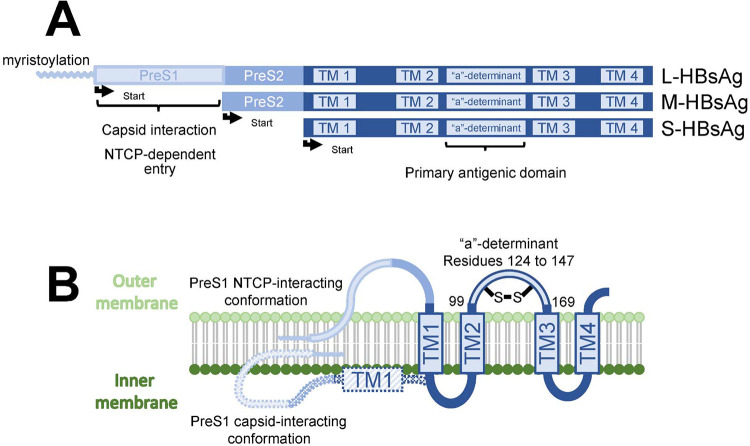
Hepatitis B virus (HBV) vaccine escape mutants (VEM) are increasingly described, threatening progress in control of this virus worldwide. Here we studied the relationship between host genetic variation, vaccine immunogenicity and viral sequences implicating VEM emergence. In a cohort of 1,096 Bangladeshi children, we identified human leukocyte antigen (HLA) variants associated with response vaccine antigens. Using an HLA imputation panel with 9,448 south Asian individuals DPB1*04:01 was associated with higher HBV antibody responses (p=4.5×10⁻³⁰). The underlying mechanism is a result of higher affinity binding of HBV surface antigen epitopes to DPB1*04:01 dimers. This is likely a result of evolutionary pressure at the HBV surface antigen 'a-determinant' segment incurring VEM specific to HBV. Prioritizing pre-S isoform HBV vaccines may tackle the rise of HBV vaccine evasion.

Keywords: Genome-wide association studies; escape variants; hepatitis B virus; human leukocyte antigen; major histocompatibility complex; vaccination.


Conflict of interest statement

Competing interests: JBR’s institution has received investigator-initiated grant funding from Eli Lilly, GlaxoSmithKline and Biogen for projects unrelated to this research. He is the CEO of 5 Prime Sciences Inc (www.5primesciences.com).

Figures

Fig. 1:
Fig. 1:
Schematic of the three HBsAg protein isoforms. A) The most restricted S-HBsAg isoform is used in yeast recombinant vaccine production. The pre-S1 segment is crucial for capsid interaction during virion assembly, and for hepatocyte viral entry in the first phase of HBV infection when it interacts with sodium taurocholate co-transporting polypeptide (NTCP). B) Representation of the two HBsAg conformations on the surface of HBV. The primary antigenic domain “a”-determinant (where most known vaccine escape variants are found) is located between transmembrane domains TM1 and TM2, facing the outside of the virion. Figure adapted with permission from Vaillant, 2021(63).
Fig. 2:
Fig. 2:
Manhattan and qq-plots of the single variants GWAS for each serology phenotype. The genome-wide significance line is set at 5×10⁻⁸/8. As shown by the genetic inflation factors (lambdas), there were no obvious signs of population stratification bias.
Fig. 3:
Fig. 3:
Visualization of amino acid residues (in red) associated with each serology phenotype. For each dimer, the blue chains represent the alpha subunits (i.e. DPA1, DRA, and DQA1, from left to right), the green chains represent the beta subunits (i.e. DPB1, DRB1, and DQB1, from left to right), and the yellow chains show the location of a given epitope in the dimers’ peptide binding grooves. See fig. S5 for similar visualization for DT and HLA-DRB5, and for PRN and HLA-DQ.
Fig. 4:
Fig. 4:
A) in-silico binding results from NetMHCIIPan. Points are the effect coefficients from beta regression run on the rank outputs of predicted binding of all 15-mers derived from 50 HBsAg protein sequences. In green, results are shown for the full HBsAg protein sequence. In orange, results are shown from an analysis restricted only to 15-mers within the S isoform of HBsAg. The S isoform is the one used in making yeast recombinant vaccines, such as those given to our participants. Coefficients and 95 % CIs are shown. The more they are positioned to the left, the higher average in-silico binding across the antigen. Dimers in bold contain the DPB1*04:01 allele, to better compare with the HBsAg antibody association tests in the right panel. B) Results from the HLA allele association studies using linear regression with antibody responses and imputed HLA alleles from the Bangladeshi datasets. Again, coefficients and 95% CIs are shown. Values to the right on the x-axis indicate greater HBsAg antibody measures. The DPB1*04:01 allele is shown in bold to better compare with the in-silico results in the left panel.
Fig. 5:
Fig. 5:
Difference in HBsAg binding affinity between the dimer with highest predicted binding affinity to HBsAg peptides (DPA1*01:03-DPB1*04:01) and the dimer with the lowest (DPA1*01:03-DPB1*26:01). The x-axis represents bins of 1 amino-acid across the entire HBsAg sequence. The y-axis represents the difference in binding affinity between the top-binding 15-mers binding in this region for both dimers. Positive values indicate greater binding for DPA1*01:03-DPB1*04:01 than for DPA1*01:03-DPB1*26:01. Negative values indicate the opposite. The colours of the vertical bars refer to the rank of affinity of binding of the 15-mer to either DPA1*01:03-DPB1*04:01 (positive values) or DPA1*01:03-DPB1*26:01 (negative values). Small blue bars mean that both dimers are predicted to bind with high affinity (top 10% quantile) at this position. Long red bars mean that the difference in dimer binding to 15-mer is predicted to be large, but weak for both. A long dark blue bar means that the expected binding difference is large and that one of the dimers bind strongly to the 15-mer. For example, 15-mers around position 300 of the HBsAg protein are expected to bind strongly to DPA1*01:03-DPB1*26:01 and to bind much better than to DPA1*01:03-DPB1*04:01. Bottom colored horizontal bar indicates the different section of the HBsAg protein.

References

    1. Hossain M. G., Ueda K., A meta-analysis on genetic variability of RT/HBsAg overlapping region of hepatitis B virus (HBV) isolates of Bangladesh. Infect. Agent. Cancer. 14, 33 (2019). - PMC - PubMed
    1. Cremer J., Hofstraat S. H. I., van Heiningen F., Veldhuijzen I. K., van Benthem B. H. B., Benschop K. S. M., Genetic variation of hepatitis B surface antigen among acute and chronic hepatitis B virus infections in The Netherlands. J. Med. Virol. 90, 1576–1585 (2018). - PMC - PubMed
    1. Munshi S. U., Tran T. T. T., Vo T. N. T., Tabassum S., Sultana N., Nguyen T. H., Jahan M., Le C. N., Baker S., Rahman M., Molecular characterization of hepatitis B virus in Bangladesh reveals a highly recombinant population. PLoS One. 12, e0188944 (2017). - PMC - PubMed
    1. Inoue T., Tanaka Y., Cross-Protection of Hepatitis B Vaccination among Different Genotypes. Vaccines. 8 (2020), doi:10.3390/vaccines8030456. - DOI - PMC - PubMed
    1. Chang M.-H., Breakthrough HBV infection in vaccinated children in Taiwan: surveillance for HBV mutants. Antivir. Ther. 15, 463–469 (2010). - PubMed

Publication types