[Preprint]. 2023 Jun 27:2023.06.26.545779.
doi: 10.1101/2023.06.26.545779.

Focusing antibody responses to the fusion peptide in rhesus macaques

Affiliations

Focusing antibody responses to the fusion peptide in rhesus macaques

Christopher A Cottrell et al. bioRxiv. .

Update in

  • Priming antibody responses to the fusion peptide in rhesus macaques.
    Cottrell CA, Pratap PP, Cirelli KM, Carnathan DG, Enemuo CA, Antanasijevic A, Ozorowski G, Sewall LM, Gao H, Allen JD, Nogal B, Silva M, Bhiman J, Pauthner M, Irvine DJ, Montefiori D, Crispin M, Burton DR, Silvestri G, Crotty S, Ward AB. Cottrell CA, et al. NPJ Vaccines. 2024 Jul 12;9(1):126. doi: 10.1038/s41541-024-00918-9. NPJ Vaccines. 2024. PMID: 38997302 Free PMC article.

Abstract

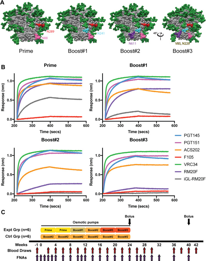
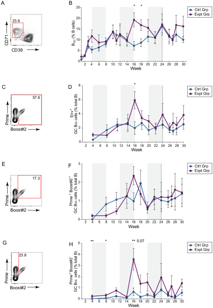
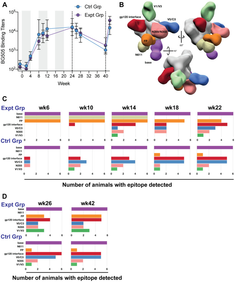
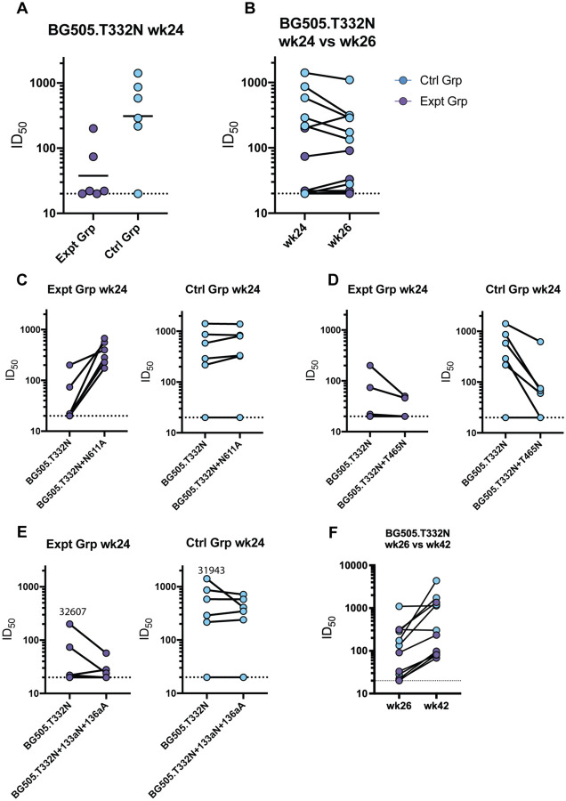
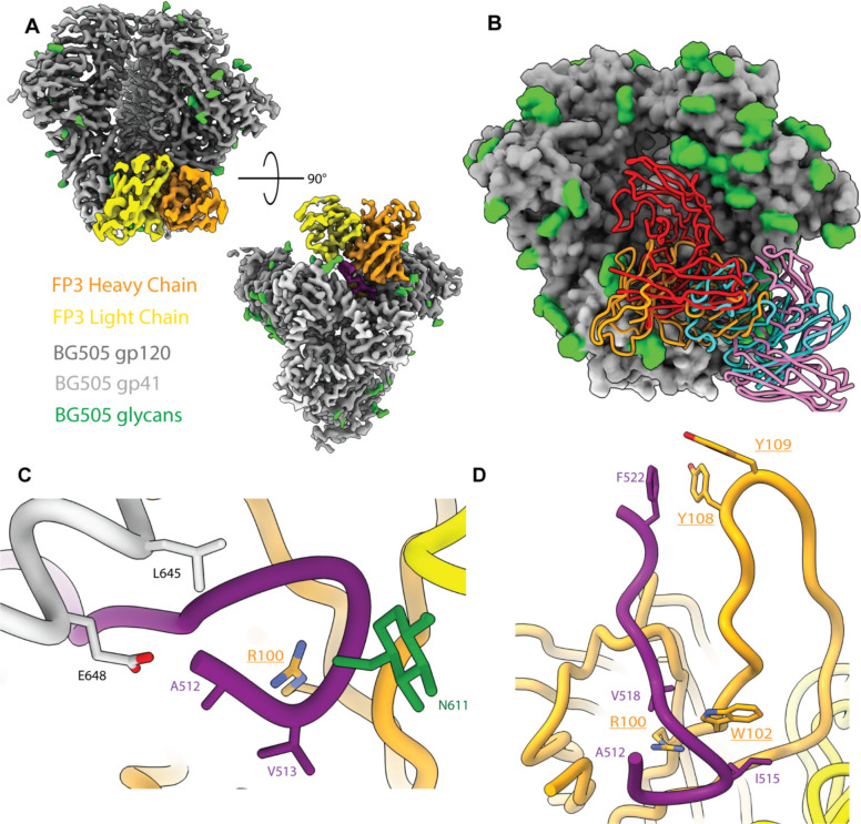
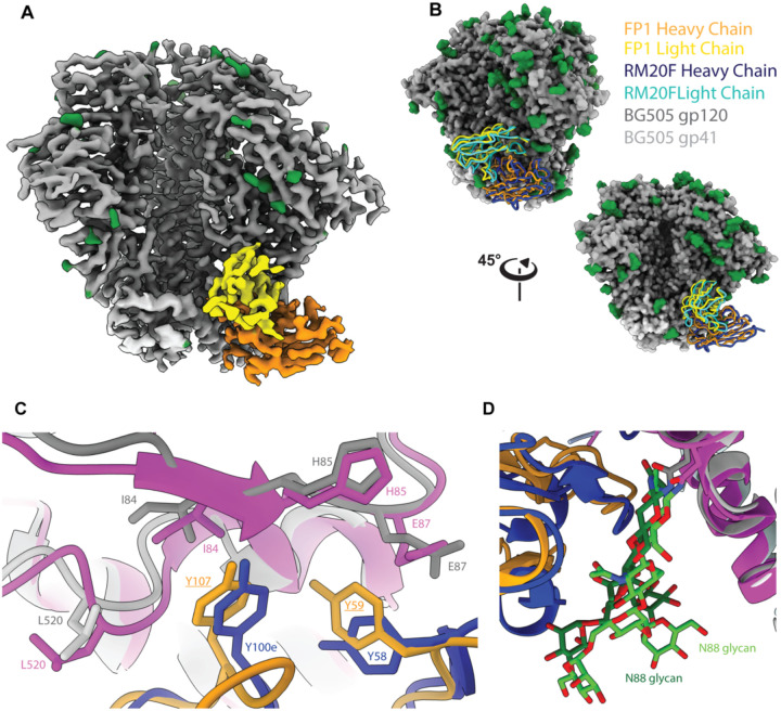
Immunodominance of antibodies targeting non-neutralizing epitopes and the high level of somatic hypermutation within germinal centers (GCs) required for most HIV broadly neutralizing antibodies (bnAbs) are major impediments to the development of an effective HIV vaccine. Rational protein vaccine design and non-conventional immunization strategies are potential avenues to overcome these hurdles. Here, we report using implantable osmotic pumps to continuously deliver a series of epitope-targeted immunogens to rhesus macaques over the course of six months to elicit immune responses against the conserved fusion peptide. Antibody specificities and GC responses were tracked longitudinally using electron microscopy polyclonal epitope mapping (EMPEM) and lymph node fine-needle aspirates, respectively. Application of cryoEMPEM delineated key residues for on-target and off-target responses that can drive the next round of structure-based vaccine design.


Conflict of interest statement

Conflicts of Interest None.

Figures

Figure 1.
Figure 1.. FP focused immunogen design and immunization scheme.
(A) Surface representations of the BG505 SOSIP.v5.2 immunogen with mutations designed to focus antibody responses to the FP epitope. N88 glycan in pink. N611 glycan in purple. Introduced N289 glycan in red. Introduced N241 glycan in turquoise. Consensus mutations around the FP epitope shown in yellow. (B) Antigenicity was assessed using BLI on panel of anti-Env mAbs: PGT145 (apex), PGT151 (FP), ACS202 (FP), VRC34 (FP), RM20F (FP), iGL-RM20F (FP), and F105 (anti-gp120, non-Nab). (C) Immunization scheme.
Figure 2.
Figure 2.. Sustained delivery immunization results in enduring GC responses.
(A) Representative BGC cell flow cytometry. (B) Quantification of BGC cell kinetics as a percentage of total CD20+ B cells. (C) Representative total Env trimer-specific binding flow cytometry, gated on BGC cells. (D) Quantification of total Env trimer-specific BGC cells kinetics as a percentage of total CD20+ B cells. (E) Representative Prime and Boost#2 dual-specific binding flow cytometry, gated on BGC cells. (F) Quantification of Prime and Boost#2-specific cell kinetics as a percentage of total CD20+ B cells. (G) Representative Prime immunogen-specific flow cytometry, gated on BGC cells. (H) Quantification of Prime immunogen-specific BGC cell kinetics as a percentage of total CD20+ B cells. Plot shows the mean of left and right side FNAs (analyzed separately) with standard error of means (SEM), * p ≤0.05, **p ≤0.01.
Figure 3.
Figure 3.. Longitudinal monitoring of the humoral immune response by ELISA and negative stain EMPEM.
(A) Plasma IgG binding ELISA of each group over the duration of the study. Six animals per group, showing geometric mean titers ± geometric SD. (B) Composite 3D map representing the epitopes observed in the longitudinal negative stain EMPEM analysis. (C) Longitudinal negative stain EMPEM analysis of the continuous immunogen delivery phase of the study. Bar graphs show how many animals per group made antibodies directed to each specific epitope. Colored to match (B). (D) Longitudinal negative stain EMPEM analysis 2 weeks after each bolus immunization, colored to match (B).
Figure 4.
Figure 4.. Serum neutralization activity.
(A) Week 24 serum neutralization activity against the BG505.T332N pseudovirus. Dotted lines indicate detection limit for the assay. (B) Comparison of BG505.T332N pseudovirus neutralization activity against before (week 24) and after (week 26) bolus nanoparticle immunization. Connecting lines indicate specific animal response differences seen between the two time points. (C) Week 24 serum neutralization activity against the BG505.T332N+N611A pseudovirus. (D) Week 24 serum neutralization activity against the BG505.T332N+T465N pseudovirus. (E) Week 24 serum neutralization activity against the BG505.T332N+133aN_136aA pseudovirus. (F) Week 26 versus week 42 serum neutralization activity against BG505.T332N pseudovirus.
Figure 5.
Figure 5.. CryoEMPEM Analysis of FP3
A) Boost #2 in complex with the predicted heavy chain (orange) and light chain (yellow) of FP3 antibody targets the fusion peptide epitope area. B) Alignment of FP3 (orange) compared to known human bnAbs PGT151 (red), VRC34.01 (cyan), and ACS202 (pink). C. Fusion peptide is fully resolved in FP3 due to stabilization of the N-terminus by the antibody (underlined residues are inferred from density; residues not underlined are sequence verified). D) The CDRH3 of FP3 antibody interacts with FP by creating hydrophobic pockets around the N-terminus as well as around F522 with the presence of hydrophobic aromatic residues.
Figure 6.
Figure 6.. CryoEMPEM Analysis of FP1
A) Boost #2 in complex with predicted heavy chain (orange) and predicted light chain (yellow) of FP1. B) Alignment of FP1 (orange and yellow) with RM20F (dark and light blue) with two views. C) Both RM20F HCDR3 (dark blue) and FP1 predicted HCDR3 (orange) interact with I84 and H85 (grey for Boost #2 orientation for FP1 interactions; pink for BG505 residue orientation for RM20F (6VN0)) with a hydrophobic residue Y100e for RM20F and predicted Y106 (Y100c; Kabat) of FP1. CDRH2 of both FP1 and RM20F interact with H85 and E87 with hydrophobic residues at position 59 and 58, respectively. D) Heavy chains from both antibodies interact and stabilize the N88 glycan (dark green – FP1; neon green – 6VN0) for their respective antigen, allowing for branched resolution of the glycan.

Similar articles

References

    1. National Center for Immunization and Respiratory Diseases. General recommendations on immunization --- recommendations of the Advisory Committee on Immunization Practices (ACIP). MMWR Recomm Rep. 2011;60(2):1–64. - PubMed
    1. Watson JC, Pearson JA, Markowitz LE, et al. An evaluation of measles revaccination among school-entry-aged children. Pediatrics. 1996;97(5):613–618. - PubMed
    1. Poland GA, Jacobson RM, Thampy AM, et al. Measles reimmunization in children seronegative after initial immunization. JAMA. 1997;277(14):1156–1158. - PubMed
    1. Banatvala JE, Van Damme P. Hepatitis B vaccine -- do we need boosters? J Viral Hepat. 2003;10(1):1–6. doi:10.1046/j.1365-2893.2003.00400.x - DOI - PubMed
    1. Miner MD, Corey L, Montefiori D. Broadly neutralizing monoclonal antibodies for HIV prevention. J Int AIDS Soc. 2021;24 Suppl 7(Suppl 7):e25829. doi:10.1002/jia2.25829 - DOI - PMC - PubMed

Publication types