[Preprint]. 2023 Jun 26:2023.06.26.546601.
doi: 10.1101/2023.06.26.546601.

Target-directed microRNA degradation regulates developmental microRNA expression and embryonic growth in mammals

Affiliations

Target-directed microRNA degradation regulates developmental microRNA expression and embryonic growth in mammals

Benjamin T Jones et al. bioRxiv. .

Update in

Abstract

MicroRNAs (miRNAs) are post-transcriptional regulators of gene expression that play critical roles in development and disease. Target-directed miRNA degradation (TDMD), a pathway in which miRNAs that bind to specialized targets with extensive complementarity are rapidly decayed, has emerged as a potent mechanism of controlling miRNA levels. Nevertheless, the biological role and scope of miRNA regulation by TDMD in mammals remains poorly understood. To address these questions, we generated mice with constitutive or conditional deletion of Zswim8 , which encodes an essential TDMD factor. Loss of Zswim8 resulted in developmental defects in heart and lung, growth restriction, and perinatal lethality. Small RNA sequencing of embryonic tissues revealed widespread miRNA regulation by TDMD and greatly expanded the known catalog of miRNAs regulated by this pathway. These experiments also uncovered novel features of TDMD-regulated miRNAs, including their enrichment in co-transcribed clusters and examples in which TDMD underlies 'arm switching', a phenomenon wherein the dominant strand of a miRNA precursor changes in different tissues or conditions. Importantly, deletion of two miRNAs, miR-322 and miR-503, rescued growth of Zswim8 null embryos, directly implicating the TDMD pathway as a regulator of mammalian body size. These data illuminate the broad landscape and developmental role of TDMD in mammals.


Conflict of interest statement

Competing interest statement

J.T.M is a scientific advisor for Ribometrix, Inc. and owns equity in Orbital Therapeutics, Inc. The other authors declare no competing interests.

Figures

Figure 1.
Figure 1.. Loss of ZSWIM8 results in perinatal lethality in mice.
(A) Genome editing strategy used to generate Zswim8−/− (∆exon 2–7) and Zswim8F/F (floxed) mice. Upper, schematic of ZSWIM8 protein with approximate locations of BC-box (red), Cul2-box (green), and SWIM domain (blue) indicated. Lower, depiction of mouse Zswim8 genomic locus with exons encoding each domain in appropriate color as defined in upper panel, scissors showing CRISPR-Cas9 targeting sites, and DNA segment with orange triangles indicating donor sequence containing loxP sites. (B) Frequency of genotypes of offspring produced from Zswim8+/‒ intercrosses at the indicated time-points. (C) Western blot of ZSWIM8 protein in E18.5 brain from embryos of the indicated genotypes.
Figure 2.
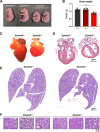
Figure 2.. ZSWIM8 deficiency leads to growth restriction and defective heart and lung development.
(A) Images of E18.5 mice of the indicated genotypes. (B) Body weights of E18.5 mice of the indicated genotypes. n=25 (Zswim8+/+), 47 (Zswim8+/−), and 30 (Zswim8−/−). Data are represented as mean ± SD with individual data points shown. ****p<0.0001 (unpaired t-test). (C) Images of E18.5 hearts of the indicated genotypes. (D-F) Representative hematoxylin and eosin (H&E)-stained sections of E18.5 hearts (D) and lungs (E-F) of the indicated genotypes.
Figure 3.
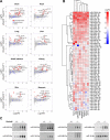
Figure 3.. The landscape of TDMD-regulated miRNAs in mouse embryonic tissues.
(A) Small RNA sequencing of E18.5 tissues. Red dots show miRNAs that were significantly upregulated in Zswim8−/− relative to Zswim8+/+ tissues (p<0.05; FDR<0.05) without a corresponding increase in the opposite strand derived from the same precursor (labeled as blue dots). n=3 biological replicates per genotype. LogFC, log2 fold change; LogCPM, log2 counts per million in Zswim8+/+ tissue. (B) Heat map showing log2 fold change of all TDMD-regulated miRNAs (Zswim8−/−/Zswim8+/+), defined as those exhibiting significant upregulation in at least one Zswim8−/− tissue (p<0.05; FDR<0.05) without a corresponding increase in the opposite strand derived from the same precursor. (C) Northern blot analysis of miRNA expression in E18.5 hearts from mice of the indicated genotypes. n=3 biological replicates per genotype. miR-16-5p served as a loading control.
Figure 4.
Figure 4.. TDMD regulates non-dominant miRNA strands, arm switching, and clustered miRNAs.
(A) Stacked bar graphs showing the relative abundance of 5p and 3p strands of the indicated miRNAs in E18.5 tissues from Zswim8+/+ or Zswim8−/− mice. (B) Schematic representation of miRNA clusters encoding TDMD-regulated miRNAs. Heat maps display log2 fold change of miRNA expression (Zswim8−/−/Zswim8+/+) for each cluster member across tissues. miRNAs labeled in red text are TDMD substrates in at least one tissue.
Figure 5.
Figure 5.. Tailing and trimming of TDMD-regulated miRNAs in mouse tissues.
(A,B) Violin plots showing the fold change (Zswim8−/−/Zswim8+/+) of tailing (A) or trimming (B) of miRNAs in E18.5 tissues. ns, not significant; ****p<0.0001; ***p<0.001; **p<0.01 (unpaired t-test). (C) Scatter plots showing the fold change of tailing or trimming of each TDMD-regulated miRNA relative to its fold change in abundance (Zswim8−/−/Zswim8+/+) in each tissue.
Figure 6.
Figure 6.. Loss of miR-322 and miR-503 rescues embryonic growth in Zswim8−/− mice.
(A) Genome editing strategy used to generate miR-322/503−/− mice. Scissors depict approximate location of CRISPR-Cas9 targeting sites. (B) Northern blot analysis of miRNA expression in E18.5 hearts of the indicated genotypes. (C) Images of E18.5 hearts of the indicated genotypes. (D,E) H&E-stained sections of E18.5 hearts (D) and lungs (E) of the indicated genotypes. VSD in Zswim8−/−; miR-322/503+/Y heart in (D) indicated with arrowhead. (F) Images of E18.5 mice of the indicated genotypes. (G,H) Body weights of E18.5 female (G) and male (H) mice of the indicated genotypes. n = 7–15 mice per genotype. Data are represented as mean ± SD with individual data points shown. ns, not significant; ****p<0.0001; **p<0.01; *p<0.05 (unpaired t-test).

References

    1. Ameres SL, Horwich MD, Hung JH, Xu J, Ghildiyal M, Weng ZP, Zamore PD. 2010. Target RNA-Directed Trimming and Tailing of Small Silencing RNAs. Science 328: 1534–1539. doi:10.1126/science.1187058 - DOI - PMC - PubMed
    1. Aqeilan RI, Calin GA, Croce CM. 2010. miR-15a and miR-16–1 in cancer: discovery, function and future perspectives. Cell Death Differ 17: 215–220. doi:10.1038/cdd.2009.69 - DOI - PubMed
    1. Bail S, Swerdel M, Liu H, Jiao X, Goff LA, Hart RP, Kiledjian M. 2010. Differential regulation of microRNA stability. RNA 16: 1032–1039. doi:10.1261/rna.1851510 - DOI - PMC - PubMed
    1. Bartel DP. 2018. Metazoan MicroRNAs. Cell 173: 20–51. doi:10.1016/j.cell.2018.03.006 - DOI - PMC - PubMed
    1. Bitetti A, Mallory AC, Golini E, Carrieri C, Carreno Gutierrez H, Perlas E, Perez-Rico YA, Tocchini-Valentini GP, Enright AJ, Norton WHJ et al. 2018. MicroRNA degradation by a conserved target RNA regulates animal behavior. Nat Struct Mol Biol 25: 244–251. doi:10.1038/s41594-018-0032-x - DOI - PubMed

Publication types