[Preprint]. 2023 Jun 29:2023.06.23.23291827.
doi: 10.1101/2023.06.23.23291827.

Increased circulating fibronectin, depletion of natural IgM and heightened EBV, HSV-1 reactivation in ME/CFS and long COVID

Affiliations

Increased circulating fibronectin, depletion of natural IgM and heightened EBV, HSV-1 reactivation in ME/CFS and long COVID

Zheng Liu et al. medRxiv. .

Abstract

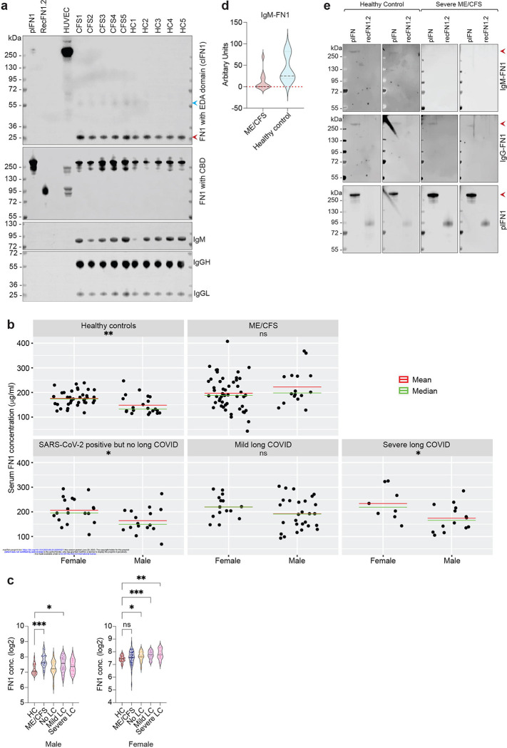
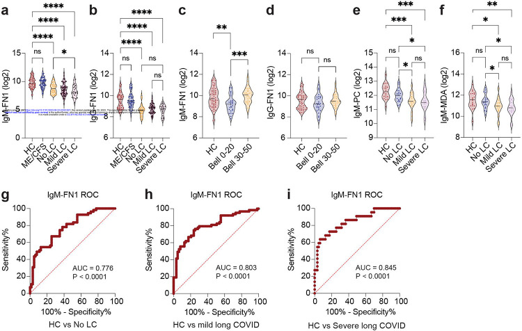
Myalgic Encephalomyelitis/ Chronic Fatigue syndrome (ME/CFS) is a complex, debilitating, long-term illness without a diagnostic biomarker. ME/CFS patients share overlapping symptoms with long COVID patients, an observation which has strengthened the infectious origin hypothesis of ME/CFS. However, the exact sequence of events leading to disease development is largely unknown for both clinical conditions. Here we show antibody response to herpesvirus dUTPases, particularly to that of Epstein-Barr virus (EBV) and HSV-1, increased circulating fibronectin (FN1) levels in serum and depletion of natural IgM against fibronectin ((n)IgM-FN1) are common factors for both severe ME/CFS and long COVID. We provide evidence for herpesvirus dUTPases-mediated alterations in host cell cytoskeleton, mitochondrial dysfunction and OXPHOS. Our data show altered active immune complexes, immunoglobulin-mediated mitochondrial fragmentation as well as adaptive IgM production in ME/CFS patients. Our findings provide mechanistic insight into both ME/CFS and long COVID development. Finding of increased circulating FN1 and depletion of (n)IgM-FN1 as a biomarker for the severity of both ME/CFS and long COVID has an immediate implication in diagnostics and development of treatment modalities.

Keywords: (n)IgM; EBV; Fibronectin; HHV-6; HSV-1; ME/CFS; dUTPase; long COVID.


Conflict of interest statement

Competing Interests: The authors declare that they have no competing interests.

Figures

Extended Fig. 1:
Extended Fig. 1:. Herpesvirus reactivation in healthy population, ME/CFS, long COVID patients.
a. Demography of distribution of patients according to the age and gender. b. Likert chart showing percentage of positivity for antibodies against EBV, HSV-1 and HHV-6 dUTPase within healthy controls (HC), ME/CFS, Covid-19 PCR positive but without long COVID (No LC), mild LC and severe LC patients. Kruskall Wallis (and its higher order equivalent, ScheirerRayHare Test) rank sum test for antibody state. HC, *P = 0.019; ME/CFS, *P = 0.021. Different amounts IgG levels in patient serum was arbitrarily divided into 4 groups (0, absent; 1, low; 2, moderate; 3, high).
Extended Fig. 2:
Extended Fig. 2:. Role of viral dUTPase proteins in mitochondrial dysfunction.
a. Confocal images showing hyperpolarization of mitochondria in U2-OS cells under transient expression of HSV-1, HHV-6 and EBV dUTPases. Mock vector was used as a control. b. Average mitochondrial surface area was compared between mock vector and EBV dUTPase transfected U2-OS cells. Data from three independent biological replicates. n=3. Unpaired two tailed non-parametric t-test. *P = 0.0278. c. Immunoblot analysis shows no major changes in dynamin related protein 1 (Drp1), p53, mitofusin1 (Mfn2), mitoguardin1 (Miga1) protein levels in presence of herpesvirus dUTPases. GAPDH staining was used as a loading control. d. TMRE dye was used to study mitochondrial membrane potential and OXPHOS in HEK293 cells. Cells were transiently transfected with HHV-6, HSV-1 dUTPases or a mock vector for 48 h. Non-transfected cells are also used as a control. Trypsinized cells were stained with TMRE dye and were used for flow cytometry. Oligomycin was used to inhibit ATP synthase. Data from 3 independent experiments. n=3. e. Data from above experiment was used to compare hyperpolarization status of mitochondria between mock transfected and EBV dUTPase transfected cells. Data from 3 independent experiments. n=3. f. Confocal images showing fragmentation of mitochondria in U2-OS cells in presence of recombinant EBV dUTPases. Same amounts of heat inactivated recombinant protein was used as a control. Average mitochondrial surface area was compared between control and recombinant EBV dUTPase exposed U2-OS cells. Data from four independent biological replicates. n=4. Unpaired two tailed non-parametric t-test. **P = 0.0032. g. Immunoblot analysis shows upregulation of dynamin related protein 1 (Drp1) levels in U2-OS cells exposed to recombinant EBV dUTPase for 24 h in a dose dependent manner. GAPDH staining was used as a loading control. Drp1 levels are quantified from three independent biological replicates and are shown in the form of a scatter plot. n=3. Unpaired two tailed non-parametric t-test. 0 vs 2 μg, *P = 0.0229; 0 vs 5 μg, *P = 0.0360; 0 vs 10 μg, *P = 0.0201. h. TMRE dye was used to study mitochondrial membrane potential and OXPHOS in HEK293 cells exposed to recombinant EBV dUTPase. Trypsinized cells were stained with TMRE dye and were used for flow cytometry. Oligomycin was used to inhibit ATP synthase. Data from 3 independent experiments. n=3. i. Coomassie dye stained polyacrylamide gel with input and immunoprecipitated samples shows efficient pull down of Halo-tagged herpesvirus dUTPases. Potential protein bands coimmunoprecipitated along with EBV and HSV-1 dUTPase protein is indicated. j. Normalized log2 ratio of LFQ (label-free quantitation) intensities of proteins. Fold change of proteins in EBV vs Mock was plotted against the same in HHV-6 vs Mock to highlight proteins that were specifically enriched in EBV dUTPase expressing co-IP. k. Normalized log2 ratio of LFQ (label-free quantitation) intensities of proteins. Fold change of proteins in HSV-1 vs Mock was plotted against the same in HHV-6 vs Mock to highlight proteins that were specifically enriched in HSV-1 dUTPase expressing co-IP. Circles indicate identified cellular proteins; circle size correlates with the number of peptides used for quantification. Significantly enriched proteins that are potential interaction partners of EBV (g) and HSV-1 (h) dUTPases are displayed in red. l. Immunoblot analysis to validate potential herpesvirus dUTPase interacting partners identified from co-IP.
Extended Fig. 3:
Extended Fig. 3:. Immune modulations in ME/CFS.
a. Immunoblot analysis shows validation of enrichment IgG against EBV dUTPase within purified IgG fractions. Recombinant dUTPase proteins are used as a bait. Left panel shows specific detection of EBV dUTPase using an antibody raised against it. Middle panel shows lack of specific signal against EBV dUTPase using a patient serum negative for EBV. Right panel confirms the absence of the specific signal against EBV dUTPase within the purified IgG. GAPDH staining was used as a loading control. b. Immunoblot analysis shows positive validation of enrichment IgG against EBV dUTPase within purified IgG fractions. Recombinant dUTPase proteins are used as a bait. Left panel shows specific detection of EBV dUTPase using an antibody raised against it. Middle panel shows specific signal against EBV dUTPases using a EBV positive patient serum. Right panel confirms the presence of the specific signal against EBV dUTPase within the purified IgG. Desired band is indicated with an arrow. GAPDH staining was used as a loading control. c. A heat map of proteins identified by mass spectrometry within the purified immunoglobulin complexes. Each row represents a specific protein, and each column represents a specific patient sample. The lower panel of the heat map from upper panel is enlarged in the lower panel along with the protein names. d. Log10 values of normalized LFQ intensities of three altered proteins (serotrasferrin, alpha2 microglobulin and fibronectin) comparing healthy controls (n=12) with ME/CFS patients (n=15).
Extended Fig. 4:
Extended Fig. 4:. Autoimmune signatures in ME/CFS.
a. The Variables Factor map (Biplot) for the Principal Components (for patients and healthy Controls combined data) shows the projection of the top 10 Autoantigen variables projected onto the plane spanned by the first two Principal Components. b. Biplot of PCA analysis showing variation condensed to key dimensions (dim reduction) for the data set shown in Fig. 2e–f.
Extended Fig. 5:
Extended Fig. 5:. Circulating fibronectin in ME/CFS.
a. Immunoblot analysis shows overall increase in both plasma fibronectin (plFN1) and cellular fibronectin (clFN1) in ME/CFS patients. Equal amounts of serum proteins from 5 ME/CFS patients with higher serum fibronectin levels were run in parallel with the same from 5 healthy controls. Purified plFN1, recombinant fibronectin isoform FN1.2 and HUVEC cell lysate expressing only cellular fibronectin were used as positive controls. Two different antibodies raised against Extra domain A (EDA) domain and CBD domain of fibronectin was used to differentiate plFN1 from clFN1 as plFN1 lacks EDA domain. b. Distribution of circulating FN1 concentrations in different groups of patients separated by gender. Two-tailed parametric t-test. Healthy control (HC) male vs female, **P = 0.0014; No LC male vs female, *P = 0.0204; severe LC male vs female, *P = 0.0347. c. Comparison of gender-specific circulating FN1 concentrations (log2 values) among different patient groups. Two-tailed parametric t-test. Healthy control (HC) male vs ME/CFS male, ***P = 0.0001; HC male vs mild LC male, *P = 0.012; HC female vs no LC female, *P = 0.0193; HC female vs mild LC female, ***P = 0.0006; HC female vs severe LC female, **P = 0.0016. d. IgM antibody levels against fibronectin (FN1) in ME/CFS patients and healthy controls in the form of a violin plot. IgM levels were determined from microarray studies where each antigen was tested twice separately. Normalized values against signal to noise ratio and net fluorescence intensity is used. e. Immunoblot analysis comparing the various species of circulating fibronectin (FN1) proteins in healthy controls and ME/CFS patient sera. Recombinant FN1.2 (recFN1.2) protein lacking extra domain-A (EDA domain) and HEK293 cell lysate serves as control. Three different antibodies detecting specific protein domains (EDA domain, CBD (cell binding domain) and cellular FN (clFN)) of FN1 was used against the same samples to compare the different species of proteins in the sera.
Extended Fig. 6:
Extended Fig. 6:. IgM-PC and IgM-MDA distributions among different groups of patients.
a. Distribution of serum IgM-PC concentrations in different groups of patients separated by gender. b. Distribution of serum IgM-MDA concentrations in different groups of patients separated by gender.
Extended Fig. 7:
Extended Fig. 7:. IgM-FN1 and IgG-FN1 distributions among different groups of patients.
a. Distribution of serum IgM-FN1 concentrations in different groups of patients separated by gender. Two-tailed parametric t-test. ME/CFS male vs female, *P = 0.0398. b. Distribution of serum IgG-FN1 concentrations in different groups of patients separated by gender. c. Comparison of gender-specific IgM-FN1 concentrations (log2 values) among different patient groups. Two-tailed parametric t-test. Healthy control (HC) male vs No LC male, **P = 0.003; HC male vs mild LC male, **P = 0.0018; HC male vs severe LC male, ***P = 0.0007; HC female vs no LC female, HC female vs mild LC female, HC female vs severe LC female, ****P < 0.0001.
Extended Fig. 8:
Extended Fig. 8:. Graphical abstract summarizing potential overlap between ME/CFS and long COVID pathogenesis.
Both ME/CFS and long COVID possibly originates as a post viral illness. SARS-CoV-2 infection is the major infection behind long COVID. However, heightened reactivation of herpesviruses like HSV-1 and EBV can potentially play a role in development of ME/CFS. Similar increase in herpesvirus reactivations including those of HSV-1, HHV-6 and EBV are also observed in ME/CFS. Virus-induced direct changes in cellular physiology are expected to be the major driver for the disease development. Subsequently, chronic tissue inflammation could lead to changes in secondary tissue homeostasis where increase in circulating fibronectin levels can play a key role in TLR2/TLR4-mediated innate immune response, cytokine production, Platelet activation, mast cell activation and alterations in clot homeostasis. Major cellular alterations within primary and secondary hematopoietic tissues might lead to substantial decrease in natural IgM production, which subsequently drive the autoimmune feature of both ME/CFS and long COVID. Changed autoimmune signature in the form of autoantibodies could then cause mitochondrial dysfunction, endothelial cell damage initiating a vicious cycle of events that can lead to severe forms of both ME/CFS and long COVID.
Fig. 1:
Fig. 1:. Reactivation of EBV in ME/CFS and long COVID patients and the role of viral dUTPase protein in cellular manipulation.
a. Immunoblot based detection of IgG against herpesvirus dUTPase in human serum. Virus-specific recombinant protein bands detected by IgG are indicated. b. Likert chart showing percentage of positivity for antibodies against EBV, HSV-1 and HHV-6 dUTPase within healthy controls (HC), ME/CFS, Covid-19 PCR positive but without long COVID (No LC), mild LC and severe LC patients. Kruskall Wallis (and its higher order equivalent, ScheirerRayHare Test) rank sum test for antibody state. EBV, *P = 0.015; HSV-1, *P = 0.068; HHV-6, **P = 0.002. Mann-Whitney non-parametric test for EBV dUTPase HC vs ME/CFS, ***P = 0.0008; for HSV-1 dUTPase HC vs severe LC, P = 0.051; for HSV-1 dUTPase No LC vs severe LC, *P = 0.013. Different amounts IgG levels in patient serum was arbitrarily divided into 4 groups (0, absent; 1, low; 2, moderate; 3, high). c. Confocal images shows hyperpolarization of mitochondria in HEK293 cells under transient expression of HSV-1, HHV-6 and EBV dUTPases. Mock vector backbone was used as a control. d. Average mitochondrial surface area from 5 biological replicates are plotted in the form of a scatter plot. n=5. Unpaired two-tailed non-parametric t-test. Mock vs EBV dUTPase, *P = 0.0414; Mock vs HHV-6 dUTPase, **P = 0.0077; Mock vs HSV-1 dUTPase, *P = 0.0479. e. Immunoblot analysis shows increase in mitofusin1 (Mfn1) and decrease in LC3β protein levels in presence of herpesvirus dUTPases. GAPDH staining was used as a loading control. Mean Mfn1 and LC3β protein levels are presented as scatter plots. Data from 3 independent experiments. n=3. Unpaired two-tailed non-parametric t-test. For Mfn1, *P = 0.03 (Mock vs HHV-6); *P = 0.01 (Mock vs HSV-1). For LC3β, *P = 0.01 (Mock vs EBV); *P = 0.05 (Mock vs HHV-6). f. EBV dUTPase interferes with autophagosome assembly. g. TMRE dyes were used to study mitochondrial membrane potential and OXPHOS in HEK293 cells. Cells were transiently transfected with herpesvirus dUTPases or a mock vector for 48 h. Trypsinized cells were stained with TMRE dye and were used for flow cytometry. Oligomycin was used to inhibit ATP synthase. Data from 3 independent experiments. n=3. MFI, mean fluorescence intensity. h. Normalized log2 ratio of LFQ (label-free quantitation) intensities of proteins. Fold change of proteins in HSV-1 vs Mock was plotted against the same in EBV vs Mock to highlight proteins that were common and were enriched in both sample sets. Circles indicate identified cellular proteins; circle size correlates with the number of razor and unique peptides used for quantification. Significantly enriched proteins that are potential interaction partners of EBV and HSV-1 dUTPases are displayed in red. i. Immunoblot analysis to validate potential herpesvirus dUTPase interacting partners identified from co-IP. GAPDH staining was used as a negative control.
Fig. 2:
Fig. 2:. Autoimmunity, mitochondrial alterations and circulating Fibronectin levels in ME/CFS.
a. Confocal images show mitochondrial architecture in primary HUVEC cells exposed to 1μg of purified IgG from patient sera. Two different representative images for each condition is shown. b. Average mitochondrial surface area in primary HUVEC cells exposed to 1μg of purified IgG from patient sera is quantified and compared between healthy controls, mild/moderate, severe ME/CFS. Data from three independent experiments from each serum sample is shown as a violin plot. n=3. Two-tailed non-parametric test. Healthy control vs mild/moderate ME/CFS, *P = 0.0329. Healthy control vs severe ME/CFS, **P = 0.0046. Mild/moderate vs severe ME/CFS, ****P < 0.0001. c. Immunoblot analysis shows decrease in mitofusin1 (Mfn1) and PLD6 protein levels in HUVEC cells exposed to 1μg of purified IgG from patient sera for 12 h. Actin staining was used as a loading control. Fold change values were derived from densitometric analysis of bands after normalization with the same for actin. n=2. HD, healthy donors; CFS, severe CFS patients. d. Heat map of log2 fold LFQ intensities of proteins detected within purified immune complexes from patient sera. Three proteins that showed differential protein levels between healthy controls and ME/CFS patients are shown. e. Multivariate analysis of clusters based on distance metrics derived from IgM antibody levels for a panel of autoantigens. Log-transformed scaled data showing relative differences between different variable in both healthy controls and patients. f. The Variables Factor map for the Principal Components (for Patients and Healthy Controls combined data) shows the projection of the top 10 Autoantigen variables projected onto the plane spanned by the first two Principal Components. g. Serum fibronectin (FN1) levels in patient sera. Log2 values of FN1 are presented as a violin plot. Two-tailed parametric t-test. Healthy control (HC) vs ME/CFS, **P = 0.005. h. Kernel density plot showing the bivariate serum FN1 distributions among healthy controls and ME/CFS patients. FN1 concentrations on both X- and Y-axis are presented as μg/ml. i. Circulating fibronectin (FN1) levels correlates with ME/CFS severity associated Bell score. Log2 fold FN1 vales are presented as a violin plot. Two-tailed parametric t-test. HC vs Bell 020, ****P < 0.0001. Bell 0–20 vs Bell 30–50, **P = 0.0032. j. AUROC analysis for circulating FN1 levels in healthy controls (HC) vs severe ME/CFS patients. k. Serum fibronectin (FN1) levels in different patient groups post SARS-CoV-2 infection. Log2 values of FN1 are presented as a violin plot. Two-tailed parametric t-test. Healthy control (HC) vs mild LC, **P = 0.0032. HC vs severe LC, *P = 0.0488. ns, not significant.
Fig. 3:
Fig. 3:. IgM and IgG levels against Fibronectin in ME/CFS and long COVID.
a. IgM levels against fibronectin (IgM-FN1) in patient sera. Log2 fold IgM-FN1 amounts are presented as a violin plot. Two-tailed parametric t-test. Healthy control (HC) vs SARS CoV-2 positive but without long COVID (No LC), HC vs mild LC, HC vs severe LC, ****P < 0.00001. No LC vs severe LC, *P = 0.0376. ns, not significant. b. IgG levels against fibronectin (IgM-FN1) in patient sera. Log2 fold IgG-FN1 amounts are presented as a violin plot. Two-tailed parametric t-test. Healthy control (HC) vs SARS CoV-2 positive but without long COVID (No LC), HC vs mild LC, HC vs severe LC, ****P < 0.00001. ns, not significant. c. IgM-fibronectin (FN1) levels correlates with ME/CFS severity associated Bell scores. Log2 fold IgM-FN1 vales are presented as a violin plot. Two-tailed parametric t-test. HC vs Bell 0–20, **P = 0.0046. Bell 0–20 vs Bell 30–50, ***P = 0.0002. d. IgG-fibronectin (FN1) levels does not correlate with ME/CFS severity associated Bell scores. Log2 fold IgG-FN1 vales are presented as a violin plot. Two-tailed parametric t-test. Ns, not significant. e. IgM levels against phosphorylcholine (IgM-PC) in patient sera. Log2 fold IgM-PC amounts are presented as a violin plot. Two-tailed parametric t-test. Healthy control (HC) vs mild LC, ***P = 0.0002. HC vs severe LC, ***P = 0.0006. No LC vs severe LC, *P = 0.0216. No LC vs mild LC, *P = 0.0111. ns, not significant. f. IgM levels against malondialdehyde (IgM-MDA) in patient sera. Log2 fold IgM-MDA amounts are presented as a violin plot. Two-tailed parametric t-test. Healthy control (HC) vs mild LC, *P = 0.0175. HC vs severe LC, **P = 0.0068. No LC vs severe LC, *P = 0.0209. No LC vs mild LC, *P = 0.0525. ns, not significant. g. AUROC analysis for IgM-FN1 levels in healthy controls (HC) vs SARS CoV-2 positive but without long COVID (No LC) patients. h. AUROC analysis for IgM-FN1 levels in healthy controls (HC) vs mild LC patients. i. AUROC analysis for IgM-FN1 levels in healthy controls (HC) vs severe LC patients.
Fig. 4:
Fig. 4:. IgM-FN1 and circulating fibronectin as a biomarker in ME/CFS and long COVID.
a. Multi-variate AUROC analysis for circulating FN1 and IgM against fibronectin (IgM-FN1) in healthy controls (HC) vs severe ME/CFS patients. Multiple logistic regression. ****P < 0.0001. b. Multi-variate AUROC analysis for circulating FN1 and IgM against fibronectin (IgM-FN1) in healthy controls (HC) vs severe LC patients. Multiple logistic regression. ****P < 0.0001. c. Multiple variable bubble plot with circulating FN1 levels plotted against IgM-FN1 for healthy controls (HC) and severe ME/CFS patients. d. Multiple variable bubble plot with circulating FN1 levels plotted against IgM-FN1 for healthy controls (HC) and total long COVID patients (mild LC + severe LC). e. Multiple variable bubble plot with circulating FN1 levels plotted against IgM-FN1 for ME/CFS and total long COVID patients (mild LC + severe LC). f. Multiple variable bubble plot with circulating FN1 levels plotted against IgM-FN1 for severe ME/CFS (with Bell 0–20) and severe long COVID patients.

References

References for Methods:

    1. Bahmer T. et al. Severity, predictors and clinical correlates of Post-COVID syndrome (PCS) in Germany: A prospective, multi-centre, population-based cohort study. EClinicalMedicine 51, 101549, doi: 10.1016/j.eclinm.2022.101549 (2022). - DOI - PMC - PubMed
    1. Horn A. et al. Long-term health sequelae and quality of life at least 6 months after infection with SARS-CoV-2: design and rationale of the COVIDOM-study as part of the NAPKON population-based cohort platform (POP). Infection 49, 1277–1287, doi: 10.1007/s15010-021-01707-5 (2021). - DOI - PMC - PubMed
    1. Wagner M. et al. Characteristics and Course of Heart Failure Stages A-B and Determinants of Progression - design and rationale of the STAAB cohort study. Eur J Prev Cardiol 24, 468–479, doi: 10.1177/2047487316680693 (2017). - DOI - PubMed
    1. Morbach C. et al. Prevalence and determinants of the precursor stages of heart failure: results from the population-based STAAB cohort study. Eur J Prev Cardiol 28, 924–934, doi: 10.1177/2047487320922636 (2021). - DOI - PubMed
    1. Hennig T. et al. Selective inhibition of miRNA processing by a herpesvirus-encoded miRNA. Nature 605, 539–544, doi: 10.1038/s41586-022-04667-4 (2022). - DOI - PubMed

References

    1. Davis H. E., McCorkell L., Vogel J. M. & Topol E. J. Long COVID: major findings, mechanisms and recommendations. Nat Rev Microbiol 21, 133–146, doi: 10.1038/s41579-022-00846-2 (2023). - DOI - PMC - PubMed
    1. Banko A., Miljanovic D. & Cirkovic A. Systematic review with meta-analysis of active herpesvirus infections in patients with COVID-19: Old players on the new field. Int J Infect Dis 130, 108–125, doi: 10.1016/j.ijid.2023.01.036 (2023). - DOI - PMC - PubMed
    1. Klein J. et al. Distinguishing features of Long COVID identified through immune profiling. medRxiv, doi: 10.1101/2022.08.09.22278592 (2022). - DOI - PMC - PubMed
    1. Rasa S. et al. Chronic viral infections in myalgic encephalomyelitis/chronic fatigue syndrome (ME/CFS). J Transl Med 16, 268, doi: 10.1186/s12967-018-1644-y (2018). - DOI - PMC - PubMed
    1. Kasimir F. et al. Tissue specific signature of HHV-6 infection in ME/CFS. Front Mol Biosci 9, 1044964, doi: 10.3389/fmolb.2022.1044964 (2022). - DOI - PMC - PubMed

Publication types