. 2023 Jun 30;15(6):3089-3105.
doi: 10.21037/jtd-22-1593. Epub 2023 May 10.

Isoproterenol pre-treatment improve the therapeutic efficacy of cardiosphere-derived cells transplantation for myocardial infarction

Affiliations

Isoproterenol pre-treatment improve the therapeutic efficacy of cardiosphere-derived cells transplantation for myocardial infarction

Xiaoying Duan et al. J Thorac Dis. .

Abstract

Background: This study aimed to investigate the effect of isoproterenol pre-treatment on the therapeutic efficacy of cardiosphere-derived cells (CDCs) transplantation for myocardial infarction (MI).

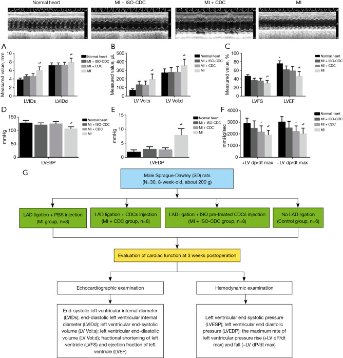
Methods: Thirty 8-week-old male Sprague-Dawley (SD) rat model of MI was generated by ligation of the left anterior descending artery. The MI rats were treated with PBS (MI group, n=8), CDCs (MI + CDC group, n=8) and isoproterenol pre-treated CDCs (MI + ISO-CDC group, n=8), respectively. In the MI + ISO-CDC group, CDCs were pre-treated by 10-6 M isoproterenol and the cultured for additional 72 h, then injected to the myocardial infraction area like other groups. At 3 weeks after the operation, echocardiographic, hemodynamic, histological assessments and Western blot were performed to compare the CDCs differentiation degree and therapeutic effect.

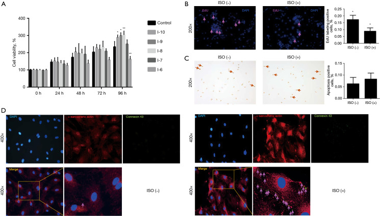
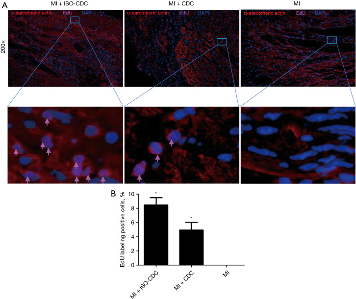
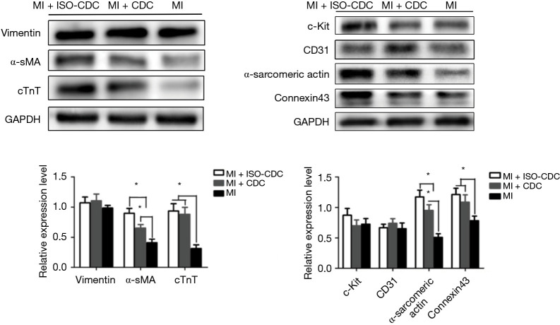
Results: Isoproterenol treatment (10-6 M) simultaneously inhibited proliferation and induced apoptosis of CDCs, up-regulated proteins of vimentin, cTnT, α-sarcomeric actin and connexin 43, and down-regulated c-Kit proteins (all P<0.05). The echocardiographic and hemodynamic analysis demonstrated that the MI rats in the two CDCs transplantation groups had significantly better recovery of cardiac function than the MI group (all P<0.05). MI + ISO-CDC group had better recovery of cardiac function than the MI + CDC group, although the differences did not reach significant. Immunofluorescence staining showed that the MI + ISO-CDC group had more EdU-positive (proliferating) cells and cardiomyocytes in the infarct area than the MI + CDC group. MI + ISO-CDC group had significantly higher protein levels of c-Kit, CD31, cTnT, α-sarcomeric actin and α-SMA in the infarct area than the MI + CDC group.

Conclusions: These results suggested that in CDCs transplantation, isoproterenol pre-treated CDCs can provide a better protective effect against MI than the untreated CDCs.

Keywords: Myocardial infarction (MI); cardiosphere-derived cells (CDCs); cell transplantation; isoproterenol.


Conflict of interest statement

Conflicts of Interest: All authors have completed the ICMJE uniform disclosure form (available at https://jtd.amegroups.com/article/view/10.21037/jtd-22-1593/coif). The authors have no conflicts of interest to declare.

Figures

Figure 1
Figure 1
Isolation and expansion of CDCs. (A) Cardiac explant tissue was cultured in CEM medium. After 1 week of culture, mesenchymal-like cells (CDCs) grew around the cardiac explant tissue (200× magnification). (B) Microscopic image of passage 1 CDCs (200× magnification). (C) Microscopic image of passage 2 CDCs (200× magnification). (D) The surface markers of the CDCs. Immunofluorescence images of P3 CDCs stained by c-Kit, Sca-1, cTnT, Nkx-2.5, GATA4, and CD34 respectively. The cell nucleus stained with DAPI exhibited blue color (400× magnification). DAPI, 4’,6-diamidino-2-phenylindole; CDC, cardiosphere-derived cell; CEM, complete explant medium.
Figure 2
Figure 2
Effects of isoproterenol on the proliferation rate of CDCs. Cell viability was determined by the MTT assay. A total of 103 P3 CDCs were plated on 96-well plates (n=6 for each group) and treated with drugs as following: control group (without drug treatment), I−10(10−10 M isoproterenol), I−9(10−9 M isoproterenol), I−8(10−8 M isoproterenol), I−7(10−7 M isoprenaline), I−6(10−6 M isoproterenol). *, P<0.05, **, P<0.01, compared to the control group at the same time point. (B) Cell proliferation was determined by Edu assay. Positively stained cell nuclei were light purple (arrows). Edu-positive cells significantly decreased after ISO treatment, *, P<0.05. (C) TUNEL staining was performed to assess the apoptosis of isoproterenol-treated CDCs. TUNEL-positive cell nuclei were dark brown (arrows). There was no statistical difference between these two groups. (D) Immunofluorescent staining between control group and 10−6 M isoproterenol group at the 72 h. It showed the CDCs have a better differentiation ability under the 10−6 M isoproterenol pretreatment. ISO, isoproterenol; DAPI, 4’,6-diamidino-2-phenylindole; CDC, cardiosphere-derived cell; MTT, 3-(4,5-Dimethylthiazol-2-yl)-2,5-diphenyltetrazolium bromide; TUNEL, terminal deoxynucleotidyl transferase dUTP nick-end labeling.
Figure 3
Figure 3
Effects of isoproterenol on the differentiation of CDCs. (A) Passage 3 CDCs on 3.5 cm plate were treated with or without isoproterenol (10−6 M) for 72 h. Protein levels of vimentin, α-SMA, cTnT, c-Kit, CD31, α-sarcomeric actin, connexin 43 were determined by Western blot. (B) Protein levels of p38α and p-p38α. P3 CDCs were treated with each designated drug for 72 h. For CDCs treated with a combination of ISO and other drugs, ISO was added to medium 2 h after treatment with other drug(s). PRO, a non-selective β-AR blocker, was used to block the effects of isoproterenol. The intensity of band was quantitated and presented as the ratio of p38α/p-p38α. The intensity of the band was quantitated and normalized to internal control GAPDH. *, P<0.05. CDC, cardiosphere-derived cell; ISO, isoproterenol; PRO, propranolol; GAPDH, glyceraldehyde 3-phosphate dehydrogenase.
Figure 4
Figure 4
Evaluation of cardiac function among groups at 3 weeks after operation. The MI rats were treated with PBS (MI group), CDCs (MI + CDC group) or isoproterenol pre-treated CDCs (MI + ISO-CDC group). All animals received echocardiographic examination, and the following echocardiographic parameters were compared: (A) LVIDs, LVIDd, (B) LV Vol;s, LV Vol;d, (C) LVFS, and LVEF. The following hemodynamic parameters were compared: (D) LVESP, (E) LVEDP, (F) +LV dP/dt max and −LV dP/dt max, (G) the experimental schema panel of this study. *, P<0.05 compared with the Normal heart group. #, P<0.05 compared with the MI group. MI, myocardial infarction; ISO, isoproterenol; CDC, cardiosphere-derived cell; LVIDs, end-systolic left ventricular internal diameter; LVIDd, end-diastolic left ventricular internal diameter; LV Vol;s, left ventricular end-systolic volume; LV Vol;d, left ventricular end-diastolic volume; LVFS, fractional shortening of left ventricle; LVEF, ejection fraction of left ventricle; LVESP, left ventricular end systolic pressure; LVEDP, left ventricular end diastolic pressure; +LV dP/dt max, the maximum rate of left ventricular pressure rise; −LV dP/dt max, the maximum rate of left ventricular pressure fall; LAD, left anterior descending artery; PBS, phosphate-buffered saline.
Figure 5
Figure 5
Masson staining in the myocardial infarct zone at 3 weeks after operation. The MI rats were treated with PBS (MI group), CDCs (MI + CDC group) or isoproterenol pre-treated CDCs (MI + ISO-CDC group). Collagen fibers were stained blue; muscle fibers and erythrocytes exhibited red; the nucleus exhibited blue-brown. Images were taken at ×200. For each group, 8 animals were used and 8 slides were analyzed. The bar chart showed quantitative results of interstitial fibrosis area. *, P<0.05 compared with the MI group. MI, myocardial infarction; ISO, isoproterenol; CDC, cardiosphere-derived cell; PBS, phosphate-buffered saline.
Figure 6
Figure 6
Immunofluorescence staining in the myocardial infarct zone at 3 weeks after operation. (A) The MI rats were treated with PBS (MI group), CDCs (MI + CDC group) or isoproterenol pre-treated CDCs (MI + ISO-CDC group). The nucleus was stained blue; Edu-positive cell nuclei was light purple (arrows); α-sarcomeric actin was stained red. Images were taken at ×200 and ×400 magnifications, respectively. (B) Quantitatively result of Edu-positive cells. *, P<0.05 compared with the MI group. MI, myocardial infarction; ISO, isoproterenol; CDC, cardiosphere-derived cell; EdU, 5-ethynyl-2’-deoxyuridine; DAPI, 4’,6-diamidino-2-phenylindole; PBS, phosphate-buffered saline.
Figure 7
Figure 7
Protein expressions of in the myocardial infarct zone at 3 weeks after operation. Protein levels of vimentin, cTnT, connexin 43, c-Kit, CD31, α-SMA, α-sarcomeric actin were determined by Western blot. The MI rats were treated with PBS (MI group), CDCs (MI + CDC group) or isoproterenol pre-treated CDCs (MI + ISO-CDC group). The intensity of the band was quantitated and normalized to internal control GAPDH. *, P<0.05. MI, myocardial infarction; ISO, isoproterenol; CDC, cardiosphere-derived cell; PBS, phosphate-buffered saline; GAPDH, glyceraldehyde 3-phosphate dehydrogenase.

Similar articles

References

    1. Ashur C, Frishman WH. Cardiosphere-Derived Cells and Ischemic Heart Failure. Cardiol Rev 2018;26:8-21. 10.1097/CRD.0000000000000173 - DOI - PubMed
    1. Thomas H, Diamond J, Vieco A, et al. Global Atlas of Cardiovascular Disease 2000-2016: The Path to Prevention and Control. Glob Heart 2018;13:143-63. 10.1016/j.gheart.2018.09.511 - DOI - PubMed
    1. Cahill TJ, Choudhury RP, Riley PR. Heart regeneration and repair after myocardial infarction: translational opportunities for novel therapeutics. Nat Rev Drug Discov 2017;16:699-717. 10.1038/nrd.2017.106 - DOI - PubMed
    1. Kachanova O, Lobov A, Malashicheva A. The Role of the Notch Signaling Pathway in Recovery of Cardiac Function after Myocardial Infarction. Int J Mol Sci 2022;23:12509. 10.3390/ijms232012509 - DOI - PMC - PubMed
    1. Konstam MA, Kramer DG, Patel AR, et al. Left ventricular remodeling in heart failure: current concepts in clinical significance and assessment. JACC Cardiovasc Imaging 2011;4:98-108. 10.1016/j.jcmg.2010.10.008 - DOI - PubMed