. 2023 Jun 28;29(24):3770-3792.
doi: 10.3748/wjg.v29.i24.3770.

Mechanism of ELL-associated factor 2 and vasohibin 1 regulating invasion, migration, and angiogenesis in colorectal cancer

Affiliations

Mechanism of ELL-associated factor 2 and vasohibin 1 regulating invasion, migration, and angiogenesis in colorectal cancer

Ming-Liang Feng et al. World J Gastroenterol. .

Abstract

Background: As a novel endogenous anti-angiogenic molecule, vasohibin 1 (VASH1) is not only expressed in tumor stroma, but also in tumor tissue. Moreover, studies have shown that VASH1 may be a prognostic marker in colorectal cancer (CRC). Knockdown of VASH1 enhanced transforming growth factor-β1 (TGF-β1)/Smad3 pathway activity and type I/III collagen production. Our previous findings suggest that ELL-associated factor 2 (EAF2) may play a tumor suppressor and protective role in the development and progression of CRC by regulating signal transducer and activator of transcription 3 (STAT3)/TGF-β1 signaling pathway. However, the functional role and mechanism of VASH1-mediated TGF-β1 related pathway in CRC has not been elucidated.

Aim: To investigate the expression of VASH1 in CRC and its correlation with the expression of EAF2. Furthermore, we studied the functional role and mechanism of VASH1 involved in the regulation and protection of EAF2 in CRC cells in vitro.

Methods: We collected colorectal adenocarcinoma and corresponding adjacent tissues to investigate the clinical expression of EAF2 protein and VASH1 protein in patients with advanced CRC. Following, we investigated the effect and mechanism of EAF2 and VASH1 on the invasion, migration and angiogenesis of CRC cells in vitro using plasmid transfection.

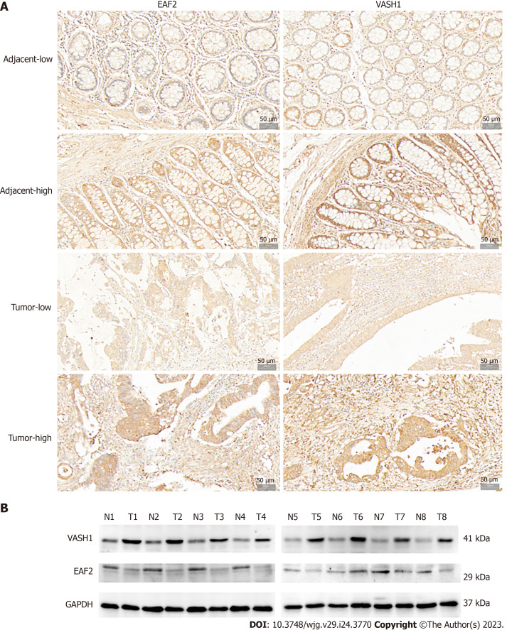
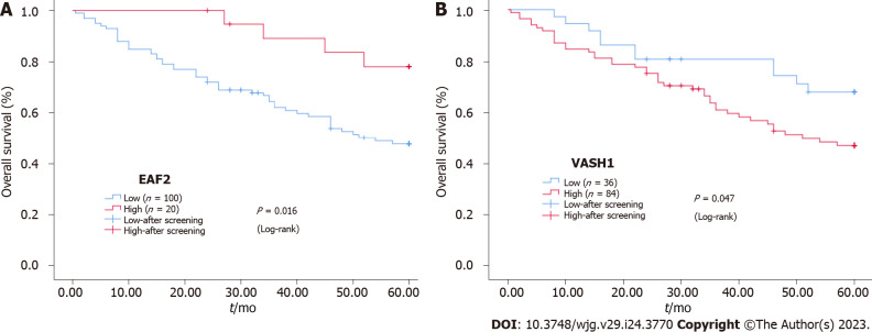
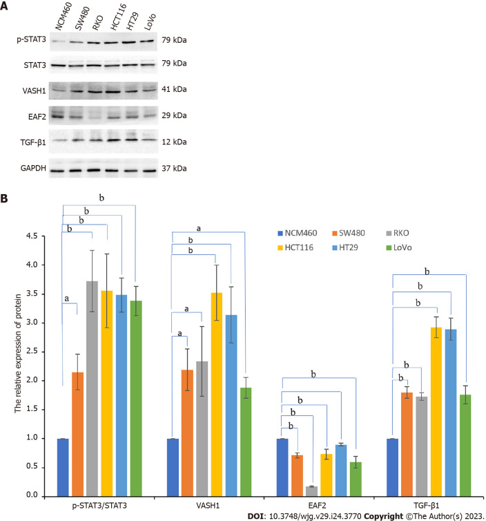
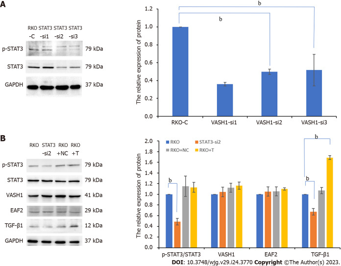
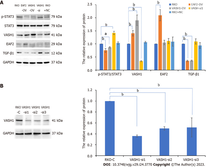
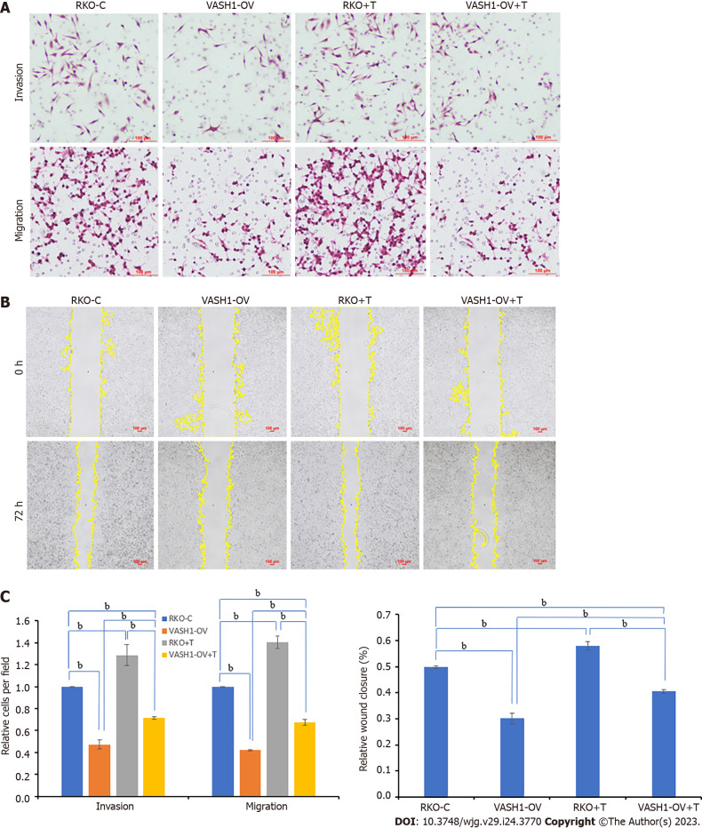
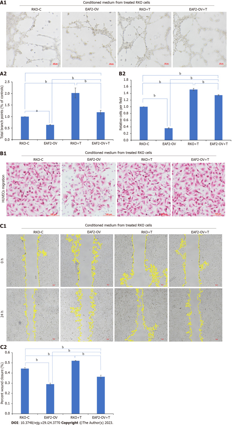
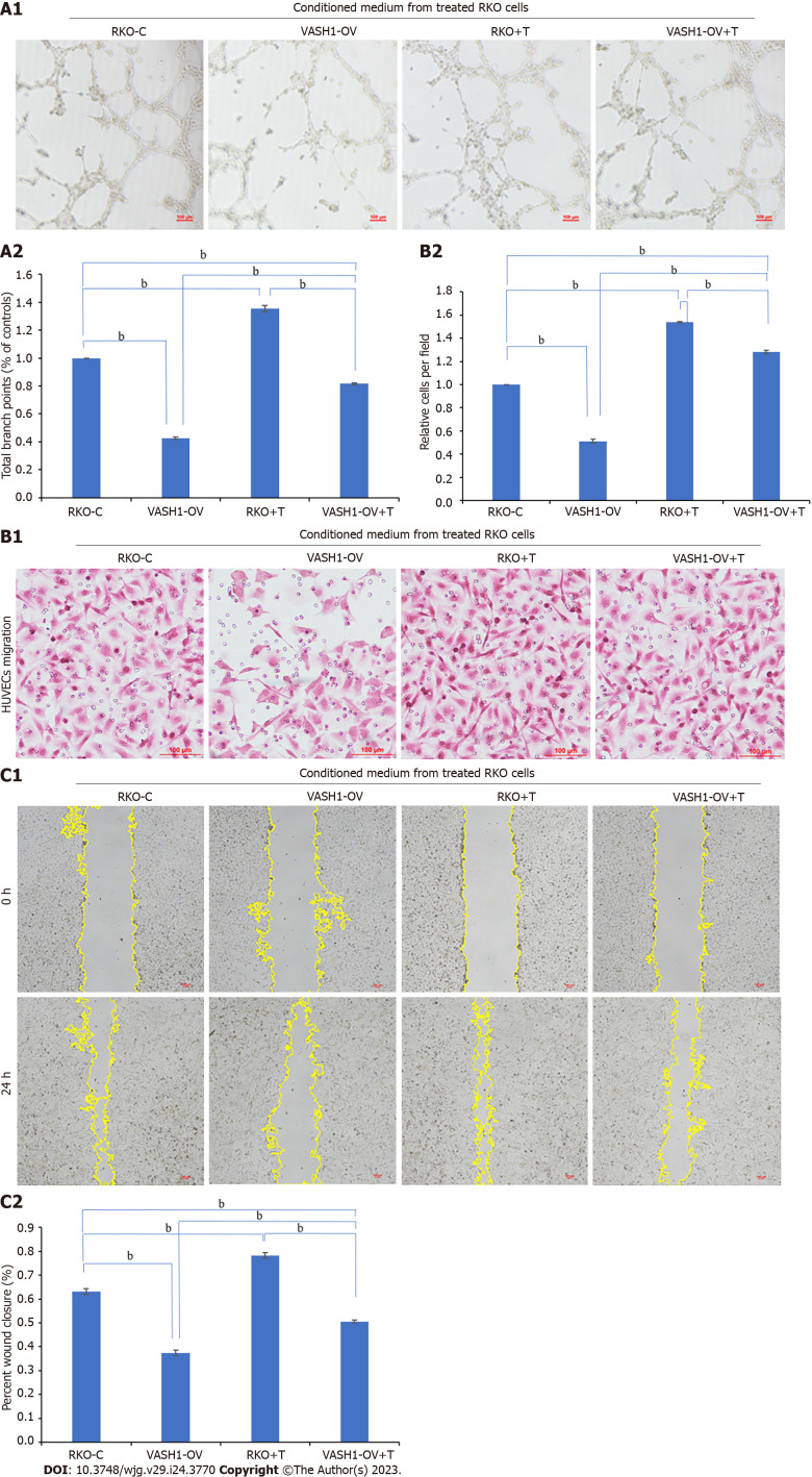
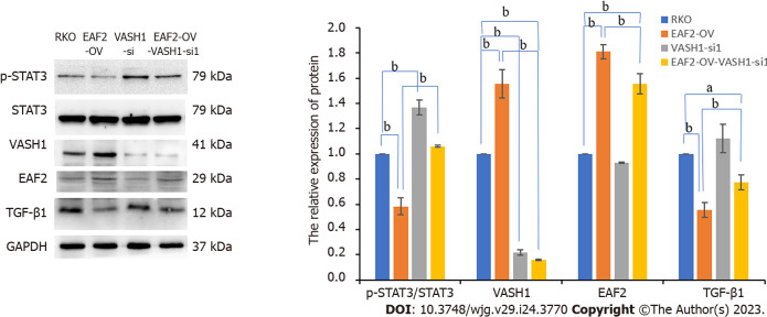
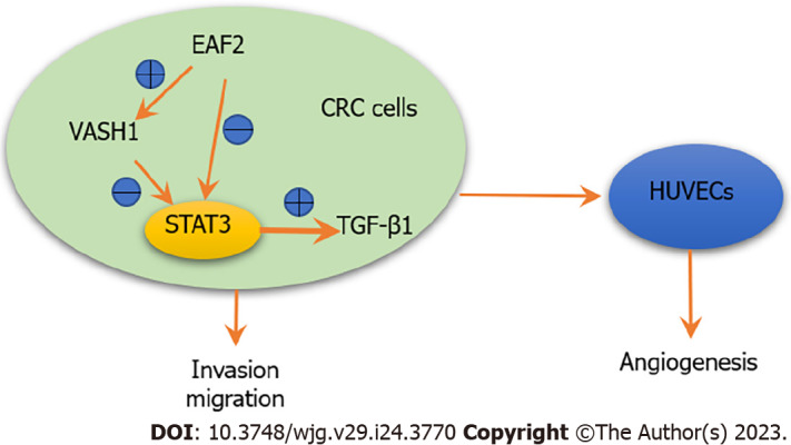
Results: Our findings indicated that EAF2 was down-regulated and VASH1 was up-regulated in advanced CRC tissue compared to normal colorectal tissue. Kaplan-Meier survival analysis showed that the higher EAF2 Level group and the lower VASH1 Level group had a higher survival rate. Overexpression of EAF2 might inhibit the activity of STAT3/TGF-β1 pathway by up-regulating the expression of VASH1, and then weaken the invasion, migration and angiogenesis of CRC cells.

Conclusion: This study suggests that EAF2 and VASH1 may serve as new diagnostic and prognostic markers for CRC, and provide a clinical basis for exploring new biomarkers for CRC. This study complements the mechanism of EAF2 in CRC cells, enriches the role and mechanism of CRC cell-derived VASH1, and provides a new possible subtype of CRC as a therapeutic target of STAT3/TGF-β1 pathway.

Keywords: Angiogenesis; Colorectal cancer; ELL-associated factor 2; Signal transducer and activator of transcription 3; Transforming growth factor-β1; Vasohibin 1.


Conflict of interest statement

Conflict-of-interest statement: The authors declare that they have no conflict of interest.

Figures

Figure 1
Figure 1
Expression of ELL-associated factor 2 and Vasohibin 1 proteins in tissues of patients with advanced colorectal cancer. A: Representative immunohistochemical staining of ELL-associated factor 2 (EAF2) and vasohibin 1 (VASH1) in colorectal adenocarcinoma tissue and the adjacent non-cancerous tissue (magnification 200 ×); B: The protein bands of EAF2 and VASH1 in 8-matched pairs of colorectal adenocarcinoma tissue and adjacent tissue were detected by Western blot assay. EAF2: ELL-associated factor 2; VASH1: Vasohibin 1; GAPDH: Glyceraldehyde-3-phosphate dehydrogenase; T: Colorectal adenocarcinoma tissue; N: Adjacent non-tumor tissue.
Figure 2
Figure 2
Kaplan-Meier survival curves. A and B: Kaplan-Meier survival curves for the protein expression of ELL-associated factor 2 and vasohibin 1 in colorectal cancer tissue. EAF2: ELL-associated factor 2; VASH1: Vasohibin 1.
Figure 3
Figure 3
Protein expression of pathway-related factors in a variety of human colorectal cancer cells and colorectal epithelial cells. A and B: The protein bands of ELL-associated factor 2, vasohibin 1, phosphorylated-signal transducer and activator of transcription 3, and transforming growth factor-β1 in colorectal cancer cell lines (SW480, RKO, HCT116, HT29, and LoVo) and normal colorectal epithelial cells (NCM460) by Western blot assay and the corresponding data. aP < 0.05, bP < 0.001. EAF2: ELL-associated factor 2; VASH1: Vasohibin 1; p-STAT3: Phosphorylated-signal transducer and activator of transcription 3; TGF-β1: Transforming growth factor-β1.
Figure 4
Figure 4
Silencing signal transducer and activator of transcription 3 phosphorylation significantly inhibits the expression of transforming growth factor-β1 protein in RKO cells. A: The protein bands of phospho-signal transducer and activator of transcription 3 (STAT3) in RKO cells transfected with STAT3 siRNA plasmids by Western blot assay and the corresponding data; B: The protein bands of pathway-related factors in RKO cells treated with STAT3 siRNA or exogenous transforming growth factor-β1 recombinant protein by Western blot assay. bP < 0.001. RKO-C: RKO cells were cultured in 1640 medium containing 10% fetal bovine serum (FBS). STAT3-si: RKO cells were transfected with STAT3 siRNA plasmids for 6 h and then cultured in 1640 medium containing 10% FBS for 24 h. RKO + NC: RKO cells were transfected empty vector for 6 h and then cultured in 1640 medium containing 10% FBS for 24 h. RKO + T: RKO cells were cultured for 24 h in 1640 medium containing 10% FBS in which 5 ng/mL TGF-β1 recombinant protein was added. VASH1: Vasohibin 1; STAT3: Signal transducer and activator of transcription 3; EAF2: ELL-associated factor 2; TGF-β1: Transforming growth factor-β1.
Figure 5
Figure 5
Overexpressing vasohibin 1 inhibits signal transducer and activator of transcription 3 phosphorylation (Tyr705) as well as transforming growth factor-β1 protein expression in RKO cells. A: The protein bands of pathway-related factors in RKO cells transfected with ELL-associated factor 2 (EAF2) overexpression plasmids, or vasohibin 1 (VASH1) overexpression plasmids, or VASH1 silencing plasmids by Western blot assay; B: The protein bands of VASH1 in RKO cells transfected with VASH1 siRNA plasmids by Western blot assay and the corresponding data. aP < 0.05, bP < 0.001. VASH1-si: RKO cells transfected with VASH1 siRNA plasmids for 6 h and then cultured in 1640 medium containing 10% fetal bovine serum (FBS) for 24 h. VASH1-OV: The group of RKO cells transfected with VASH1 overexpression plasmids for 6 h and then cultured in 1640 medium containing 10% FBS for 24 h. EAF2-OV: RKO cells transfected with EAF2 overexpression plasmids for 6 h and then cultured in 1640 medium containing 10% FBS for 24 h. VASH1: Vasohibin 1; EAF2: ELL-associated factor 2; TGF-β1: Transforming growth factor-β1.
Figure 6
Figure 6
Overexpressing ELL-associated factor 2 inhibits the invasion and migration of RKO cells by suppressing transforming growth factor-β1 related pathway. A: The invasion and migration abilities were evaluated by the transwelll assay; B: The migration ability was analyzed by wound healing assay after 72 h; C: The results of migration and invasion abilities of RKO cells were measured using ImageJ software. aP < 0.05, bP < 0.001. ELL-associated factor 2 (EAF2)-OV + T: The group of RKO cells were transfected with EAF2 overexpression plasmids for 24 h and then cultured with 5 ng/mL transforming growth factor-β1 recombinant protein for 24 h. EAF2: ELL-associated factor 2.
Figure 7
Figure 7
Overexpressing Vasohibin 1 inhibits the invasion and migration of RKO cells by suppressing transforming growth factor-β1 related pathway. A: The invasion and migration abilities were evaluated by the transwelll assay; B: The migration ability was analyzed by wound healing assay after 72 h; C: The results of migration and invasion abilities of RKO cells were measured using ImageJ software. bP < 0.001. vasohibin 1 (VASH1)-OV + T: The group of RKO cells were transfected with VASH1 overexpression plasmids for 24 h and then cultured with 5 ng/mL transforming growth factor-β1 recombinant protein for 24 h. VASH1: Vasohibin 1.
Figure 8
Figure 8
Overexpressing ELL-associated factor 2 inhibits tumor-induced angiogenesis in RKO cells via transforming growth factor-β1 related pathway. A: Conditioned medium from RKO cells transfected with ELL-associated factor 2-overexpressed plasmids or treated with transforming growth factor-β1 recombinant protein was collected to perform tube formation assay; B and C: The migration ability of Human Umbilical Vein Endothelial Cells was analyzed by transwell migration assay (B) and wound healing assay (C). And the results were measured by ImageJ software. aP < 0.05, bP < 0.001. EAF2: ELL-associated factor 2; TGF-β1: Transforming growth factor-β1.
Figure 9
Figure 9
Overexpressing Vasohibin 1 inhibits tumor-induced angiogenesis in RKO cells via transforming growth factor-β1 related pathway. A: Conditioned medium from RKO cells transfected with VASH1-overexpressed plasmids or treated with transforming growth factor-β1 recombinant protein was collected to perform tube formation assay; B and C: The migration ability of Human Umbilical Vein Endothelial Cells was analyzed by transwell migration assay (B) and wound healing assay (C). And the results were measured by ImageJ software. bP < 0.001. VASH1: Vasohibin 1; EAF2: ELL-associated factor 2.
Figure 10
Figure 10
Silencing Vasohibin 1 reverses the signal transducer and activator of transcription 3/transforming growth factor-β1 pathway inhibited by ELL-associated factor 2 overexpression. aP < 0.05, bP < 0.001. ELL-associated factor 2 (EAF2)-OV- Vasohibin 1 (VASH1-si): The group of RKO cells transfected with EAF2 overexpression plasmids and VASH1 siRNA plasmids. VASH1: Vasohibin 1; EAF2: ELL-associated factor 2.
Figure 11
Figure 11
Schematic depiction of ELL-associated factor 2 and vasohibin 1 regulating the signal transducer and activator of transcription 3/ transforming growth factor-β1 signaling pathway in this study. Overexpression of ELL-associated factor 2 may inhibit the activity of signal transducer and activator of transcription 3/transforming growth factor-β1 pathway by up-regulating the expression of vasohibin 1, thereby inhibiting cell invasion, migration and angiogenesis. EAF2: ELL-associated factor 2; TGF-β1: Transforming growth factor-β1; STAT3: Signal transducer and activator of transcription 3; CRC: Colorectal cancer; HUVECs: Human umbilical vein endothelial cells; VASH1: Vasohibin 1.

Similar articles

Cited by

References

    1. Shaukat A, Kahi CJ, Burke CA, Rabeneck L, Sauer BG, Rex DK. ACG Clinical Guidelines: Colorectal Cancer Screening 2021. Am J Gastroenterol. 2021;116:458–479. - PubMed
    1. Li X, Wu Y, Tian T. TGF-β Signaling in Metastatic Colorectal Cancer (mCRC): From Underlying Mechanism to Potential Applications in Clinical Development. Int J Mol Sci. 2022;23 - PMC - PubMed
    1. Kobayashi M, Wakabayashi I, Suzuki Y, Fujiwara K, Nakayama M, Watabe T, Sato Y. Tubulin carboxypeptidase activity of vasohibin-1 inhibits angiogenesis by interfering with endocytosis and trafficking of pro-angiogenic factor receptors. Angiogenesis. 2021;24:159–176. - PubMed
    1. Tamaki K, Moriya T, Sato Y, Ishida T, Maruo Y, Yoshinaga K, Ohuchi N, Sasano H. Vasohibin-1 in human breast carcinoma: a potential negative feedback regulator of angiogenesis. Cancer Sci. 2009;100:88–94. - PMC - PubMed
    1. Zhao G, Yang Y, Tang Y, Han R, Sun Y. Reduced expression of vasohibin-1 is associated with clinicopathological features in renal cell carcinoma. Med Oncol. 2012;29:3325–3334. - PubMed