

NEK6 Promotes the Progression of Osteosarcoma Through Activating STAT3 Signaling Pathway by Down-Regulation of miR-26a-5p
- PMID: 37426517
- PMCID: PMC10329465
- DOI: 10.2147/IJGM.S413461
NEK6 Promotes the Progression of Osteosarcoma Through Activating STAT3 Signaling Pathway by Down-Regulation of miR-26a-5p
Abstract
Background: Osteosarcoma is a malignant tumor originating from the skeletal system. There is no effective treatment other than surgery and chemotherapy, which seriously endangers the health of children and adolescents. NEK6 is a novel discovered Serine/Threonine protein kinase that can regulate cell cycle and activate several oncogenic pathways.
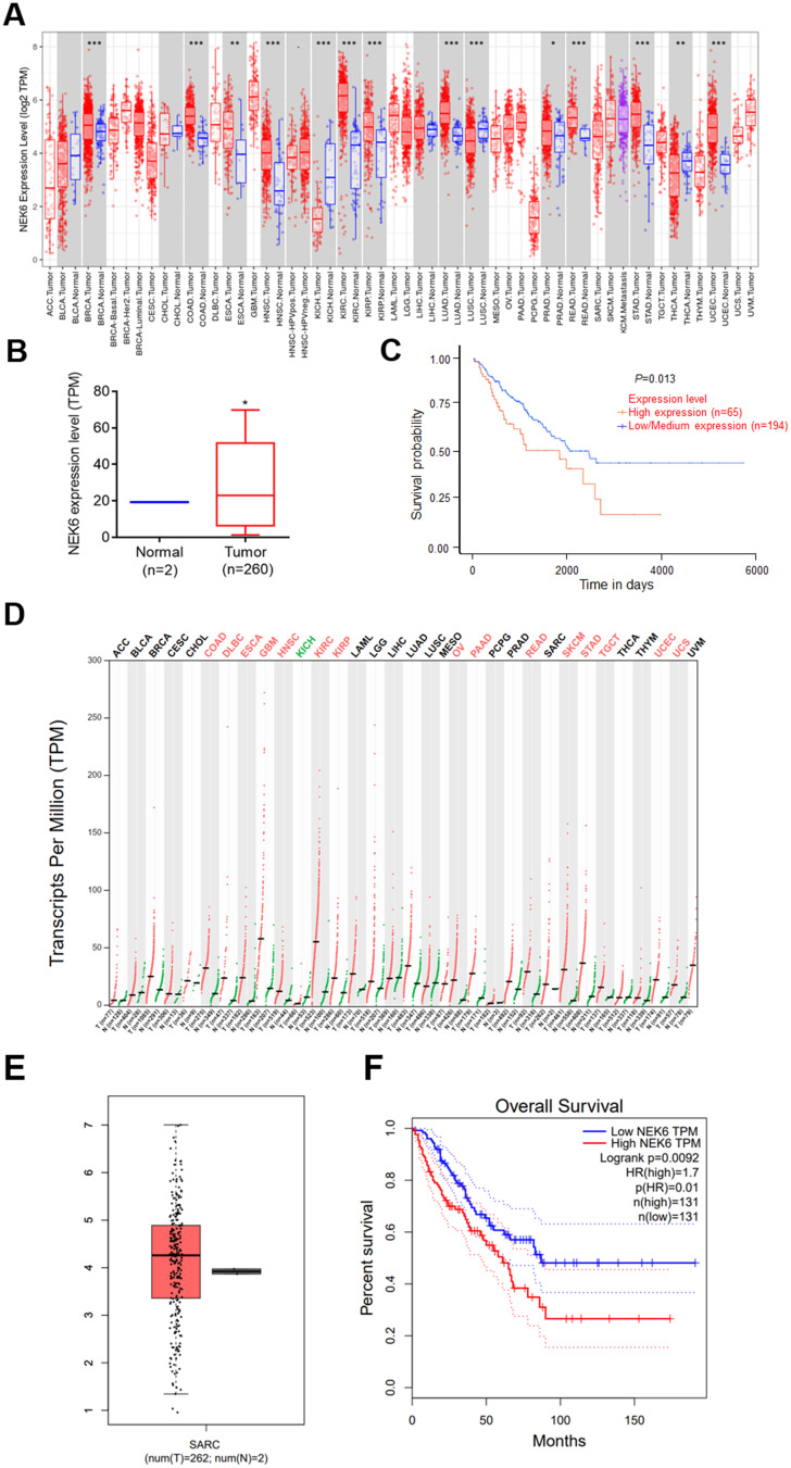
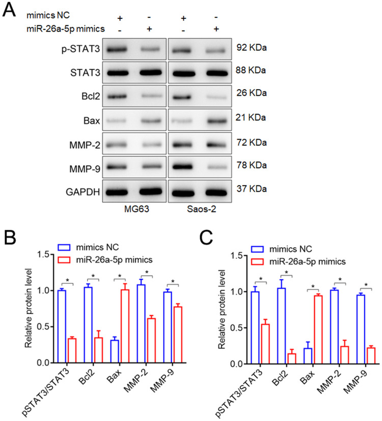
Methods: NEK6 expression in pan-cancer including sarcoma was evaluated using analysis tools of TIMER, UALCNA and GEPIA with TCGA database, and its association with overall survival in patients with sarcoma was also analyzed. TargetScan, tarbase, microT-CDS and Starbase online software were used to predict NEK6-targeted miRNAs, including miR-26a-5p. Tumor tissues from patients with osteosarcoma were collected for NEK6 and miRNA detection using RT-qPCR. NEK6 down-regulated by siRNAs or miR-26a-5p in osteosarcoma cells was detected by RT-qPCR, Western blot and Immunofluorescence staining assays. Effects of NEK6 knockdown on proliferation, migration, invasion and apoptosis of osteosarcoma cells were detected by CCK-8, wound healing, transwell and flow cytometry, respectively. The expressions of STAT3, metastasis and apoptosis-related genes were detected by Western blot.
Results: High expression of NEK6 and low expression of miR-26a-5p were lowly expressed in osteosarcoma and they were negative correlation. NEK6 has been confirmed as a direct target for miR-26a-5p. In addition, NEK6 down-regulated by siRNAs or miR-26a-5p led to inhibition of cell proliferation, migration and invasion while promoting cell apoptosis. The levels of phosphorylated STAT3 and metastasis genes (MMP-2, MMP-9) were inhibited, while apoptotic gene Bax was promoted and Bcl2 was inhibited by miR-26a-5p upregulation.
Conclusion: NEK6 can promote osteosarcoma progression via activating STAT3 signaling pathway, which is inhibited by miR-26a-5p, suggesting that NEK6 is a potential oncogene and miR-26a-5p is a suppressor of osteosarcoma. The strategy of inhibiting of NEK6 by miR-26a-5p may be an effective approach for osteosarcoma therapy.
Keywords: NEK6; STAT3 signaling pathway; miR-26a-5p; osteosarcoma.
© 2023 Zhu et al.
Conflict of interest statement
The authors declare that they have no conflicts of interest in this work.
Figures


Similar articles
-
LINC00657/miR-26a-5p/CKS2 ceRNA network promotes the growth of esophageal cancer cells via the MDM2/p53/Bcl2/Bax pathway.Biosci Rep. 2020 Jun 26;40(6):BSR20200525. doi: 10.1042/BSR20200525. Biosci Rep. 2020. PMID: 32426838 Free PMC article.
-
The regulatory effect of has-circ-0001146/miR-26a-5p/MNAT1 network on the proliferation and invasion of osteosarcoma.Biosci Rep. 2020 Jun 26;40(6):BSR20201232. doi: 10.1042/BSR20201232. Biosci Rep. 2020. PMID: 32453410 Free PMC article.
-
microRNA-26a-5p Promotes Proliferation and Migration of Osteosarcoma Cells by Targeting HOXA5 in vitro and in vivo.Onco Targets Ther. 2019 Dec 30;12:11555-11565. doi: 10.2147/OTT.S232100. eCollection 2019. Onco Targets Ther. 2019. PMID: 32021239 Free PMC article.
-
MiR-26a-5p regulates proliferation, apoptosis, migration and invasion via inhibiting hydroxysteroid dehydrogenase like-2 in cervical cancer cell.BMC Cancer. 2022 Aug 10;22(1):876. doi: 10.1186/s12885-022-09970-x. BMC Cancer. 2022. PMID: 35948893 Free PMC article.
-
MiR-26a-5p Serves as an Oncogenic MicroRNA in Non-Small Cell Lung Cancer by Targeting FAF1.Cancer Manag Res. 2020 Aug 11;12:7131-7142. doi: 10.2147/CMAR.S261131. eCollection 2020. Cancer Manag Res. 2020. PMID: 32848467 Free PMC article.
Cited by
-
The kinase NEK6 positively regulates LSD1 activity and accumulation in local chromatin sub-compartments.Commun Biol. 2024 Nov 10;7(1):1483. doi: 10.1038/s42003-024-07199-x. Commun Biol. 2024. PMID: 39523439 Free PMC article.
-
PSMD14 is a novel prognostic marker and therapeutic target in osteosarcoma.Diagn Pathol. 2024 Jun 11;19(1):79. doi: 10.1186/s13000-024-01489-y. Diagn Pathol. 2024. PMID: 38863002 Free PMC article.
-
Differential Expression of NEK Kinase Family Members in Esophageal Adenocarcinoma and Barrett's Esophagus.Cancers (Basel). 2023 Sep 30;15(19):4821. doi: 10.3390/cancers15194821. Cancers (Basel). 2023. PMID: 37835513 Free PMC article.
-
The NIMA-related kinase family and cancer.Front Oncol. 2025 Mar 27;15:1556917. doi: 10.3389/fonc.2025.1556917. eCollection 2025. Front Oncol. 2025. PMID: 40212678 Free PMC article. Review.
References
-
- Eker N, Tokuc AG, Yılmaz B, et al. Outcomes of osteosarcoma in children without high-dose methotrexate: could it be less toxic without effecting survival rates? J Adolesc Young Adult Oncol. 2022;11:252–258. - PubMed
LinkOut - more resources
Full Text Sources
Research Materials
Miscellaneous
