

Pulmonary Embolism and Giant Cavitary Lesion Developing After COVID-19 Pneumonia
- PMID: 37426844
- PMCID: PMC10327987
- DOI: 10.36518/2689-0216.1109
Pulmonary Embolism and Giant Cavitary Lesion Developing After COVID-19 Pneumonia
Abstract
Introduction: The clinical manifestations of the worldwide pandemic, which began in mainland China in December 2019, were very similar to viral pneumonia and defined as Coronavirus disease 2019 (COVID-19). Complications such as acute respiratory distress syndrome (ARDS), acute cardiac tissue damage, secondary infections, isolated coagulopathy and pulmonary embolism have been reported with COVID-19 disease.
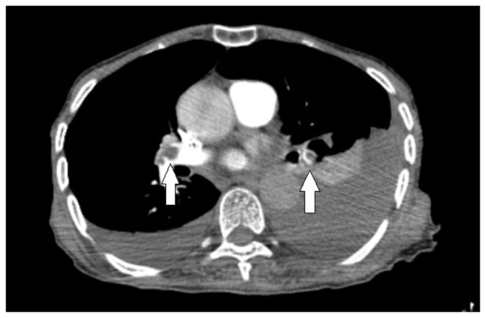
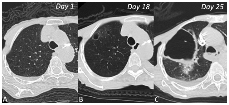
Clinical findings: A 79-year-old woman admitted to the emergency room (ER) had complaints of fever and cough. The patient was admitted to the ER with the suspicion of COVID-19. Samples were collected with a nasopharyngeal swab and confirmed as COVID-19. In addition, a chest CT examination was performed. In the first evaluation after admittance, the D-dimer value was measured as 450 μg/L. In the follow-up of the patient, on the 18th day, increased respiratory distress and high D-dimer level (7893 μg/L) were detected in the laboratory findings.
Outcomes: A chest CT scan had ground-glass opacities compatible with COVID-19 pneumonia. A giant cavitary lesion was detected following the development of pulmonary embolism after COVID-19 disease.
Conclusions: In rare cases of COVID-19 cavitation development may occur after pulmonary infarction. In addition, it should be remembered that emphysema, giant bulla and pneumothorax may develop in COVID-19 pneumonia cases undergoing HFNC oxygen therapy. We present a case of a giant cavitary lesion that developed following a COVID-19-related pulmonary embolism.
Keywords: COVID-19; SARS-Cov-2; cavitation, d-dimer; coronavirus infections/complications; lung diseases; pulmonary embolism.
© 2020 HCA Physician Services, Inc. d/b/a Emerald Medical Education.
Conflict of interest statement
Conflicts of Interest The authors declare they have no conflicts of interest.
Figures
Similar articles
-
Simultaneous Giant cavity pulmonary lesion and pneumothorax following COVID-19 pneumonia.Radiol Case Rep. 2021 Sep;16(9):2534-2536. doi: 10.1016/j.radcr.2021.06.026. Epub 2021 Jun 14. Radiol Case Rep. 2021. PMID: 34149974 Free PMC article.
-
Mediastinal Emphysema, Giant Bulla, and Pneumothorax Developed during the Course of COVID-19 Pneumonia.Korean J Radiol. 2020 May;21(5):541-544. doi: 10.3348/kjr.2020.0180. Epub 2020 Mar 20. Korean J Radiol. 2020. PMID: 32207255 Free PMC article.
-
NEUROPROTECTIVE AND ANTIOXIDANT POTENTIAL OF MONTELUKAST-ACETYLCYSTEINE COMBINATION THERAPY FOR BRAIN PROTECTION IN PATIENTS WITH COVID-19 INDUCED PNEUMONIA.Georgian Med News. 2023 Feb;(335):111-118. Georgian Med News. 2023. PMID: 37042600
-
[Follow-up of patients after COVID-19 pneumonia. Pulmonary sequelae].Rev Alerg Mex. 2020 Oct-Dec;67(4):350-369. doi: 10.29262/ram.v67i4.847. Rev Alerg Mex. 2020. PMID: 33631903 Review. Spanish.
-
Similarities and Differences of Early Pulmonary CT Features of Pneumonia Caused by SARS-CoV-2, SARS-CoV and MERS-CoV: Comparison Based on a Systemic Review.Chin Med Sci J. 2020 Sep 30;35(3):254-261. doi: 10.24920/003727. Chin Med Sci J. 2020. PMID: 32972503 Free PMC article.
Cited by
-
Evaluating the Cavitary Lung Lesions on CT Scan of COVID-19 Patients: A Retrospective Study.J Community Hosp Intern Med Perspect. 2024 Nov 2;14(6):23-29. doi: 10.55729/2000-9666.1411. eCollection 2024. J Community Hosp Intern Med Perspect. 2024. PMID: 39839166 Free PMC article.
-
Simultaneous Giant cavity pulmonary lesion and pneumothorax following COVID-19 pneumonia.Radiol Case Rep. 2021 Sep;16(9):2534-2536. doi: 10.1016/j.radcr.2021.06.026. Epub 2021 Jun 14. Radiol Case Rep. 2021. PMID: 34149974 Free PMC article.
References
Publication types
LinkOut - more resources
Full Text Sources
Miscellaneous