

Deconvoluting human Brodmann area 8 based on its unique structural and functional connectivity
- PMID: 37426900
- PMCID: PMC10323427
- DOI: 10.3389/fnana.2023.1127143
Deconvoluting human Brodmann area 8 based on its unique structural and functional connectivity
Abstract
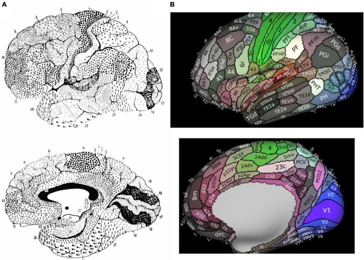
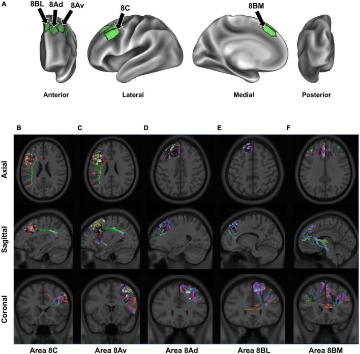
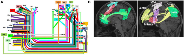
Brodmann area 8 (BA8) is traditionally defined as the prefrontal region of the human cerebrum just anterior to the premotor cortices and enveloping most of the superior frontal gyrus. Early studies have suggested the frontal eye fields are situated at its most caudal aspect, causing many to consider BA8 as primarily an ocular center which controls contralateral gaze and attention. However, years of refinement in cytoarchitectural studies have challenged this traditional anatomical definition, providing a refined definition of its boundaries with neighboring cortical areas and the presence of meaningful subdivisions. Furthermore, functional imaging studies have suggested its involvement in a diverse number of higher-order functions, such as motor, cognition, and language. Thus, our traditional working definition of BA8 has likely been insufficient to truly understand the complex structural and functional significance of this area. Recently, large-scale multi-modal neuroimaging approaches have allowed for improved mapping of the neural connectivity of the human brain. Insight into the structural and functional connectivity of the brain connectome, comprised of large-scale brain networks, has allowed for greater understanding of complex neurological functioning and pathophysiological diseases states. Simultaneously, the structural and functional connectivity of BA8 has recently been highlighted in various neuroimaging studies and detailed anatomic dissections. However, while Brodmann's nomenclature is still widely used today, such as for clinical discussions and the communication of research findings, the importance of the underlying connectivity of BA8 requires further review.
Keywords: Brodmann area 8; cognition; connectivity; fMRI; network; neuroimaging.
Copyright © 2023 Dadario, Tanglay and Sughrue.
Conflict of interest statement
ND has no disclosures. OT was an employee of Omniscient Neurotechnology. MS was a co-founder of Omniscient Neurotechnology. No aspects related to these products were discussed in the current work. The remaining author declares that the research was conducted in the absence of any commercial or financial relationships that could be construed as a potential conflict of interest.
Figures
Similar articles
-
Parcellation-based modeling of the dorsal premotor area.J Neurol Sci. 2020 Aug 15;415:116907. doi: 10.1016/j.jns.2020.116907. Epub 2020 May 17. J Neurol Sci. 2020. PMID: 32526524
-
Medial reward and lateral non-reward orbitofrontal cortex circuits change in opposite directions in depression.Brain. 2016 Dec;139(Pt 12):3296-3309. doi: 10.1093/brain/aww255. Epub 2016 Oct 14. Brain. 2016. PMID: 27742666
-
A Connectomic Atlas of the Human Cerebrum-Chapter 1: Introduction, Methods, and Significance.Oper Neurosurg. 2018 Dec 1;15(suppl_1):S1-S9. doi: 10.1093/ons/opy253. Oper Neurosurg. 2018. PMID: 30260422 Free PMC article.
-
The role of ventral premotor cortex in action execution and action understanding.J Physiol Paris. 2006 Jun;99(4-6):396-405. doi: 10.1016/j.jphysparis.2006.03.005. Epub 2006 May 24. J Physiol Paris. 2006. PMID: 16723210 Review.
-
The primary motor and premotor areas of the human cerebral cortex.Neuroscientist. 2006 Apr;12(2):143-52. doi: 10.1177/1073858405284255. Neuroscientist. 2006. PMID: 16514011 Review.
Cited by
-
A Dual Role for the Dorsolateral Prefrontal Cortex (DLPFC) in Auditory Deviance Detection.Brain Sci. 2024 Sep 29;14(10):994. doi: 10.3390/brainsci14100994. Brain Sci. 2024. PMID: 39452008 Free PMC article.
-
Harnessing the frontal aslant tract's structure to assess its involvement in cognitive functions: new insights from 7-T diffusion imaging.Sci Rep. 2024 Jul 29;14(1):17455. doi: 10.1038/s41598-024-67013-w. Sci Rep. 2024. PMID: 39075100 Free PMC article.
-
Revised cytoarchitectonic mapping of the human premotor cortex identifies seven areas and refines the localisation of frontal eye fields.Commun Biol. 2025 Aug 1;8(1):1143. doi: 10.1038/s42003-025-08528-4. Commun Biol. 2025. PMID: 40751095 Free PMC article.
-
Deconstructing a common pathway concept for Deep Brain Stimulation in the case of Obsessive-Compulsive Disorder.Mol Psychiatry. 2025 Sep;30(9):4274-4285. doi: 10.1038/s41380-025-03008-x. Epub 2025 Apr 6. Mol Psychiatry. 2025. PMID: 40189699 Free PMC article.
-
Role for Left Dorsomedial Prefrontal Cortex in Self-Generated, but not Externally Cued, Language Production.Neurobiol Lang (Camb). 2025 Jun 12;6:nol_a_00166. doi: 10.1162/nol_a_00166. eCollection 2025. Neurobiol Lang (Camb). 2025. PMID: 40528833 Free PMC article.
References
-
- Bailey P. (1951). The isocortex of man. Urbana, IL: University of Illinois Press.
Publication types
LinkOut - more resources
Full Text Sources