

Radius of curvature of the radial head matches the capitellum: a magnetic resonance imaging analysis
- PMID: 37426921
- PMCID: PMC10328745
- DOI: 10.1016/j.jseint.2023.02.009
Radius of curvature of the radial head matches the capitellum: a magnetic resonance imaging analysis
Abstract
Background: The purpose of this study is to utilize elbow magnetic resonance imaging (MRI) to compare the radius of curvature (ROC) of the radial head peripheral cartilaginous rim and the cartilage contour of the capitellum to evaluate if the radial head could be a suitable osteochondral autograft for capitellar pathology.
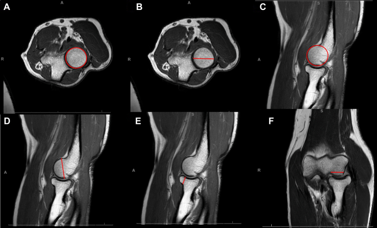
Methods: All patients who underwent an MRI of the elbow over a three-year period were reviewed. Patients with the following diagnoses were excluded: osteochondritis dissecans, osteomyelitis, tumor, and osteoarthritis. The radius of curvature of the radial head (RhROC) was measured on the axial oblique MRI sequence. The radius of curvature of the capitellum (CapROC) was measured on sagittal oblique MRI sequences, the width of the articular surface of the capitellum on coronal MRI sequences and the radial head height (RhH) and capitellar vertical height on sagittal oblique sequences. All measurements were obtained at the midpoint of the radiocapitellar joint. Spearman's coefficient was used to assess the correlation between ROC measurements.
Results: Eighty-three patients were included with a mean age of 43 +/- 17 years (57 males and 26 females, 51 right and 32 left elbows). The median RhROC and CapROC measurements were 12.3 mm (interquartile range [IQR] 1.6) and 11.9 mm (IQR 1.7), respectively. The median difference was 0.3 mm (IQR = 0.6; CI 95% = [0.24, 0.46]; P < .001). A strong positive correlation was found between RhROC and CapROC (ρ = 0.89; r2 = 0.819; P < .001). Ninety-four percent (78/83) of patients had a median difference between the RhROC and CapROC of less than or equal to 1 mm, and 63% (52/83) were within 0.5 mm. The inter-rater and intra-rater reliability for RhROC and CapROC was good, intraclass correlation coefficient (ICC) = 0.89, ICC = 0.87, and ICC = 0.96, ICC = 0.97, respectively. RhH was 10.6 ± 1.3 mm, and the width of the articular surface of the capitellum was found to be 13.8 ± 1.6 mm.
Conclusion: The ROC of the convex peripheral cartilaginous rim of the radial head is similar to the ROC of the capitellum. In addition, the RhH was approximately 78% of the capitellar articular width. Based on this imaging analysis, the radial head could prove to be a robust local osteochondral autograft with a similar cartilage contour in the reconstruction of the capitellum in complex intra-articular distal humerus fractures with associated radial head fractures and in the setting of "kissing lesions" of the radiocapitellar joint. Furthermore, an osteochondral plug harvested from the "safe zone" of the peripheral cartilaginous rim of the radial head could be utilized to treat isolated osteochondral lesions of the capitellum.
Keywords: Capitellum; Distal humerus; Elbow; Imaging; Radial head; Radius of curvature.
Figures

 ) of the ulna articulation with the capitellum.
References
-
- Arner O., Ekengren K., Von Schreeb T. Fractures of the head and neck of the radius; a clinical and roentgenographic study of 310 cases. Acta Chir Scand. 1957;112:115–134. - PubMed
-
- Davidson P.A., Moseley J.B., Jr., Tullos H.S. Radial head fracture. A potentially complex injury. Clin Orthop Relat Res. 1993:224–230. - PubMed
LinkOut - more resources
Full Text Sources