

Role of Imaging in Cardiomyopathies
- PMID: 37427006
- PMCID: PMC10326670
- DOI: 10.15420/cfr.2022.26
Role of Imaging in Cardiomyopathies
Abstract
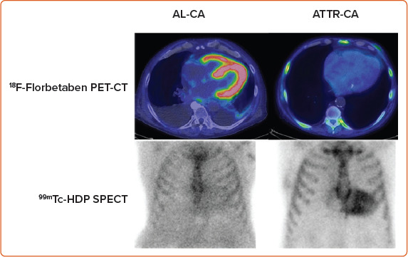
Imaging has a central role in the diagnosis, classification, and clinical management of cardiomyopathies. While echocardiography is the first-line technique, given its wide availability and safety, advanced imaging, including cardiovascular magnetic resonance (CMR), nuclear medicine and CT, is increasingly needed to refine the diagnosis or guide therapeutic decision-making. In selected cases, such as in transthyretin-related cardiac amyloidosis or in arrhythmogenic cardiomyopathy, the demonstration of histological features of the disease can be avoided when typical findings are observed at bone-tracer scintigraphy or CMR, respectively. Findings from imaging techniques should always be integrated with data from the clinical, electrocardiographic, biomarker, genetic and functional evaluation to pursue an individualised approach to patients with cardiomyopathy.
Keywords: Imaging; cardiac magnetic resonance; cardiomyopathies; echocardiography; nuclear medicine.
Copyright © 2023, Radcliffe Cardiology.
Conflict of interest statement
Disclosure: The authors have no conflicts of interest to declare.
Figures
References
-
- Cardim N, Galderisi M, Edvardsen T et al. Role of multimodality cardiac imaging in the management of patients with hypertrophic cardiomyopathy: an expert consensus of the European Association of Cardiovascular Imaging endorsed by the Saudi Heart Association. Eur Heart J Cardiovasc Imaging. 2015;16:280. doi: 10.1093/ehjci/jeu291. - DOI - PubMed
Publication types
LinkOut - more resources
Full Text Sources
Research Materials
