. 2023 Jun 28;26(2):350.
doi: 10.3892/ol.2023.13936. eCollection 2023 Aug.

Are there reliable multiparametric MRI criteria for differential diagnosis between intracranial meningiomas and solitary intracranial dural metastases?

Affiliations

Are there reliable multiparametric MRI criteria for differential diagnosis between intracranial meningiomas and solitary intracranial dural metastases?

Hongjie Wu et al. Oncol Lett. .

Abstract

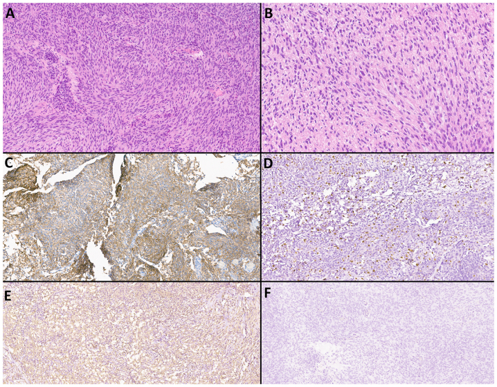
Intracranial meningiomas are the most common tumors of the central nervous system (CNS). Meningiomas account for up to 36% of all brain tumors. The incidence of metastatic brain lesions has not been determined. Up to 30% of adult patients with cancer of one localization or another suffer from a secondary tumor lesion of the brain. The vast majority of meningiomas have meningeal localization; >90% are solitary. The incidence of intracranial dural metastases (IDM) is 8-9% of cases, while in 10% of cases, the brain is the only localization, and in 50% of cases the metastases are solitary. Typically, the task of distinguishing between meningioma and dural metastasis does not involve difficulties. Periodically, there is a situation when the differential diagnosis between these tumors is ambiguous, since meningiomas and solitary IDM may have similar characteristics, in particular, a cavity-less solid structure, limited diffusion of water molecules, the presence of extensive peritumoral edema, and an identical contrast pattern. The present study included 100 patients with newly diagnosed tumors of the CNS, who subsequently underwent examination and neurosurgical treatment at the Federal Center for Neurosurgery with histological verification between May 2019 and October 2022. Depending on the histological conclusion, two study groups of patients were distinguished: The first group consisted of patients diagnosed with intracranial meningiomas (n=50) and the second group of patients were diagnosed with IDM (n=50). The study was performed using a magnetic resonance imaging (MRI) General Electric Discovery W750 3T before and after contrast enhancement. The diagnostic value of this study was estimated using Receiver Operating Characteristic curve and area under the curve analysis. Based on the results of the study, it was found that the use of multiparametric MRI (mpMRI) in the differential diagnosis of intracranial meningiomas and IDM was limited by the similarity of the values of the measured diffusion coefficient. The assumption, previously put forward in the literature, regarding the presence of a statistically significant difference in the apparent diffusion coefficient values, which make it possible to differentiate tumors, was not confirmed. When analyzing perfusion data, IDM showed higher cerebral blood flow (CBF) values compared with intracranial meningiomas (P≤0.001). A threshold value of the CBF index was revealed, which was 217.9 ml/100 g/min, above which it is possible to predict IDM with a sensitivity and specificity of 80.0 and 86.0%, respectively. Diffusion-weighted images are not reliable criteria for differentiating intracranial meningiomas from IDM and should not influence the diagnosis suggested by imaging. The technique for assessing the perfusion of a meningeal lesion makes it possible to predict metastases with a sensitivity and specificity close to 80-90% and deserves attention when making a diagnosis. In the future, in order to reduce the number of false negative and false positive results, mpMRI would require additional criteria to be included in the protocol. Since IDM differs from intracranial meningiomas in the severity of neoangiogenesis and, accordingly, in greater vascular permeability, the technique for assessing vascular permeability (wash-in parameter with dynamic contrast enhancement) may serve as a refining criterion for distinguishing between dural lesions.

Keywords: MR perfusion; biomarkers; diffusion coefficient measurement; intracranial dural metastasis; intracranial meningioma; mpMRI; neuroimaging.


Conflict of interest statement

The authors declare that they have no competing interests.

Figures

Figure 1.
Figure 1.
Brain magnetic resonance imaging of a patient with meningothelial meningioma (WHO Grade 1). Supratentorially, in the left hemisphere of the frontal region, against the background of moderate vasogenic edema, a clearly demarcated extracerebral mass is visible, characterized by a hypointense signal on T2-WI, intense and homogeneous accumulation of a contrast agent, diffusion restriction with corresponding areas of increased values of volumetric and velocity cerebral blood flow, as well as prolongation of blood transit time. SWAN indicates the presence of cortical draining veins in the formation structure. (A) T2-WI; (B and C) DWI and ADC; (D) SWAN; (E) T1-WI with contrast; (F) CBV; (G) CBF; and (H) MTT. SWAN, susceptibility-weighted angiography; T1-WI, T1-weighted image; T2-WI, T2-weighted image; DWI, diffusion-weighted imaging; ADC, apparent diffusion coefficient; CBV, cerebral blood volume; CBF, cerebral blood flow; MTT, mean transit time.
Figure 2.
Figure 2.
Brain magnetic resonance imaging with solitary intracranial dural metastasis of lung adenocarcinoma. In the parasagittal parts of the left frontal area, there is an extracerebral mass with a base on the falciform process of the dura mater, surrounded by a moderately pronounced zone of perifocal edema, relatively homogeneously accumulating contrast throughout the volume, with limited diffusion according to DWI and ADC, the presence of artifacts from blood decay products in the tumor structure, as well as high values of rCBV and rCBF, and lengthening of MTT. (A) T2-WI; (B and C) DWI and ADC; (D) SWAN; (E) T1-WI with contrast; (F) CBV; (G) CBF; and (H) MTT. SWAN, susceptibility-weighted angiography; T1-WI, T1-weighted image; T2-WI, T2-weighted image; DWI, diffusion-weighted imaging; ADC, apparent diffusion coefficient; CBV, cerebral blood volume; CBF, cerebral blood flow; r, relative; MTT, mean transit time.
Figure 3.
Figure 3.
Brain magnetic resonance imaging of a patient with atypical meningioma (WHO Grade 2). In the frontal region of the left hemisphere, against the background of perifocal edema, an extracerebral tumor is visible with an intense and homogeneous accumulation of a contrast agent, the ‘dural tail’ phenomenon, diffusion limitation, an increase in volumetric and velocity cerebral blood flow, and a prolongation of blood transit time. The SWAN demonstrates the presence of peripheral draining veins around the mass. (A) T2-WI; (B and C) DWI and ADC; (D) SWAN; (E) T1-WI with contrast; (F) CBV; (G) CBF; and (H) MTT. SWAN, susceptibility-weighted angiography; T1-WI, T1-weighted image; T2-WI, T2-weighted image; DWI, diffusion-weighted imaging; ADC, apparent diffusion coefficient; CBV, cerebral blood volume; CBF, cerebral blood flow; MTT, mean transit time.
Figure 4.
Figure 4.
Brain magnetic resonance imaging with solitary intracranial dural metastasis of prostate adenocarcinoma. In the occipital region of the left hemisphere, an extracerebral formation is visible with heterogeneous contrast enhancement, and restriction of diffusion, surrounded by a pronounced zone of edema. SWAN indicates the presence of point artifacts of magnetic susceptibility due to hemorrhages and intratumoral vascular shunts. According to the results of MRI perfusion, high values of relative cerebral blood volume (rCBV) and relative cerebral blood flow (rCBF) in the tumor structure are determined. The mean transit time (MTT) indicator is extended. (A) T2-WI; (B and C) DWI and ADC; (D) SWAN; (E) T1-WI with contrast; (F) CBV; (G) CBF; and (H) MTT. SWAN, susceptibility-weighted angiography; T1-WI, T1-weighted image; T2-WI, T2-weighted image; DWI, diffusion-weighted imaging; ADC, apparent diffusion coefficient; CBV, cerebral blood volume; CBV, cerebral blood flow; r, relative; MTT, mean transit time.
Figure 5.
Figure 5.
Comparison of some characteristics of patients with intracranial meningiomas and solitary IDM. The incidence of (A) bone invasion and (B) the severity of perifocal edema among patients with intracranial meningiomas and solitary IDM. The prevalence of bone invasion was shown grater in cases with meningiomas than in cases with IDM (P<0.001), as well as a greater severity of perifocal edema in patients with metastases (P<0.001). IDM, intracranial dural metastasis.
Figure 6.
Figure 6.
Demonstration of evaluating the results of the values of the CBV, rCBV, CBF, and rCBF. Graphs of (A) CBV, (B) normalized rCBV, (C) CBF velocity, and (D) normalized rCBF velocity for intracranial meningiomas and solitary IDM. (A) The median CBV was significantly higher for IDM than for intracranial meningiomas (P≤0.001). The Y-axis plots the values of the CBV in ml/100 ml; (B) the median increase in rCBV was significantly higher for IDM than for intracranial meningiomas (P≤0.001). The Y-axis plots the ratio of rCBV in the ROI to the normal white matter of the semioval center, representing the normalized cerebral blood flow volume; (C) Median CBF was significantly higher for IDM than for intracranial meningiomas (P≤0.001). The Y-axis shows the values of CBF velocity in ml/100 g/min. (D) The median rCBF was significantly higher for IDM than for intracranial meningiomas (P≤0.001). The Y-axis plots the ratio of rCBF velocity in the ROI to the normal white matter of the semioval center, representing the normalized rCBF velocity. ROI, region of interest; IDM, intracranial dural metastasis; CBV, cerebral blood volume; CBV, cerebral blood flow; r, relative.
Figure 7.
Figure 7.
The ROC curve and the AUC analysis. (A) The AUC corresponding to CBV for differentiating intracranial meningiomas and solitary IDM was 0.805±0.44 [95% CI, 0.719-0.890] (P≤0.001) with sensitivity and specificity values of 76.5 and 78.0%, respectively; (B) The AUC corresponding to rCBV for differentiating intracranial meningiomas and IDM was 0.811±0.46 [95% CI, 0.722-0.900] (P≤0.001) with sensitivity and specificity values of 74.5 and 82.0%, respectively. (C) The AUC corresponding to CBF for differentiating intracranial meningiomas and IDM was 0.8±0.48 [95% CI, 0.706-0.894] (P<0.001) with sensitivity and specificity values of 80.4 and 86.0%, respectively. (D) The AUC corresponding to rCBF for differentiating intracranial meningiomas and IDM was 0.79±0.5 [95% CI, 0.692-0.888] (P≤0.001) with sensitivity and specificity values of 82.4 and 76.0%, respectively. If the values of CBV, rCBV, CBF, and rCBF were ≤ threshold value, the patient was predicted to have an intracranial meningioma. The threshold value of CBV was 28.25 ml/100 g, for rCBV it was 5.4, for CBF it was 217.9 ml/100 g/min, and for rCBF was 5.6. ROC, receiver operating characteristic; AUC, area under the curve; IDM, intracranial dural metastasis; CBV, cerebral blood volume; CBV, cerebral blood flow; r, relative.
Figure 8.
Figure 8.
Patient with atypical meningioma (WHO Grade 2). (A and B) Examination of a series of prepared slides stained with hematoxylin and eosin reveals tumor tissues built from arachnoid endothelial cells. Cellularity was increased. The nuclei were predominantly round-oval or slightly elongated with a small centrally located nucleolus. The cytoplasm varies in tinctorial properties from optically transparent to light oxyphilic. The cells were located in continuous fields with the formation of multidirectional vortices. In most cellular zones, there were mitotic figures up to 4–5 figures/10 points (magnification, ×40). The stroma was unevenly expressed and rich in collagen. On immunohistochemical examination, tumor cells expressed (C) Vimentin, (D) with a Ki-67 proliferation index of 10%, (E) non-uniformly diffusely anti-EMA, or (F) complete absence of pankeratin AE1/AE3.
Figure 9.
Figure 9.
A patient with metastatic adenocarcinoma of the sigmoid colon. (A and B) When examining a series of prepared slides stained with hematoxylin and eosin, tumor tissue was determined based on the presence of atypical epithelial cells with manifestations of pronounced nuclear polymorphisms. The nuclear-cytoplasmic ratio was increased. The chromatin pattern was heterogeneous and smeared. There were numerous pathological mitoses, 1–2 figures in each field of view (magnification ×40). Cells form cribriform and tubular glandular structures. Stroma with myxoid changes, poorly expressed, exhibited infiltration of small lymphocytes, and extensive necrosis in all fields of vision. (C) Tumor cells were strongly stained with anti-EMA, (D) Ki-67 proliferative activity index of 50%, (E) Vimentin expression in the stromal component, and (F) pankeratin AE1/AE3.
Figure 10.
Figure 10.
Microscopic examination of histological preparations from a patient with intracranial meningioma and solitary IDM (metastasis of clear cell renal cell carcinoma). (A) Hematoxylin and eosin stained clear cell renal cell carcinoma metastasis (magnification, ×20). (B) Hematoxylin and eosin stained clear cell meningioma (magnification, ×20). The cells possessed a similar morphology in shape and size, the cytoplasm in both cases was optically transparent, cells were located in continuous fields, and in certain regions, the stromal component outlined small lobed structures. IDM, intracranial dural metastasis.
Figure 11.
Figure 11.
Microscopic examination of histological preparations from a patient with intracranial meningioma and solitary intracranial dural metastasis (metastasis of acinar adenocarcinoma of the prostate). (A) Metastasis of acinar adenocarcinoma of the prostate, stained with hematoxylin and eosin, (magnification, ×20). Cells of a large size with glandular morphology, nuclei polymorphic in shape and size, with a cytoplasm that appeared outlined, optically light, varied in volume, with moderate lymphocytic infiltration, and continuous fields of necrosis on the left and right. (B) Meningothelial meningioma, stained with hematoxylin and eosin (magnification, ×20). Cells were of a medium size, with a similar structure, and an arachnoid endothelial appearance, forming typical microconcentric structures; the nuclei were round-oval and monomorphic; the stroma was fibrous and well expressed.

Similar articles

Cited by

References

    1. Dolecek TA, Propp JM, Stroup NE, Kruchko C. CBTRUS statistical report: Primary brain and central nervous system tumors diagnosed in the United States in 2005–2009. Neuro Oncol. 2012;14((Suppl 5)):v1–49. doi: 10.1093/neuonc/nos218. - DOI - PMC - PubMed
    1. Louis DN, Perry A, Wesseling P, Brat DJ, Cree IA, Figarella-Branger D, Hawkins C, Ng HK, Pfister SM, Reifenberger G, et al. The 2021 WHO classification of tumors of the central nervous system: A summary. Neuro Oncol. 2021;23:1231–1251. doi: 10.1093/neuonc/noab106. - DOI - PMC - PubMed
    1. Olar A, Wani KM, Sulman EP, Mansouri A, Zadeh G, Wilson CD. Mitotic index is an independent predictor of recurrence-free survival in meningioma. Brain Pathol. 2015;25:266–275. doi: 10.1111/bpa.12174. - DOI - PMC - PubMed
    1. Perry A, Scheithauer BW, Stafford SL, Lohse CM, Wollan PC. ‘Malignancy’ in meningiomas: A clinicopathologic study of 116 patients, with grading implications. Cancer. 1999;85:2046–2056. doi: 10.1002/(SICI)1097-0142(19990501)85:9<2046::AID-CNCR23>3.0.CO;2-M. - DOI - PubMed
    1. Sughrue ME, Sanai N, Shangari G, Parsa AT, Berger MS, McDermott MW. Outcome and survival following primary and repeat surgery for World Health Organization Grade III meningiomas. J Neurosurg. 2010;113:202–209. doi: 10.3171/2010.1.JNS091114. - DOI - PubMed