. 2023 Aug 22;148(8):661-678.
doi: 10.1161/CIRCULATIONAHA.122.063402. Epub 2023 Jul 10.

Multiomics of Tissue Extracellular Vesicles Identifies Unique Modulators of Atherosclerosis and Calcific Aortic Valve Stenosis

Affiliations

Multiomics of Tissue Extracellular Vesicles Identifies Unique Modulators of Atherosclerosis and Calcific Aortic Valve Stenosis

Mark C Blaser et al. Circulation. .

Abstract

Background: Fewer than 50% of patients who develop aortic valve calcification have concomitant atherosclerosis, implying differential pathogenesis. Although circulating extracellular vesicles (EVs) act as biomarkers of cardiovascular diseases, tissue-entrapped EVs are associated with early mineralization, but their cargoes, functions, and contributions to disease remain unknown.

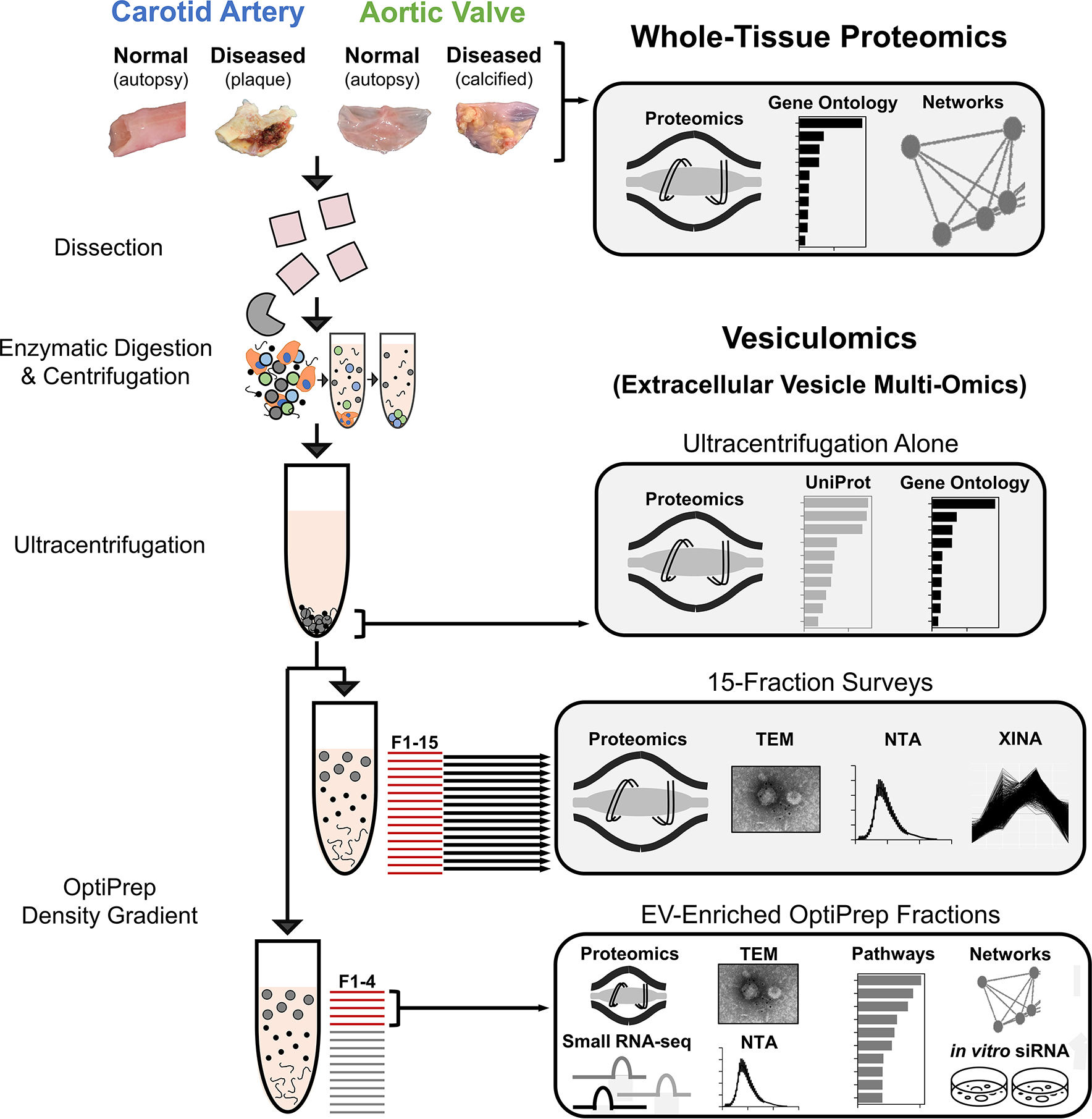
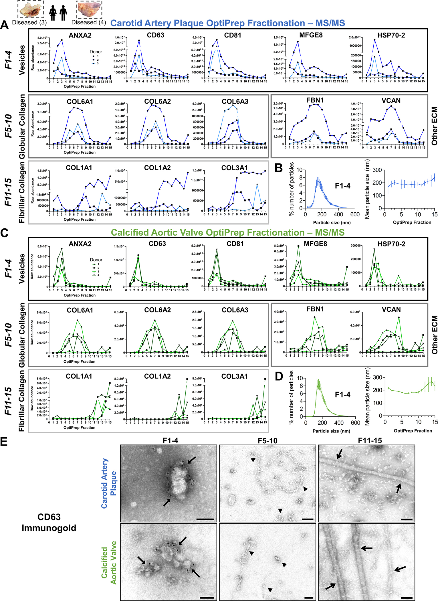
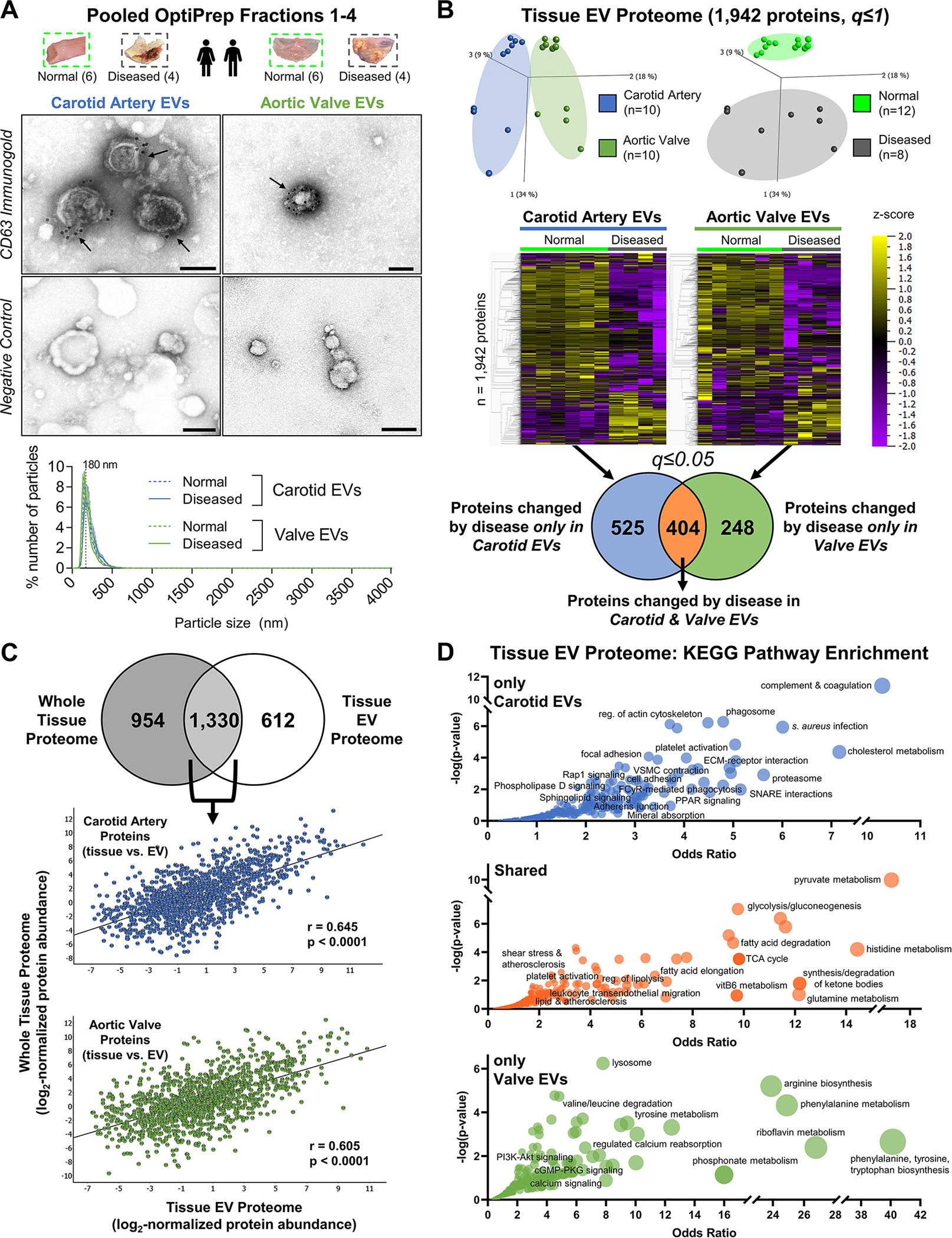
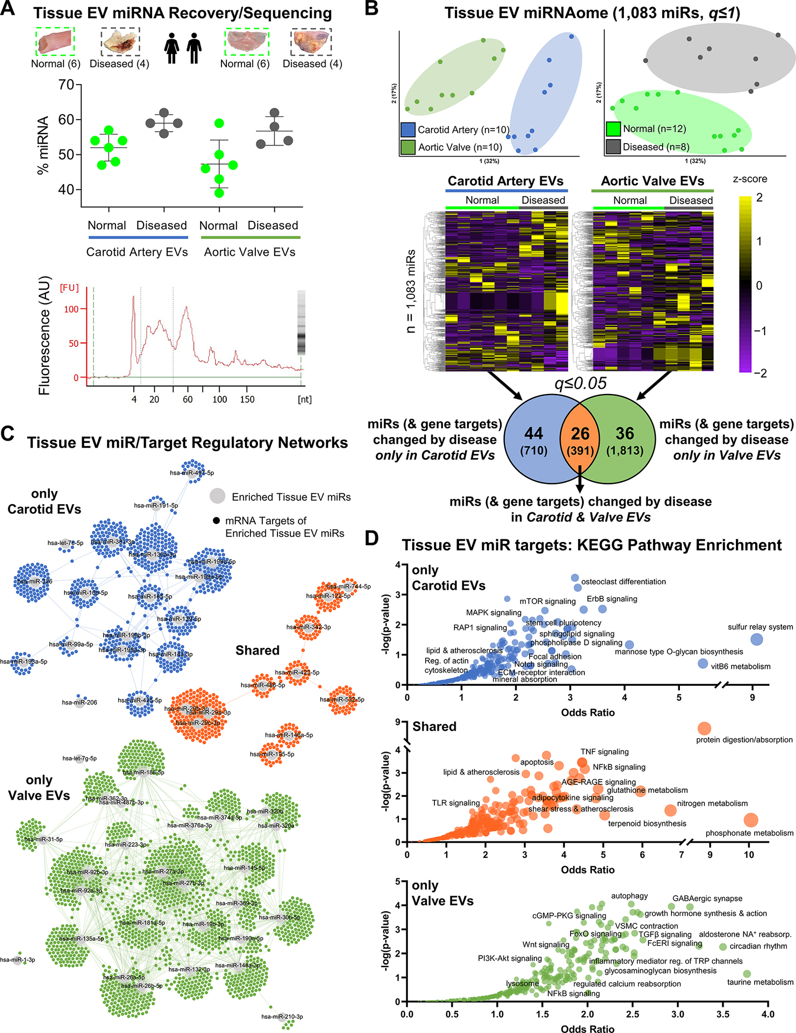
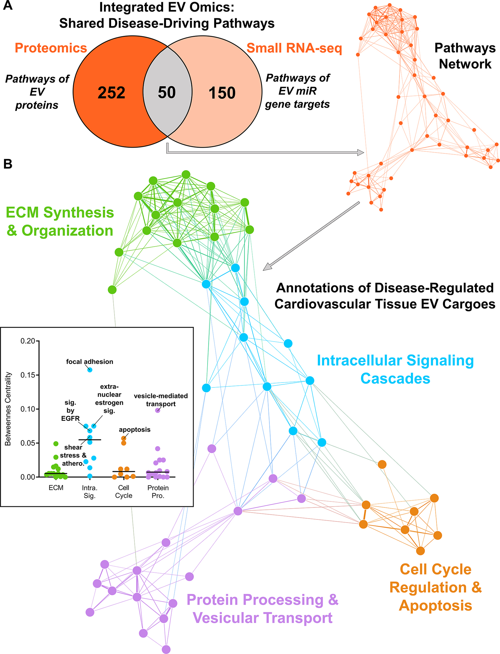
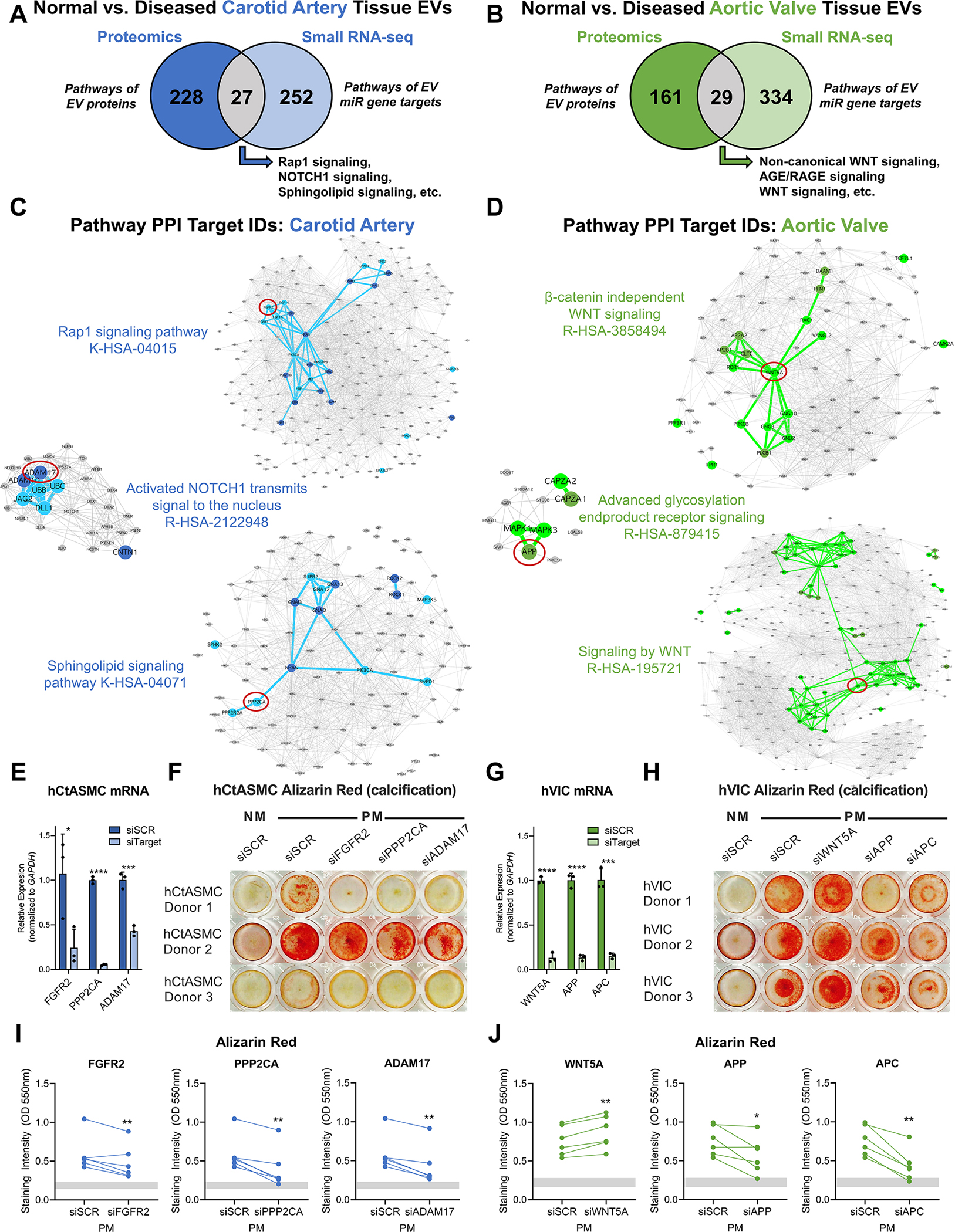
Methods: Disease stage-specific proteomics was performed on human carotid endarterectomy specimens (n=16) and stenotic aortic valves (n=18). Tissue EVs were isolated from human carotid arteries (normal, n=6; diseased, n=4) and aortic valves (normal, n=6; diseased, n=4) by enzymatic digestion, (ultra)centrifugation, and a 15-fraction density gradient validated by proteomics, CD63-immunogold electron microscopy, and nanoparticle tracking analysis. Vesiculomics, comprising vesicular proteomics and small RNA-sequencing, was conducted on tissue EVs. TargetScan identified microRNA targets. Pathway network analyses prioritized genes for validation in primary human carotid artery smooth muscle cells and aortic valvular interstitial cells.

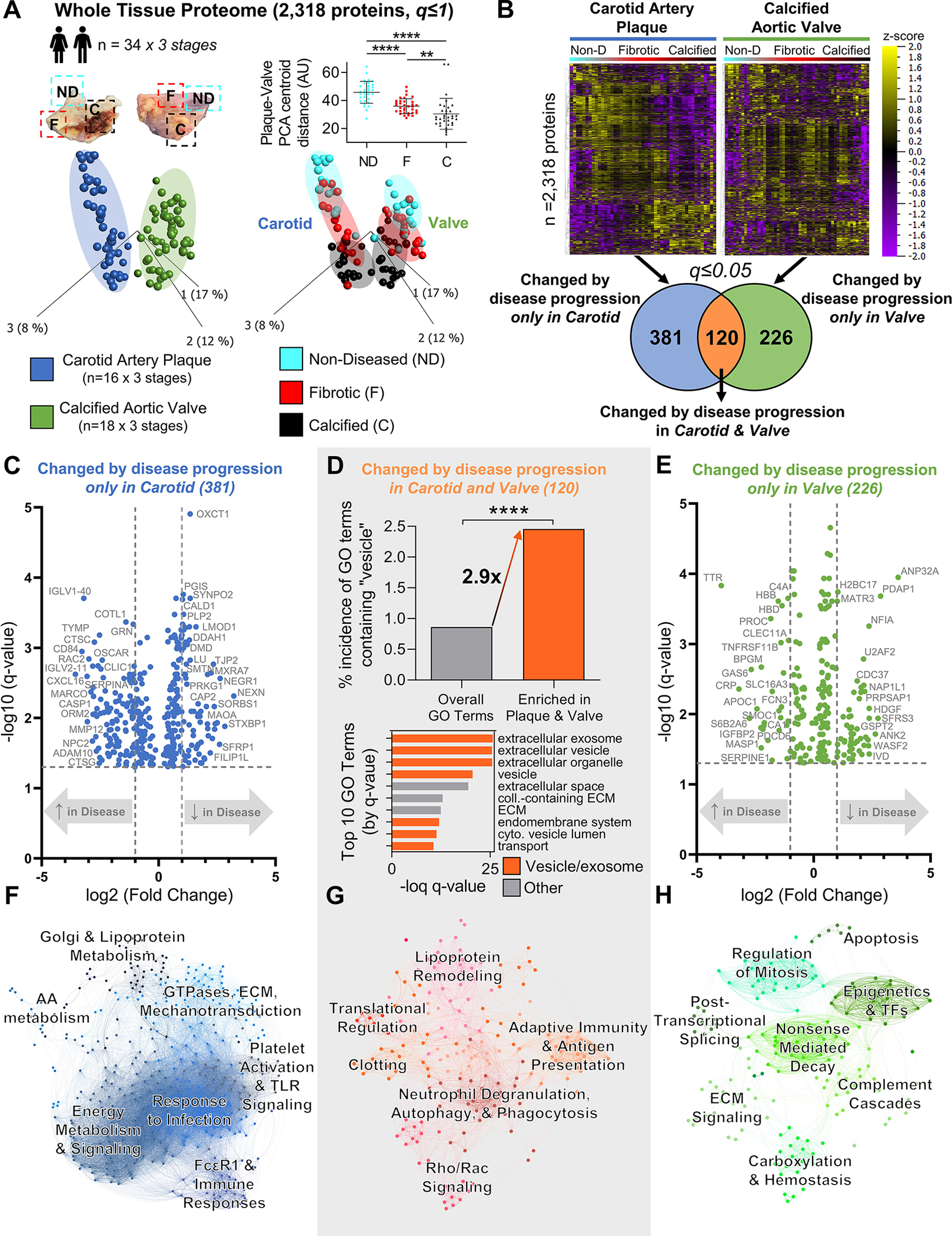
Results: Disease progression drove significant convergence (P<0.0001) of carotid artery plaque and calcified aortic valve proteomes (2318 proteins). Each tissue also retained a unique subset of differentially enriched proteins (381 in plaques; 226 in valves; q<0.05). Vesicular gene ontology terms increased 2.9-fold (P<0.0001) among proteins modulated by disease in both tissues. Proteomics identified 22 EV markers in tissue digest fractions. Networks of proteins and microRNA targets changed by disease progression in both artery and valve EVs revealed shared involvement in intracellular signaling and cell cycle regulation. Vesiculomics identified 773 proteins and 80 microRNAs differentially enriched by disease exclusively in artery or valve EVs (q<0.05); multiomics integration found tissue-specific EV cargoes associated with procalcific Notch and Wnt signaling in carotid arteries and aortic valves, respectively. Knockdown of tissue-specific EV-derived molecules FGFR2, PPP2CA, and ADAM17 in human carotid artery smooth muscle cells and WNT5A, APP, and APC in human aortic valvular interstitial cells significantly modulated calcification.

Conclusions: The first comparative proteomics study of human carotid artery plaques and calcified aortic valves identifies unique drivers of atherosclerosis versus aortic valve stenosis and implicates EVs in advanced cardiovascular calcification. We delineate a vesiculomics strategy to isolate, purify, and study protein and RNA cargoes from EVs entrapped in fibrocalcific tissues. Integration of vesicular proteomics and transcriptomics by network approaches revealed novel roles for tissue EVs in modulating cardiovascular disease.

Keywords: aortic valve stenosis; atherosclerosis; extracellular vesicles; microRNAs; proteomics.


Conflict of interest statement

Disclosures H. Higashi is an employee of Kowa Company, Ltd and was a visiting scientist at Brigham and Women’s Hospital during this study. Kowa had no role in study design, data collection and analysis or manuscript preparation. Dr E. Aikawa is a member of the scientific board of Elastrin Therapeutics Inc. Dr Camussi is a member of the scientific board of Unicyte AG. Dr Libby is an unpaid consultant to and is involved in clinical trials for Amgen, AstraZeneca, Esperion Therapeutics, Ionis Pharmaceuticals, Kowa Pharmaceuticals, Novartis, Pfizer, Sanofi-Regeneron, and XBiotech, Inc. and a member of scientific advisory boards for Amgen, Corvidia Therapeutics, DalCor Pharmaceuticals, IFM Therapeutics, Kowa Pharmaceuticals, Olatec Therapeutics, Medimmune, Novartis, and XBiotech, Inc. He serves on the board of XBiotech, Inc. Dr Libby’s laboratory has received research funding in the past 2 years from Novartis. The other authors report no conflicts.

Figures

Figure 1:
Figure 1:. Experimental Overview – Isolation and Analysis of Cardiovascular Tissue-Entrapped Extracellular Vesicles.
Label-free proteomics was conducted on whole-tissue samples of human normal carotid arteries, diseased carotid artery atherosclerotic plaques (from carotid endarterectomies), normal aortic valves, and diseased calcified aortic valves (from valve replacement surgeries for aortic valve stenosis). These sample types also underwent enzymatic digestion and serial low- and high-speed centrifugation. The high-speed supernatant then underwent ultracentrifugation to wash/pellet EVs. 15-fraction survey experiments utilized OptiPrep density gradient separation in concert with mass spectrometry, transmission electron microscopy (TEM), nanoparticle tracking analysis (NTA), and coabundance profiling (XINA) to identify which fractions were enriched in EVs and which contained non-EV contaminants. Enrichment performance was compared to that of ultracentrifugation alone by performing mass spectrometry on the pellet resulting from ultracentrifuged tissue digests. Tissue EV cargoes were then assessed by pooling EV-enriched OptiPrep fractions (F1–4) together and employing mass spectrometry, transcriptomics, TEM, NTA, and bioinformatics approaches to interrogate and classify these vesicles.
Figure 2:
Figure 2:. Whole-Tissue Proteomics Finds Conservation of Extracellular Vesicle-Associated Processes Between Vascular and Valvular Disease Progression.
A, Principal component analysis of 2,318 proteins (q≤1) quantified by disease stage-specific whole-tissue proteomics of human carotid artery plaques and calcified aortic valves revealed significant convergence of carotid and valve tissues during disease pathogenesis; n=16 carotid artery plaque and 18 calcified aortic valve donors x 3 stages of disease (non-diseased, fibrotic, calcified) per donor; mean±SD; *p<0.05, ****p<0.0001. B, Unfiltered heat map analyses (q≤1; ordered by hierarchical clustering) demonstrated disease stage-specific alterations of protein abundances in carotid artery plaques (left) and calcified aortic valves (right). Enrichment analysis identified 381 proteins whose abundances were significantly differentially-enriched only by disease progression in carotid artery plaques, 226 proteins in calcified aortic valves, and 120 proteins in both tissue types (significantly-enriched proteins filtered at q≤0.05). C and E, Volcano plots of proteins significantly differentially-enriched by disease progression only in carotid artery plaques (381 proteins) or only in calcified aortic valves (226 proteins); cutoffs at a fold-change of 2 and a q-value of 0.05. D, In proteins significantly differentially-enriched in both carotid artery plaques and calcified aortic valves (120 proteins), we found a found a statistically significant 2.9-fold increase in the incidence of vesicle-associated GO terms vs. the total GO term database (****p<0.0001). In addition, 7 of the top 10 most-significant GO terms were associated with extracellular vesicles, exosomes, exocytosis, or secretion (orange bars). F-H, Networks based on KEGG, Reactome, and BioCarta pathway enrichment among proteins significantly differentially-enriched by disease progression only in carotid artery plaques (381 proteins, F), in both carotid artery plaques and calcified aortic valves (120 proteins, G), or only in calcified aortic valves (226 proteins, H) with pathways as the nodes (node size corresponds to -log(q-value)) and shared detected proteins between pathways as the edges (edge thickness matches the Jaccard index of overlap between detected proteins of the two connected pathway nodes). Unbiased clustering of pathways into real network communities by the Louvain method revealed shared- and tissue-specific drivers of disease pathogenesis.
Figure 3:
Figure 3:. Tissue EVs are Highly Enriched in the Least-Dense Fractions from Density Gradient Separation.
A and C, From 15-fraction survey experiments, raw abundance by mass spectrometry of selected proteins from intact human carotid artery plaques and calcified aortic valves demonstrated that extracellular vesicle marker proteins (Annexin A2, CD63, CD81, MFGE8 [lactadherin], HSP70–2) were highly enriched in the four least-dense fractions of both tissue types; n=3 carotid artery plaque and 4 calcified aortic valve donors. Enrichment of globular collagens (Collagen VIA1, VIA2, VIA3), fibrillar collagens (Collagen IA1, IA2, IIIA1), and other components of the extracellular matrix (ECM; FBN1, VCAN) was identified in denser fractions. B and D, Nanoparticle tracking analysis confirmed the characteristic presence of particles ~150–200 nm in diameter in fractions 1–4 of both intact carotid artery plaques and calcified aortic valves (n=3 per tissue type), while mean particle size remained consistent between fractions (mean±SEM). E, Representative CD63-labelled immunogold transmission electron microscopy (TEM) identified CD63+ membrane-bound EVs in fractions 1–4 (arrows) from intact carotid artery plaques (top) and calcified aortic valves (bottom); bar=100 nm. Consistent with mass spectrometry-derived protein abundance in A/C, TEM showed abundant globular collagens in fractions 5–10 (arrowheads) and fibrillar collagens in the most-dense fractions (open arrows).
Figure 4:
Figure 4:. Proteomics of EV-Enriched Pooled Fractions to Quantify EV Protein Cargoes in Normal and Diseased Vascular and Valvular Tissue.
A, Tissue EVs were isolated by density gradient separation from intact normal carotid arteries (n=6 donors), intact diseased carotid artery atherosclerotic plaques (n=4), intact normal aortic valves (n=6), and intact diseased calcified aortic valves (n=4). Representative CD63-labelled immunogold transmission electron microscopy (TEM) and negative control images from pooled fractions 1–4 of intact human carotid artery (left) and aortic valve (right) demonstrated EV enrichment and purification; bar=100 nm. Nanoparticle tracking analysis (bottom, all donors, mean±SEM) found that EVs from intact normal and diseased carotid arteries and aortic valves all had similar mean diameters with a single peak around 200nm, and were isolated without co-enrichment of larger microparticles or apoptotic bodies. B, Isolated tissue EVs from every donor underwent both proteomics and small RNA-seq. The tissue EV proteome was composed of 1,942 proteins. Unfiltered principal component analyses (q≤1) identified tissue- (left) and disease state-specific (right) clustering. Unfiltered heat map analyses (q≤1; ordered by hierarchical clustering) illustrated alterations in individual tissue EV protein abundances between normal and diseased carotid arteries and aortic valves. Abundances of 525 tissue EV proteins were significantly differentially-enriched only between normal and diseased carotid arteries, 248 tissue EV proteins differed only between normal and diseased aortic valves, and 404 tissue EV proteins were significantly altered by disease pathogenesis in both tissue types (significantly-enriched proteins filtered at q≤0.05). C, 68.5% of the tissue EV proteome (1,330 proteins) was also found in the whole-tissue proteome; linear regression identified moderate and statistically significant correlations of per-protein abundances between the whole-tissue and diseased tissue EV proteomes in both carotid artery (Pearson’s r = 0.645, p<0.0001) and aortic valve (Pearson’s r = 0.605, p<0.0001). D, Bubble plots of KEGG pathways that were significantly-enriched amongst tissue EV proteins changed by disease only in carotid arteries (top, 525 proteins), in both carotid arteries and aortic valves (middle, 404 proteins), or only in aortic valves (bottom, 248 proteins) identified shared- and tissue-specific roles of disease-altered cardiovascular tissue EV cargoes. Bubble size corresponds to the percentage of differentially-enriched proteins amongst all pathway constituents.
Figure 5:
Figure 5:. Sequencing Reveals the Human Tissue EV non-coding miRNAome from Vessels and Valves.
A, Along with proteomics, small RNA-seq was also performed on those tissue EVs collected from intact normal carotid arteries (n=6 donors), intact diseased carotid artery atherosclerotic plaques (n=4), intact normal aortic valves (n=6), and intact diseased calcified aortic valves (n=4). Top: RNA fragment analysis found that density gradient-enriched EVs contained enhanced levels of miRNA (mean miRNA content=53.0% of all small RNA; mean±SD). Bottom: representative fragment analysis tracing showing large miRNA-associated fragment peak at 24 nucleotides (nt). B, Unfiltered principal component analyses (q≤1) identified tissue- (left) and disease state-specific (right) clustering of 1,083 miRs sequenced in the tissue EV miRNAome. Unfiltered heat map analyses (q≤1; ordered by hierarchical clustering) characterized tissue EV miR cargoes altered between normal and diseased carotid arteries and aortic valves: 44 tissue EV miRs (with 710 unique high-confidence gene targets) were significantly differentially-enriched by disease pathogenesis only in carotid arteries, 36 tissue EV miRs (1,813 unique targets) changed only in aortic valves, and 26 tissue EV miRs (391 unique targets) were altered in both tissues (significantly-enriched miRs filtered at q≤0.05). C, miR/target regulatory networks of TargetScan-predicted gene targets (≥95th percentile weighted context++ score) of tissue EV miRs altered by disease only in carotid arteries (top; blue), in both carotid arteries and aortic valves (middle; orange), or only in aortic valves (bottom; green). D, Bubble plots of KEGG pathways significantly-enriched in the unique gene targets of tissue EV miRs changed by disease only in carotid arteries (top), in both carotid arteries and aortic valves (middle) or only in aortic valves (bottom) reveals the regulatory landscape associated with disease-altered tissue EV miR cargoes. Bubble size corresponds to the percentage of unique targets of differentially-enriched miRs amongst all pathway constituents.
Figure 6:
Figure 6:. Pathway Networks Shared Across Omics Layers Identify Conserved Roles of Cardiovascular Tissue EV Cargoes Altered by Disease.
A, The network of 50 overlapping KEGG, Reactome, and BioCarta pathways that were significantly-enriched in the proteome and gene targets of miRs altered by disease in both intact carotid artery and aortic valve tissue EVs (n=6 normal carotid arteries, n=4 diseased carotid artery atherosclerotic plaques, n=6 normal aortic valves, n=4 diseased calcified aortic valves). Pathways are nodes (node size corresponds to -log(q-value)) and shared detected genes between pathways are edges (edge thickness matches the Jaccard index of overlap between detected genes of the two connected pathway nodes). B, Louvain clustering revealed 4 distinct annotations shared by disease-altered cardiovascular tissue-derived EV cargoes, including modulation of intracellular signaling cascades and cell cycle regulation and apoptosis. Pathway network betweenness-centrality scores (inset) implicate estrogen/epidermal growth factor signaling as a key common molecular constituent of tissue EVs.
Figure 7:
Figure 7:. Integration of EV Multi-Omics Identifies Modulators of Calcification.
A and B, Overlap of KEGG, Reactome, and BioCarta pathways enriched amongst tissue EV proteins and unique gene targets of tissue EV miRs that were differentially-enriched between intact normal and diseased carotid arteries (A) and aortic valves (B). N=6 normal carotid arteries, n=4 diseased carotid artery atherosclerotic plaques, n=6 normal aortic valves, n=4 diseased calcified aortic valves. C and D,Protein-protein interaction networks further prioritized candidate EV-derived calcification modulators: constituents of selected overlapping carotid artery (C, blue nodes; FGFR2, PPP2CA, ADAM17) and aortic valve pathways (D, green nodes; WNT5A, APP, APC) that were significantly altered by disease progression in EV multi-omics had high betweenness-centrality scores in their respective networks. Node diameter corresponds to node degree. E and G, Relative mRNA expression levels of FGFR2, PPP2CA, ADAM17, WNT5A, APP, and APC vs. GAPDH in primary human carotid artery smooth muscle cells (hCtASMCs, E) and human aortic valvular interstitial cells (hVICs, G) after 6 days in normal medium (NM) incubated with scrambled siRNA (siSCR) or siRNA targeting FGFR2, PPP2CA, ADAM17, WNT5A, APP, and APC (siFGFR2, siPPP2CA, siADAM17, siWNT5A, siAPP, siAPC) demonstrated robust knockdown of target gene expression; n=1 donor per cell type, triplicate wells per donor, duplicate qPCR reactions averaged per well; mean±SD; *p<0.05, ***p<0.001, ****p<0.0001. F and H, Representative Alizarin red staining of target knockdown in hCtASMCs (F) and hVICs (H) at days 14–21 in NM or pro-calcifying medium (PM) culture. I and J, Quantification of solubilized Alizarin red stain in hCtASMCs (I) and hVICs (J) treated as in F and H for 14 and 21 days confirmed that inhibition of molecules identified by integration of EV multi-omics in A-D significantly modulated calcification of human vascular and valvular cells; n=3 hCtASMC donors and 3 hVIC donors, duplicate wells averaged per timepoint per donor; *p<0.05, **p<0.01. Grey bars indicate the minimum to maximum intensity range of the NM siSCR condition.

References

    1. Chen J, Budoff MJ, Reilly MP, Yang W, Rosas SE, Rahman M, Zhang X, Roy JA, Lustigova E, Nessel L, et al. Coronary Artery Calcification and Risk of Cardiovascular Disease and Death Among Patients With Chronic Kidney Disease. JAMA cardiology. 2017;2:635–643. doi: 10.1001/jamacardio.2017.0363 - DOI - PMC - PubMed
    1. Rapp AH, Hillis LD, Lange RA, Cigarroa JE. Prevalence of coronary artery disease in patients with aortic stenosis with and without angina pectoris. The American journal of cardiology. 2001;87:1216–1217; A1217. doi: 10.1016/s0002-9149(01)01501-6 - DOI - PubMed
    1. Otto CM, Kuusisto J, Reichenbach DD, Gown AM, O’Brien KD. Characterization of the early lesion of ‘degenerative’ valvular aortic stenosis. Histological and immunohistochemical studies. Circulation. 1994;90:844–853. doi: 10.1161/01.cir.90.2.844 - DOI - PubMed
    1. Rossebo AB, Pedersen TR, Boman K, Brudi P, Chambers JB, Egstrup K, Gerdts E, Gohlke-Barwolf C, Holme I, Kesaniemi YA, et al. Intensive lipid lowering with simvastatin and ezetimibe in aortic stenosis. N Engl J Med. 2008;359:1343–1356. doi: 10.1056/NEJMoa0804602 - DOI - PubMed
    1. Itoh S, Mizuno K, Aikawa M, Aikawa E. Dimerization of sortilin regulates its trafficking to extracellular vesicles. The Journal of biological chemistry. 2018;293:4532–4544. doi: 10.1074/jbc.RA117.000732 - DOI - PMC - PubMed

Publication types

Supplementary concepts