. 2023 Jul 10;8(13):e167768.
doi: 10.1172/jci.insight.167768.

ADCC-activating antibodies correlate with decreased risk of congenital human cytomegalovirus transmission

Affiliations

ADCC-activating antibodies correlate with decreased risk of congenital human cytomegalovirus transmission

Eleanor C Semmes et al. JCI Insight. .

Abstract

Human cytomegalovirus (HCMV) is the most common vertically transmitted infection worldwide, yet there are no vaccines or therapeutics to prevent congenital HCMV (cCMV) infection. Emerging evidence indicates that antibody Fc effector functions may be a previously underappreciated component of maternal immunity against HCMV. We recently reported that antibody-dependent cellular phagocytosis (ADCP) and IgG activation of FcγRI/FcγRII were associated with protection against cCMV transmission, leading us to hypothesize that additional Fc-mediated antibody functions may be important. In this same cohort of HCMV-transmitting (n = 41) and nontransmitting (n = 40) mother-infant dyads, we report that higher maternal sera antibody-dependent cellular cytotoxicity (ADCC) activation is also associated with lower risk of cCMV transmission. We investigated the relationship between ADCC and IgG responses against 9 viral antigens and found that ADCC activation correlated most strongly with sera IgG binding to the HCMV immunoevasin protein UL16. Moreover, we determined that higher UL16-specific IgG binding and FcγRIII/CD16 engagement were associated with the greatest risk reduction in cCMV transmission. Our findings indicate that ADCC-activating antibodies against targets such as UL16 may represent an important protective maternal immune response against cCMV infection that can guide future HCMV correlates studies and vaccine or antibody-based therapeutic development.

Keywords: Adaptive immunity; Immunoglobulins; Immunology; Infectious disease; NK cells.


Conflict of interest statement

Conflict of interest: SRP is a consultant for Moderna, Merck, Pfizer, GSK, Dynavax, and Hoopika CMV vaccine programs and leads sponsored research programs with Moderna, Merck, and Dynavax. She also serves on the board of the National CMV Foundation and as an educator on CMV for Medscape. KMW has a sponsored research project from Moderna on immune correlates of congenital CMV infection.

Figures

Figure 1
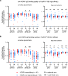
Figure 1. HCMV-specific ADCC and FcγRIII/CD16 activating antibodies in HCMV transmitting versus nontransmitting mother-infant dyads.
HCMV-specific ADCC and FcγRIII IgG activation was measured using maternal (M) and cord blood (CB) sera from HCMV transmitting (red circles, n = 41) and nontransmitting (blue diamonds, n = 40) mother-infant dyads. Antibody responses were compared between and within dyads. (A) NK cell degranulation (% CD107a+ NK cells; gating strategy in Supplemental Figure 2) was quantified as a read-out of ADCC using a flow-based assay. Primary NK cells were isolated from PBMCs by negative selection with magnetic beads prior to coincubation with HCMV-infected and mock-infected cells. Cytogam (light green), HCMV seropositive (light and dark blue), and HCMV seronegative (gray) sera samples were included as controls. (B) HCMV-specific antibody ADCC responses in transmitting and nontransmitting dyads. (C) Flow cytometry of FcγR-CD3ζ BW cells showing unstained (red), isotype control (orange), anti-FcγRI/CD64 (light green), anti-FcγRII/CD32 (blue), and anti-FcγRIII/CD16 (purple) PE–conjugated antibody staining. (D) Anti–HCMV IgG FcγRIII activation in transmitting and nontransmitting dyads. (E) Scatterplots showing Spearman correlations between HCMV-specific ADCC and FcγRIII IgG activation. IgG transfer ratio equals paired cord blood/maternal sera responses. Horizontal black bars denote median. (A and D) FDR-corrected P values for Mann-Whitney U test (left) or Wilcoxon signed-rank test (right). *P < 0.05, **P < 0.01, ***P < 0.001.
Figure 2
Figure 2. HCMV-specific NK cell ADCC and FcγRIII/CD16 activating antibodies cluster with anti-UL16 and anti-UL141 IgG responses.
Hierarchical clustering was performed on Spearman correlation coefficients to group strongly correlated immune variables. Matrix of maternal sera antibody responses showing Spearman correlation coefficients from –1.0 (blue) to +1.0 (red). Nonsignificant correlations (P > 0.05) shown in white. MFI, mean florescent intensity; level, total antigen-specific IgG binding measured by a binding antibody multiplex assay; gB-transfected cell IgG, IgG binding to cell-associated gB as measured in ref. .
Figure 3
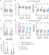
Figure 3. HCMV antigen–specific IgG binding magnitude to FcγRIII/CD16 in transmitting versus nontransmitting dyads.
HCMV antigen-specific IgG binding to FcγRIII in maternal (M) and cord blood (CB) sera was measured using a binding antibody multiplex assay with a biotinylated FcγR and streptavidin-PE detection antibody. HCMV antigen–specific IgG binding to FcγRIII was compared between transmitting (red circles, n = 41) and nontransmitting (blue diamonds, n = 40) mother-infant dyads. (A) HCMV antigen-specific IgG binding to FcγRIII high-affinity V158 variant. (B) HCMV antigen-specific IgG binding to FcγRIII low-affinity F158 variant. Horizontal black bars denote median. (A and B) FDR-corrected P values reported for Mann-Whitney U test. *P < 0.05, **P < 0.01, ***P < 0.001.
Figure 4
Figure 4. HCMV antigen-specific IgG binding quality to FcγRIII/CD16 in transmitting versus nontransmitting dyads.
HCMV antigen-specific IgG binding to FcγRIII in maternal (M) and cord blood (CB) sera was measured using a binding antibody multiplex assay with a biotinylated FcγR and streptavidin-PE detection antibody. Antigen-specific IgG binding to FcγRIII was normalized to total antigen-specific IgG binding (i.e., total antigen-specific IgG level) as a ratio and compared between transmitting (red circles, n = 41) and nontransmitting (blue diamonds, n = 40) mother-infant dyads. (A) Normalized HCMV antigen-specific IgG binding to FcγRIII high-affinity V158 variant. (B) Normalized HCMV antigen-specific IgG binding to FcγRIII low-affinity F158 variant. Horizontal black bars denote median. (A and B) FDR-corrected P values reported for Mann-Whitney U test. *P < 0.05, **P < 0.01, ***P < 0.001.
Figure 5
Figure 5. Anti-UL141 and anti-UL16 IgG binding in HCMV transmitting versus nontransmitting mother-infant dyads.
Anti-UL141 and anti-UL16 IgG binding was measured with a binding antibody multiplex assay using maternal (M) and cord blood (CB) sera from HCMV transmitting (red circles, n = 41) and nontransmitting (blue diamonds, n = 40) mother-infant dyads. (A) Total anti-UL141 IgG binding (i.e., level). (B) Anti-UL141 IgG binding to FcγRIII high-affinity V158 and low-affinity F158 variants before and after normalization for total anti-UL141 IgG level. (C) Total anti-UL16 IgG binding (i.e., level). (D) Anti-UL16 IgG binding to FcγRIII high-affinity V158 and low-affinity F158 variants before and after normalization for total anti-UL16 IgG level. (E) Anti–UL16 IgG FcγRIII activation measured via FcγR-CD3ζ BW cell activation assay. (A–E) FDR-corrected P values reported for Mann-Whitney U test. *P < 0.05, **P < 0.01, ***P < 0.001.
Figure 6
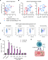
Figure 6. Anti-UL16 IgG activates NK cell ADCC in HCMV nontransmitting pregnancies.
(AD) ADCC activation was quantified as NK cell degranulation (% CD107a+ NK cells; gating strategy in Supplemental Figure 2) against fibroblasts infected with HCMV (A and B) or transduced with a recombinant adenovirus (rAd) expressing UL16 or a control rAd (C and D). Anti-UL16 IgG binding was measured with a binding antibody multiple assay. (A and B) Scatterplots of maternal sera responses from transmitting (red circles, n = 41) and nontransmitting (blue diamonds, n = 40) dyads. (A) Spearman correlation between anti-HCMV ADCC and anti-UL16 IgG levels. (B) Spearman correlation between anti-HCMV ADCC and anti-UL16 IgG binding to FcγRIII high-affinity V158 and low-affinity F158 variants. (C and D) NK cell degranulation against UL16rRAd (purple) and control rAd (black) using Cytogam (positive control), seronegative sera (negative control), and a limited subset of maternal sera samples (n = 6) selected based on sera volume availability. Dots represent biological replicates tested at 1:10 and 1:50 sera dilution. Serum samples with a range of representative anti–UL16 IgG FcγRIII binding responses were included. (E) Hypothetical model demonstrating proposed role for anti-UL16 IgG binding to FcγRIII in mediating NK cell ADCC against HCMV-infected cells. (E) FDR-corrected P values reported for Mann-Whitney U test. **P < 0.01, ***P < 0.001.

Update of

References

    1. Boppana SB, et al. Congenital cytomegalovirus infection: clinical outcome. Clin Infect Dis. 2013;57 Suppl 4(supp 4):S178–S181. - PMC - PubMed
    1. Francis SS, et al. In utero cytomegalovirus infection and development of childhood acute lymphoblastic leukemia. Blood. 2017;129(12):1680–1684. doi: 10.1182/blood-2016-07-723148. - DOI - PMC - PubMed
    1. Zuhair M, et al. Estimation of the worldwide seroprevalence of cytomegalovirus: a systematic review and meta-analysis. Rev Med Virol. 2019;29(3):e2034. doi: 10.1002/rmv.2034. - DOI - PubMed
    1. Gerna G, Lilleri D. Human cytomegalovirus (HCMV) infection/re-infection: development of a protective HCMV vaccine. New Microbiol. 2019;42(1):1–20. - PubMed
    1. Vanarsdall AL, et al. HCMV trimer- and pentamer-specific antibodies synergize for virus neutralization but do not correlate with congenital transmission. Proc Natl Acad Sci U S A. 2019;116(9):3728–3733. doi: 10.1073/pnas.1814835116. - DOI - PMC - PubMed

Publication types