. 2023 Jul 10;8(13):e166137.
doi: 10.1172/jci.insight.166137.

A nondepleting anti-CD19 antibody impairs B cell function and inhibits autoimmune diseases

Affiliations

A nondepleting anti-CD19 antibody impairs B cell function and inhibits autoimmune diseases

Jeffrey S Boyles et al. JCI Insight. .

Abstract

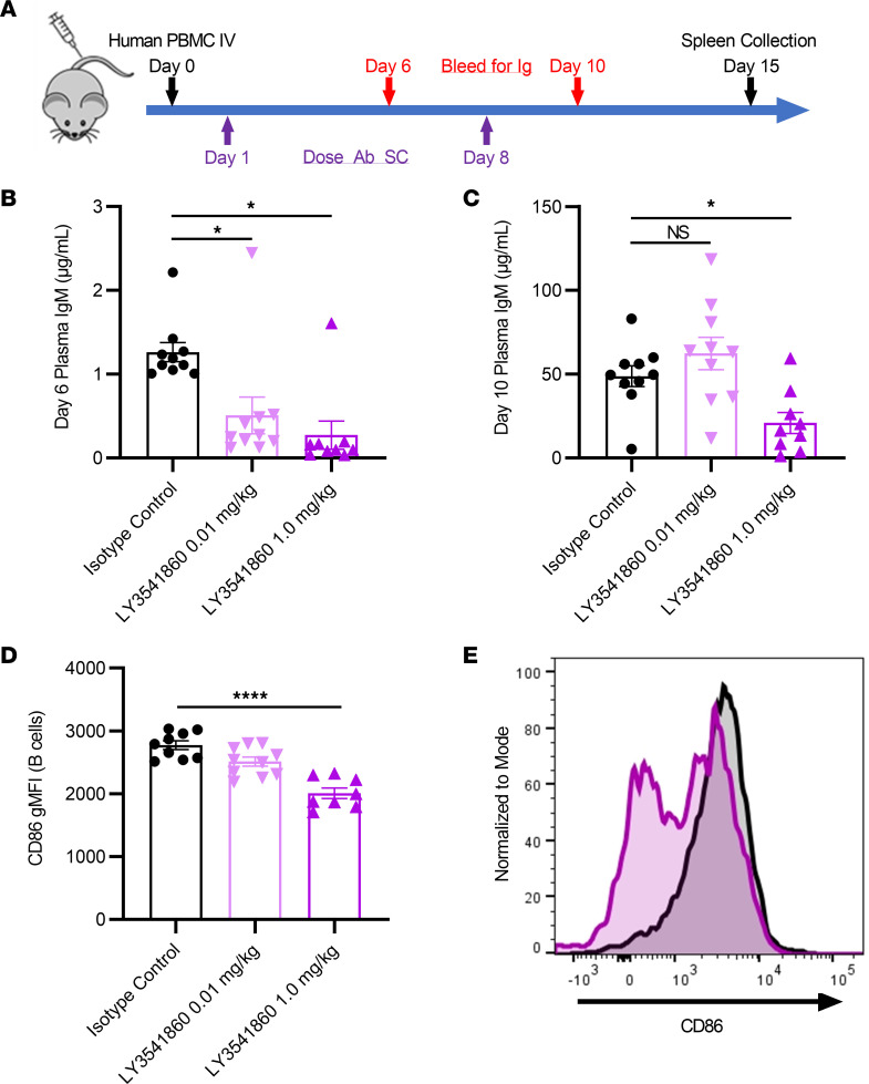
B cells contribute to multiple aspects of autoimmune disorders, and B cell-targeting therapies, including B cell depletion, have been proven to be efficacious in treatment of multiple autoimmune diseases. However, the development of novel therapies targeting B cells with higher efficacy and a nondepleting mechanism of action is highly desirable. Here we describe a nondepleting, high-affinity anti-human CD19 antibody LY3541860 that exhibits potent B cell inhibitory activities. LY3541860 inhibits B cell activation, proliferation, and differentiation of primary human B cells with high potency. LY3541860 also inhibits human B cell activities in vivo in humanized mice. Similarly, our potent anti-mCD19 antibody also demonstrates improved efficacy over CD20 B cell depletion therapy in multiple B cell-dependent autoimmune disease models. Our data indicate that anti-CD19 antibody is a highly potent B cell inhibitor that may have potential to demonstrate improved efficacy over currently available B cell-targeting therapies in treatment of autoimmune conditions without causing B cell depletion.

Keywords: Adaptive immunity; Autoimmunity; Immunoglobulins; Immunology.


Conflict of interest statement

Conflict of interest: During the execution of this work, all authors were employees and stockholders of Eli Lilly and Company.

Figures

Figure 1
Figure 1. Generation, optimization, binding affinity, kinetics, and specificity of LY3541860.
(A) Analytical HIC HPLC chromatograms of C323, C323.C1, and LY3541860 demonstrate the reduction in hybrophobicity through engineering. (B) ELISA binding to a titration of biotinylated huCD19-HSA fusion protein with C323 showing barely detectable binding under these conditions. (C) Representative SPR sensorgrams of LY3541860 Fab binding to human CD19-Fc are shown in black, and the 1:1 kinetic fit is shown in red. (D) Representative SET titrations of CD19 expressing cells at different fixed LY3541860 antibody concentrations. (E) Representative data demonstrating binding of fluorescently labeled isotype control antibody (right) or LY3541860 (left) in human whole blood (gated on viable CD45+ cells). (F) Dose response binding of fluorescently labeled LY3541860 and isotype control antibody on human B cells in whole blood (B cells gated as viable/CD45+/CD20+/CD3) obtained from 4 independent donors. Data are shown as mean gMFI ± SEM.
Figure 2
Figure 2. LY3541860 does not induce B cell apoptosis or cause ADCC/CDC in vitro.
(A) In vitro ADCC assay. The data are representative of 3 assay runs. (B) In vitro CDC assay. The data are representative of 3 assay runs. (C) Induction of B cell apoptosis in vitro. Apoptotic B cells were detected using annexin V staining (gated as live, annexin V+). The assay was repeated 2 times using B cells from 4 independent donors. Data obtained from 1 representative donor are shown. Each point represent mean ± SEM from 3 technical replicates. (D) Volcano plots showing differential expression of apoptosis pathway genes by B cells treated in vitro with LY3541860, isotype control, or obexelimab. mRNA was counted on the NanoString nCounter platform (n = 6 per groups). (E) Bar plots quantifying the number of differentially expressed apoptosis pathway genes by B cells treated in vitro with LY3541860, isotype control, or obexelimab.
Figure 3
Figure 3. LY3541860 inhibits B cell proliferation, activation, and plasmablast differentiation in vitro.
(A) Representative data showing a concentration-dependent decrease of B cell proliferation induced by anti-IgM cross-linking in vitro IC50, determined by generating dose-response curves and fitting to a 4 parameter logistic fit of the CCPM values as a function of LY3541860 concentration (mean ± SEM, n = 3). (B) Inhibition of CpG-induced upregulation of CD69 on B cells in human whole blood. Representative data showing a concentration-dependent decrease of percent of CD69+ cells on viable B cells in human whole blood after incubation with CpG for 24 hours. The IC50 was determined by generating dose response curve and fitting to a 4 parameter logistic fit of the percent of CD69+ cells as a function of LY3541860 concentration. Data are shown as mean ± SEM. (C) Inhibition of primary memory B cell differentiation into plasmablasts in the presence of BAFF/IL-21/IL-2/anti-CD40 and different concentrations of either LY3541860 or isotype control as indicated. The graph shows the percent of plasmablasts (identified as CD20lo/CD38hi) in culture. The assay was performed 3 times and data from one representative experiment is shown.
Figure 4
Figure 4. Inhibition of B cell activation in vivo in humanized NSG mice.
(A) Study design. (B and C) Concentration of human IgM in plasma of hNSG mice treated with either isotype or different doses of LY3541860 as indicated on day 6 (B) or 10 (C) after engraftment. (D) Expression of CD86 on human B cells in spleens of hNSG mice on day 10 after engraftment. *P < 0.05 versus isotype by 1-way ANOVA with Tukey’s post hoc test; data are shown as mean ± SEM. n = 9, 8, and 10 for the isotype, 1.0, and 0.1 mg/kg LY3541860 groups, respectively. (E) Representative FACS histogram demonstrating expression of CD86 on human B cells in spleens of hNSG mice on day 10 after engraftment. Isotype treated, black; 1.0 mg/kg LY 3541860 treated, magenta.
Figure 5
Figure 5. Inhibition of BCR capping and downstream BCR signaling with LY3541860 treatment.
(A) Immunofluorescence images showing primary human B cells in the absence of stimulation. CD19 is shown in red, IgM in green, and nuclear staining in blue. (B) Immunofluorescence images showing primary human B cells after the overnight isotype control treatment followed by 24-hour stimulation. CD19 is shown in red, IgM in green, and nuclear staining in blue. Yellow arrows indicate capping. Dotted line box is shown in higher magnification. . (C) Immunofluorescence images showing primary human B cells after the overnight LY3541860 treatment followed by 24-hour stimulation. CD19 is shown in red, IgM in green, and nuclear staining in blue. Yellow arrows indicate capping. Dotted line box is shown in higher magnification. Scale bars: 5 μm (insets), 10 μm (merged image). (D) Percentage of untreated unstimulated or stimulated B cells “capping” after the LY3541860 or Isotype control treatment. n = 6 for isotype control (total of 585 cells) and n = 5 for LY354186 (total of 1933 cells) of randomly imaged coverslip regions. *P = 0.0475, ****P < 0.0001; 1-way ANOVA with Tukey’s test. (E and F) gMFI of pAKT (E) and pERK (F) on B cells activated in the presence of LY3541860 or isotype control; data are shown as mean ± SEM. Experiment was performed 3 times using B cells from 5 healthy donors. Representative data from 1 donor are shown.
Figure 6
Figure 6. Improved efficacy of surrogate anti-CD19 antibody over CD20-mediated B cell depletion in mouse models of autoimmunity.
(AC) CIA model, n = 12 mice/group representative of 3 independent experiments. (A and B) Clinical score over time and histology microphotographs of the front paws and summary histology score. Arrows indicate affected joints with severe inflammation and cartilage damage, with marked pannus and bone resorption as well as moderate periosteal bone formation. (C) Anti–collagen IgG levels in CIA model (day 42). (D) Frequency of B cells (live/CD45+/B220+/CD3) in spleens, day 42. (E) NOD model (n = 10 mice/group), incidence of the disease over time (representative of 2 independent experiments). (F) EAE model (n = 15 mice/group), clinical score over time. Percent of inhibition over isotype control on the last day of study is indicated. Red asterisk indicate statistical significance over isotype control.
Figure 7
Figure 7. Reversibility of anti-CD19–induced B cell inhibition.
Expression of “free CD19” (unoccupied by the treatment), CD86, and CD80 on splenic B cells after anti-CD40–induced B cell activation in vivo. (A) Anti-CD19 antibody introduced 24 hours before anti-CD40 stimulation (before washout). (B) Anti-CD19 antibody introduced 1 month before anti-CD40 stimulation (after washout). Data are shown as mean ± SEM, n = 5, representative of 2 independent experiments.
Figure 8
Figure 8. Model of the molecular mechanism of action of LY3541860.
Left: CD19 facilitated BCR capping upon antigen recognition, leading to the formation of signalosome, which results in enhanced phosphorylation of downstream signaling components (Erk and AKT) and in enhanced transcription of BCR mediated genes and the induction of efficient B cell activation. Right: LY3541860 prevents CD19 from facilitating BCR capping, leading to the blunted phosphorylation of downstream signaling components, changes in the transcription of BCR-mediated genes, and inhibition of B cells activation.

Comment in

References

    1. Lee DSW, et al. B cell depletion therapies in autoimmune disease: advances and mechanistic insights. Nat Rev Drug Discov. 2021;20(3):179–199. doi: 10.1038/s41573-020-00092-2. - DOI - PMC - PubMed
    1. Ramwadhdoebe TH, et al. Effect of rituximab treatment on T and B cell subsets in lymph node biopsies of patients with rheumatoid arthritis. Rheumatology (Oxford) 2019;58(6):1075–1085. doi: 10.1093/rheumatology/key428. - DOI - PMC - PubMed
    1. Kavanaugh A, et al. Assessment of rituximab’s immunomodulatory synovial effects (ARISE trial). 1: clinical and synovial biomarker results. Ann Rheum Dis. 2008;67(3):402–408. doi: 10.1136/ard.2007.074229. - DOI - PMC - PubMed
    1. Vos K, et al. Early effects of rituximab on the synovial cell infiltrate in patients with rheumatoid arthritis. Arthritis Rheum. 2007;56(3):772–778. doi: 10.1002/art.22400. - DOI - PubMed
    1. Ahuja A, et al. Depletion of B cells in murine lupus: efficacy and resistance. J Immunol. 2007;179(5):3351–3361. doi: 10.4049/jimmunol.179.5.3351. - DOI - PubMed