. 2024 Jun;20(2):518-533.
doi: 10.1007/s12024-023-00662-x. Epub 2023 Jul 10.

Postmortem computed tomography assessment of skeletal and dental age in Polish children, adolescents, and young adults

Affiliations

Postmortem computed tomography assessment of skeletal and dental age in Polish children, adolescents, and young adults

Oleksiy Lopatin et al. Forensic Sci Med Pathol. 2024 Jun.

Abstract

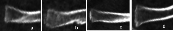
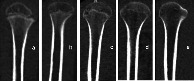
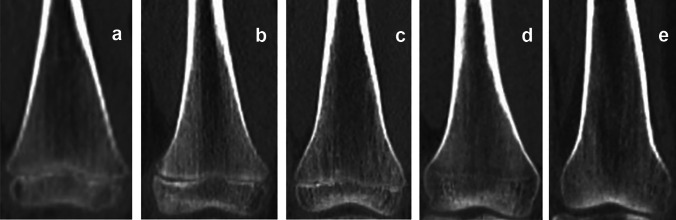
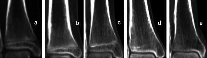
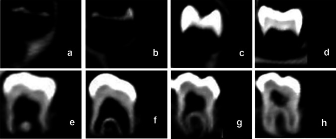
This paper presents a retrospective analysis of postmortem computed tomography (PMCT) scans of secondary ossification centers in the medial clavicular epiphysis, iliac crest apophysis, proximal humeral epiphysis, distal femoral epiphysis, proximal tibial epiphysis, and distal tibial epiphysis. At the same time, we analyzed PMCT scans of the maxillary and mandibular incisors, canines, premolars, and molars. We assessed 203 corpses, whose age ranged from 2 to 30 years, including 156 males and 47 females. The purpose of our study was to compare the processes of secondary ossification center fusion and permanent tooth maturation. Our research hypothesis was that certain stages of skeletal and dental maturation occur along consistent timelines that can be related to the chronological age. Secondary ossification center fusion was evaluated based on Kreitner and also McKern and Steward's classifications. The process of permanent tooth maturation was evaluated with Demirjian's method. Spearman's correlation coefficients (Rho) were positive in all analyses, which indicates that epiphyseal fusion progresses with age. The strongest relationship between the age and the stages of ossification was observed in the proximal tibial epiphysis (p < 0.001; Rho = 0.93) in females and in the medial clavicular epiphysis (p < 0.001; Rho = 0.77) in males. Studies show the importance of concomitant analysis of skeletal and dental maturation with a subsequent comparison of the results to achieve a greater precision in age estimation. A comparison of the results obtained in the study population of Polish children, adolescents, and young adults with the results of other studies in populations of similar ages showed a number of similarities in the time windows of dental and skeletal maturation. These similarities may help in age estimation.

Keywords: Age estimation; Dental age estimation; Permanent teeth; Postmortem computed tomography; Secondary ossification centers; Skeletal age estimation.


Conflict of interest statement

The authors declare no competing interests.

Figures

Fig. 1
Fig. 1
Computed tomography scans showing the process of ossification and subsequent fusion at the medial clavicular epiphysis based on Kreitner’s four-stage classification [7]. a Stage 1—epiphysis not ossified. b Stage 2—epiphysis ossified without any fusion. c Stage 3—partial epiphyseal fusion. d Stage 4—complete epiphyseal fusion; image source: in-house archives
Fig. 2
Fig. 2
Computed tomography images depicting the stages of ossification and fusion of the iliac crest apophysis based on Kreitner’s four-stage classification [8]. a Stage 1—no ossification of the iliac crest apophysis. b Stage 2—iliac crest apophyseal ossification without any fusion with the ilium. c Stage 3—partial fusion. d Stage 4—complete fusion; image source: in-house archives
Fig. 3
Fig. 3
The process of proximal humeral epiphyseal fusion, based on McKern and Steward’s classification [10]. a Stage 0—non-union (no epiphyseal fusion, with a complete radiolucent gap visible between the epiphysis and diaphysis). b Stage 1—beginning of union (the epiphyseal gap is no longer complete, but more than half of its length remains radiolucent). c Stage 2—active union (the terminal plate of the epiphysis can no longer be distinguished and less than half of the epiphyseal gap remains radiolucent). d Stage 3—recent union (the epiphysis and diaphysis are completely fused forming a single bone, with a metaphyseal line possibly remaining at the border between the epiphysis and diaphysis). e Stage 4—complete union (all traces of epiphyseal differentiation have disappeared); image source: in-house archives
Fig. 4
Fig. 4
The process of distal femoral epiphyseal fusion, based on McKern and Steward’s classification [10]: ae Stages 0–4, respectively; image source: in-house archives
Fig. 5
Fig. 5
The process of proximal tibial epiphyseal fusion, based on McKern and Steward’s classification [10]: ae Stages 0–4, respectively; image source: in-house archives
Fig. 6
Fig. 6
The process of distal tibial epiphyseal fusion, based on McKern and Steward’s classification [10]: ae Stages 0–4, respectively; image source: in-house archives
Fig. 7
Fig. 7
Computed tomography images depicting the process of permanent tooth maturation, classified based on Demirjian’s method [2, 12]. a Stage A—beginning of calcification (no fusion of single calcified occlusal points). b Stage B—calcification points fuse, the contour of the occlusal surface is recognizable. c Stage C—enamel formation has been completed, dentin formation has begun. d Stage D—crown formation is complete. e Stage E—root formation, the radicular bifurcation forms in molars. f Stage F—the root length is equal to or greater than the crown height. g Stage G—the roots have almost reached their full length, the apical end is partially open. h Stage H—the root apex is completely closed; image source: in-house archives

References

    1. Dedouit F, Saint-Martin P, Mokrane FZ, Savall F, Rousseau H, Crubézy E, Rougé D, Telmon N. Virtual anthropology: useful radiological tools for age assessment in clinical forensic medicine and thanatology. Radiol Med. 2015. 10.1007/s11547-015-0525-1. 10.1007/s11547-015-0525-1 - DOI - PubMed
    1. Schaefer M, Black S, Scheuer L. Juvenile osteology. Elsevier. 2009.
    1. Schmeling A, Olze A, Reisinger W, Geserick G. Forensic age diagnostics of living people undergoing criminal proceedings. Forensic Sci Int. 2004. 10.1016/j.forsciint.2004.04.059. 10.1016/j.forsciint.2004.04.059 - DOI - PubMed
    1. Woźniak K, Moskała A, Urbanik A, Kopacz P, Kłys M. Postmortem imaging studies with data processing and 3D reconstruction: a new path of development of classic forensic medicine? Arch Med Sadowej Kryminol. 2009;59(2):124–30. - PubMed
    1. Woźniak K, Moskala A, Urbanik A, Kłys M. Value of postmortem CT examinations in cases of extensive mechanical injuries causing considerable corpse destruction. Arch Med Sadowej Kryminol. 2010;60(1):38–47. - PubMed

MeSH terms

LinkOut - more resources