

Successful surgical treatment of omphalocele with umbilical evagination of the bladder: an extremely rare presentation of neonatal case
- PMID: 37428342
- PMCID: PMC10333145
- DOI: 10.1186/s40792-023-01710-y
Successful surgical treatment of omphalocele with umbilical evagination of the bladder: an extremely rare presentation of neonatal case
Abstract
Background: A few cases of small omphalocele with umbilical evagination of the bladder have been reported. However, its embryology is yet to be elucidated. Only a few reports have indicated the existence of urachal anomalies and umbilical cysts related to bladder evagination. The incidence of urachal anomalies at birth is reported to be 1 in 5000-8000 live birth, and urachal aplasia is rare. Herein, we report a rare, novel case of urachal aplasia.
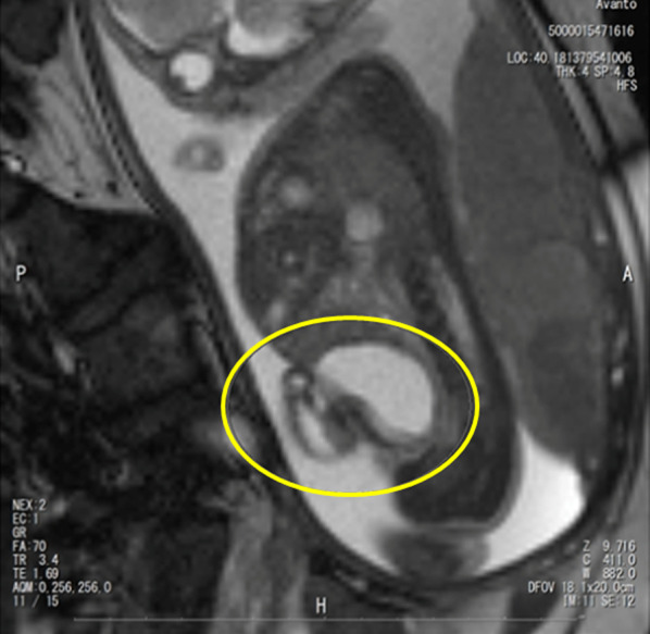
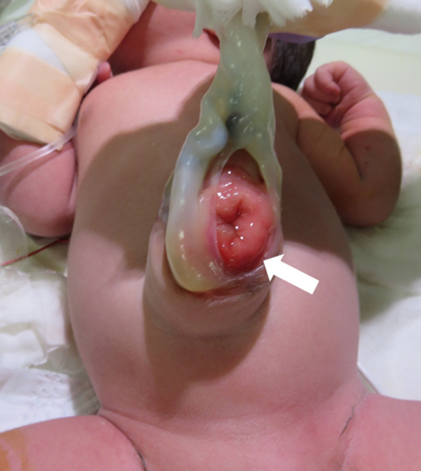
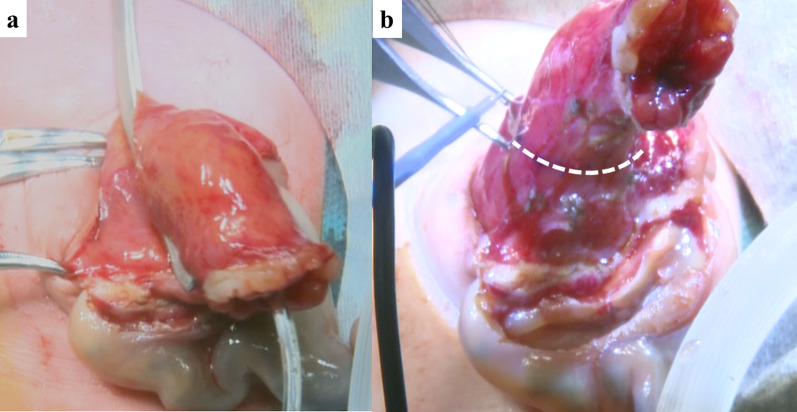
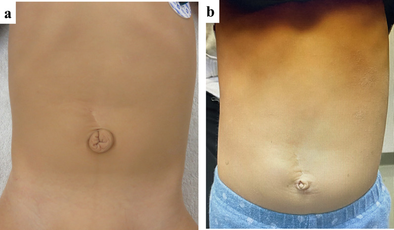
Case presentation: We encountered a small omphalocele with bladder evagination associated with urachal aplasia for which the neonate underwent surgery one day after birth. The patient was a one-day-old boy with a prenatally diagnosed omphalocele. A fetal magnetic resonance image (MRI) scan (25 weeks of gestation) revealed a 30 × 33 mm (approximately 1.3 in.) cystic lesion which was suspected to be an umbilical cyst. The baby was born vaginally at 38 weeks, weighing 2956 g. An omphalocele (hernial orifice diameter, 4 cm × 3 cm) with bladder prolapse was recognized. After sac excision, the prolapsed bladder was resected and closed with two-layer sutures. In order to secure sufficient bladder capacity, we estimated the minimum residual volume as 21 ml after bladder plasty. The remaining bladder capacity was confirmed to be 30 ml by injecting a contrast dye and saline into the bladder. The neonate had no associated cardiac urogenital or skeletal anomalies. Postoperative course was uneventful. The patient was regularly followed up for two years after surgery and underwent umbilicoplasty. He had no trouble with urinary function.
Conclusion: In this case, we experienced extremely rare condition of a small omphalocele with bladder evagination associated with urachal aplasia and reviewed 7 case reports of anomalies similar to those in the present case. Umbilical cord cysts may be an informative indicator of these symptoms in utero. Therefore, ultrasonography scans should be conducted until delivery, despite the spontaneous disappearance of cord cysts.
Keywords: Omphalocele; Umbilical evagination of the bladder; Urachal aplasia.
© 2023. The Author(s).
Conflict of interest statement
The authors have no conflicts of interest to report.
Figures
References
-
- Shimokawara I, Yamahata K, Johnishi J, Asaishi K, Kobayashi T, Totsuka M, Narimatsu E. A case of vesico-umbilical fistula combined with hernia into the umbilical cord. J Jpn Soc Pediatr Surg. 1978;14:71–76.
-
- Akashi K, Shigemoto H, Fujita W, Nishimoto T, Kataoka N, Takagi K. Congenital urinary umbilical fistula associated with a hernia into the umbilical cord: report of a case. J Jpn Soc Pediatr Surg. 1985;21:1016–1021.
-
- Takeuchi S, Nakahira M, Nagata N, Shiokawa C, Tamate S, Yamada C, Kadowaki H. Aplasia of urachus associated with hernia into the umbilical cord- report of a case. J Jpn Soc Pediatr Surg. 1972;8:225–229.
LinkOut - more resources
Full Text Sources
