. 2023 Aug;54(4):349-363.
doi: 10.1007/s10735-023-10137-6. Epub 2023 Jul 10.

Granulocyte-colony stimulating factor ameliorates di-ethylhexyl phthalate-induced cardiac muscle injury via stem cells recruitment, Desmin protein regulation, antifibrotic and antiapoptotic mechanisms

Affiliations

Granulocyte-colony stimulating factor ameliorates di-ethylhexyl phthalate-induced cardiac muscle injury via stem cells recruitment, Desmin protein regulation, antifibrotic and antiapoptotic mechanisms

Shaimaa A Abdelrahman et al. J Mol Histol. 2023 Aug.

Abstract

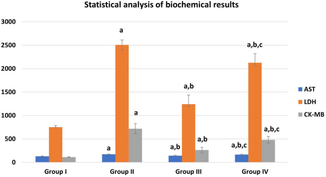
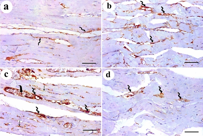
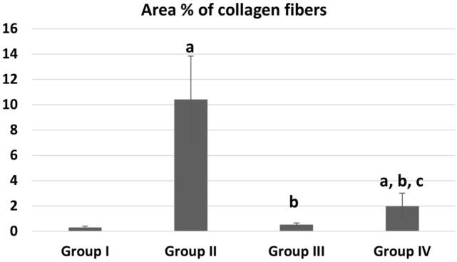
Phthalates are common plasticizers present in medical-grade plastics and other everyday products. Di-ethylhexyl phthalate (DEHP) has been noted as a causative risk factor for the initiation and augmentation of cardiovascular functional disorders. G-CSF is a glycoprotein found in numerous tissues throughout the body and is currently applied in clinical practice and has been tested in congestive heart failure. We aimed to examine in depth the effect of DEHP on the histological and biochemical structure of the cardiac muscle in adult male albino rats and the mechanisms underlying the possible ameliorative effect of G-CSF. Forty-eight adult male albino rats were divided into control group, DEHP group, DEHP+ G-CSF group and DEHP-recovery group. We measured serum levels of aspartate aminotransferase (AST), creatine kinase MB isoenzyme (CK-MB) and lactate dehydrogenase (LDH). Left ventricular sections were processed for light and electron microscope examination, and immunohistochemical staining of Desmin, activated Caspase-3 and CD34. DEHP significantly increased enzyme levels, markedly distorted the normal architecture of cardiac muscle fibers, downregulated Desmin protein levels and enhanced fibrosis, and apoptosis. G-CSF treatment significantly decreased the enzyme levels compared to DEHP group. It enhanced CD34 positive stem cells recruitment to injured cardiac muscle, therefore improved the ultrastructural features of most cardiac muscle fibers via anti-fibrotic and anti-apoptotic effects in addition to increased Desmin protein expression levels. The recovery group showed partial improvement due to persistent DEHP effect. In conclusion, administration of G-CSF effectively corrected the histopathological, immunohistochemical and biochemical alterations in the cardiac muscle after DEHP administration by stem cells recruitment, Desmin protein regulation, antifibrotic and antiapoptotic mechanisms.

Keywords: Cardiac muscle; Di-ethylhexyl phthalate; G-CSF; Histology; Immunohistochemistry.


Conflict of interest statement

There is no conflict of interests.

Figures

Fig. 1
Fig. 1
Biochemical results in different groups: a significant with control group I, b significant with DEHP group II, c significant with G-CSF group III
Fig. 2
Fig. 2
H & E-stained sections of rat cardiac muscle showing: a a longitudinal section of the control group shows branching cardiac muscle fibers with pale-stained central nuclei (arrows). Intercalated discs (green arrow) and narrow intercellular spaces (arrow heads) contain blood capillaries (C) are noticed. b A transverse section of the same group shows polygonal cardiac muscle fibers with central nucleus (arrows) and minimal intercellular spaces (arrow heads). c and d DEHP-treated group shows cardiac muscle fibers with darkly stained nuclei and cytoplasm (arrows) with disturbed myofilaments (curved arrows). Wide intercellular spaces (arrow heads) contain dilated and congested blood vessels (BV). e G-CSF-treated group shows most cardiac muscle fibers have pale stained oval nuclei (arrows) surrounded by acidophilic striated cytoplasm. A few of them show deeply stained nuclei and cytoplasm (yellow arrows). f A transverse section of the same group shows polygonal cardiac muscle fibers with central rounded nuclei and acidophilic cytoplasm (arrows). Few of them have dark nuclei (yellow arrows) and few myofilaments (curved arrows). Intercellular spaces (arrow heads) are narrow. g and h DEHP-recovery group shows deeply stained nuclei and cytoplasm (arrows), marked reduction in myofibrils (curved arrows) with complete loss of striations. Some cardiac muscle fibers have peripherally located nuclei (thick arrows) with wide intercellular spaces (arrow heads). (H&E × 400; Scale bar 30 µm)
Fig. 3
Fig. 3
Mallory’s trichrome-stained sections show a delicate collagen fibers among cardiac muscle fibers of the control group. b Abundant collagen fibers aggregation in between the cardiac muscle fibers and around the congested blood vessels in DEHP-treated group. c Mild aggregation of collagen fibers in G-CSF-treated group. d Moderate aggregation of collagen fibers in DEHP-recovery group (Curved arrows indicate collagen fibers deposition). (Mallory’s trichrome stain × 400; Scale bar 30 µm)
Fig. 4
Fig. 4
Desmin protein immunoexpression appears as dark brown color at the level of intercalated discs (thick arrows). a control group shows prominent Desmin expression. b DEHP group shows marked reduction in Desmin expression. c G-CSF-treated group shows prominent expression of Desmin while d DEHP-recovery group shows moderate Desmin expression. (Avidin biotin Peroxidase system × 400; Scale bar 30 µm)
Fig. 5
Fig. 5
Caspase-3 immunoexpression shows a negative cytoplasmic expression for caspase-3 in the control group. b DEHP-treated group shows strong positive reaction in the cytoplasm of most cardiac muscle fibers. c G-CSF-treated group shows faint brown reaction in the cytoplasm of most cardiac muscle fibers. d DEHP-recovery group shows moderate caspase-3 expression in most cardiac muscle fibers. (Avidin biotin Peroxidase system × 400; Scale bar 30 µm)
Fig. 6
Fig. 6
CD34+ ve stem cells immunoexpression shows a faint brown expression in the endothelial cells lining the blood vessels (zigzag arrow) in control group. b DEHP-treated group shows moderate immunoreaction in the endothelial cells lining the blood vessels (zigzag arrow). c G-CSF-treated group shows dark brown CD34 immunoreactions in the lumen (curved arrow) and endothelial cells lining the blood vessels (zigzag arrow) and in between the cardiac muscle fibers (arrow). d DEHP-recovery group shows moderate immunoreactions in the endothelial cells lining the blood vessels (zigzag arrow). (Avidin biotin Peroxidase system × 400; Scale bar 30 µm)
Fig. 7
Fig. 7
Transmission electron micrographs from rat cardiac muscles show a the control group shows a part of cardiac muscle cell with central oval euchromatic nucleus (N). Its sarcoplasm is packed with regular profiles of longitudinally arranged myofibrils (blue arrows) that are separated by thin layer of sarcoplasm containing rows of mitochondria (M). The myofibrils are diverted at the perinuclear area (red rectangle) leaving an obvious electron lucent area containing mitochondria. b The higher magnification of the same group shows the myofibrils are arranged in an alternative dark (A) and light (I) bands. The (A) bands have lighter (H) zones; that are bisected by dark (M) line (arrow). The (I) bands are traversed by dark Z-lines. An intercalated disc with both transverse electron dense portion (red circle) and longitudinal smooth segment (curved arrow) is also seen. c DEHP-treated group shows apart of cardiac muscle cell containing euchromatic nucleus (N) with patches of heterochromatin. Its sarcoplasm contains relatively attenuated myofibrils (green arrows). Among these myofibrils, numerous rows of variable sized mitochondria (M) are observed. d Another section of the same group shows cardiac muscle cell sarcoplasm containing widely separated Bizarre-shaped mitochondria (B) and empty electron lucent spaces in between them. Distorted attenuated myofibrils (green arrow) and corrugated sarcolemma (red arrow) are noticed. e DEHP-treated group also shows wide intercellular spaces (two headed arrow) among attenuated cardiac muscle cells containing prominent aggregations of collagen fibers (CF). f A wide intercellular space (two headed arrows) among cardiac muscle cells containing dilated congested blood vessels (BV) and g a widened distorted intercalated disc (red circle), can be observed in the same group. (TEM, Scale bars: a, c, d, e = 5 µm; b, g = 2 µm, and f = 10 µm)
Fig. 8
Fig. 8
Transmission electron micrographs from rat cardiac muscles show a G-CSF-treated rats show part of cardiac muscle cell with ovoid euchromatic nucleus (N) with irregular nuclear envelope (red arrow). Its sarcoplasm is packed with well-formed myofibrils (blue arrows) which are separated by rows of flattened mitochondria (M). b Another section of the same group shows the intercellular space contains moderate aggregation of collagen fibers (CF) and blood capillaries (BV). Few areas of focal destruction (green circle) of myofibrils are noticed. Intercalated discs (ID) appear normal. c DEHP-recovery group shows a part of cardiac muscle fiber with ovoid euchromatic nucleus (N) showing peripheral thin rim of heterochromatin and surrounded by a perinuclear space (red rectangle) filled with aggregation of mitochondria (M). The sarcoplasm contains well-formed (blue arrows) and attenuated myofibrils (green arrows) with focal areas of myofilaments destruction (green circle). Intercalated disc (ID) is noticed. d Another section of the same group shows parts of cardiac muscle fibers surrounded by wide intercellular spaces containing prominent collagen fibers aggregation (CF). Focal destruction of myofibrils (green circles) and corrugated sarcolemma (red arrow) are noticed. (TEM, Scale bars = 5 µm)
Fig. 9
Fig. 9
Area % of collagen fibers deposition in different groups. a Significant with control group I, b significant with DEHP group II, c significant with G-CSF group III
Fig. 10
Fig. 10
Area % of Desmin immunoreaction in different groups. a Significant with control group I, b significant with DEHP group II, c significant with G-CSF group III
Fig. 11
Fig. 11
Area % of Caspase 3 immunoreaction in different groups. a Significant with control group I, b significant with DEHP group II, c significant with G-CSF group III
Fig. 12
Fig. 12
Area % of CD34 immunoreaction in different groups. a Significant with control group I, b significant with DEHP group II, c significant with G-CSF group III

Similar articles

References

    1. Abdel Mohsen AF, Ahmed NAW, Altaib ZM, Zaher SM. Effect of cisplatin on cerebellar cortex of albino rat and possible protective role of granulocyte colony stimulating factor versus Citrullus lanatus juice: a histological Study. Egypt J Histol. 2020;43(3):702–717.
    1. Amara I, Timoumi R, Annabi E, Neffati F, Najjar MF, Bouaziz C, Abid-Essefi S. Di (2-ethylhexyl) phthalate induces cardiac disorders in BALB/c mice. Environ Sci Pollut Res. 2019;26(8):7540–7549. doi: 10.1007/s11356-019-04219-w. - DOI - PubMed
    1. Aschauer-Wallner S, Leis S, Bogdahn U, Johannesen S, Couillard-Despres S, Aigner L. Granulocyte colony-stimulating factor in traumatic spinal cord injury. Drug Discov Today. 2021;26(7):1642–1655. doi: 10.1016/j.drudis.2021.03.014. - DOI - PubMed
    1. Atia T, Abdel-Gawad S. Pulmonary toxicity induced by exposure to phthalates, an experimental study. Inhalation Toxicol. 2019;31(9–10):376–383. doi: 10.1080/08958378.2019.1695025. - DOI - PubMed
    1. Aung KH, Win-Shwe TT, Kanaya M, Takano H, Tsukahara S. Involvement of hemeoxygenase-1 in di (2-ethylhexyl) phthalate (DEHP)-induced apoptosis of Neuro-2a cells. J Toxicol Sci. 2014;39(2):217–229. doi: 10.2131/jts.39.217. - DOI - PubMed