. 2023 Jul 24;18(5):054101.
doi: 10.1088/1748-605X/ace5fe.

Fucoidan and microtopography on polyvinyl alcohol hydrogels guided axons and enhanced neuritogenesis of pheochromocytoma 12 (PC12) cells

Affiliations

Fucoidan and microtopography on polyvinyl alcohol hydrogels guided axons and enhanced neuritogenesis of pheochromocytoma 12 (PC12) cells

Yuan Yao et al. Biomed Mater. .

Abstract

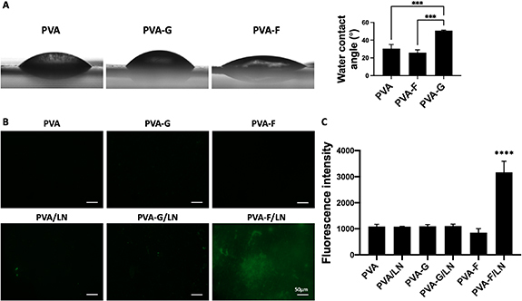
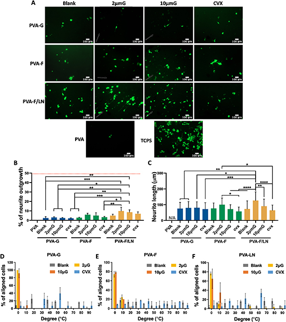
Artificial nerve grafts that support axon growth hold promises in promoting nerve regeneration and function recovery. However, current artificial nerve grafts are insufficient to regenerate axons across long nerve gaps. Specific biochemical and biophysical cues are required to be incorporated to artificial nerve grafts to promote neural cell adhesion and guide neurite outgrowth. Polyvinyl alcohol (PVA) nerve conduits have been clinically approved, but the applicability of PVA nerve conduits is limited to short injuries due to low cell binding. In this study, we explored the incorporation of biochemical cues and topographical cues for promoting neuritogenesis and axon guidance. PVA was conjugated with extracellular matrix proteins and fucoidan, a bioactive sulfated polysaccharide, to improve cell adhesion. Micro-sized topographies, including 1.8 μm convex lenses, 2 μm gratings, and 10 μm gratings were successfully fabricated on PVA by nanofabrication, and the synergistic effects of topography and biochemical molecules on pheochromocytoma 12 (PC12) neuritogenesis and neurite alignment were studied. Conjugated fucoidan promoted the percentage of PC12 with neurite outgrowth from 0% to 2.8% and further increased to 5% by presenting laminin on the surface. Additionally, fucoidan was able to bind nerve growth factor (NGF) on the surface and allow for PC12 to extend neurites in NGF-free media. The incorporation of 2 μm gratings could double the percentage of PC12 with neurite outgrowth and neurite length, and guided the neurites to extend along the grating axis. The work presents a promising strategy to enhance neurite formation and axon guidance, presenting significant value in promoting nerve regeneration.

Keywords: artificial nerve graft; axon guidance; fucoidan; neuritogenesis; polyvinyl alcohol; topography.


Figures

Figure 1.
Figure 1.
Schematic of gelatin, fucoidan, and laminin modifications on PVA through carbonyldiimidazole (CDI) reaction. PAV-G and PVA-F were prepared by activating PVA with 100 mg ml−1 CDI for 1 h at room temperature, followed by overnight incubation in 10 mg ml−1 gelatin or fucoidan solution at 37 °C. PVA-LN was prepared by conjugating poly-l-lysine (PLL) to CDI-activated PVA at 4 °C overnight, followed by incubation in 20 μg ml−1 laminin for 1 h at 37 °C.
Figure 2.
Figure 2.
Representative fluorescence microscopy images of the Live/Dead assay of PC12 cell lines on unpatterned PVA hydrogels. PVA surfaces were conjugated with gelatin (PVA-G), fucoidan (PVA-F), and PLL-laminin (PVA-LN). PC12 cell lines were seeded with maintenance media at a seeding density of 20 000 cells cm−2. Cells were cultured for 14 d and stained with calcein-AM for live cell (green fluorescence) and ethidium homodimer-1 for dead cells (red fluorescence). The scale bars in all images are 50 μm.
Figure 3.
Figure 3.
Polyvinyl alcohol (PVA) hydrogel patterning and characterization. (A) Scanning electron microscope (SEM) images of PVA films without pattern (blank) and with 2 μm gratings (2 μmG), 10 μm gratings (10 μmG), 1.8 μm convex lens (CVX). (B) PVA-F tubular grafts with luminal modification of 2 μm gratings (2 μmG). Top panel shows the lumen of PVA-F graft that was cut open longitudinally. Bottom panel shows the 2 μmG topography on the luminal surface of PVA-F grafts.
Figure 4.
Figure 4.
Neurite outgrowth of PC12 cells cultured with various compositions of media. (A) Representative phase-contrast images of PC12 cells cultured with various compositions of media. The PVA surfaces were conjugated with fucoidan (PVA-F) and the seeding density of PC12 was 20 000 cells cm−2. Scale bar = 100 μm. White arrows indicate neurite formation. (B) Percentage of PC12 cells that extended neurites. (C) Average neurite length. n = 3, * and ** indicates a significant difference using one-way ANOVA p < 0.05 and p < 0.01, respectively.
Figure 5.
Figure 5.
Laminin coating on PVA, PVA-F, and PVA-G hydrogels, (A) Contact angle measurement of PVA, PVA-F, PVA-G hydrogels. n = 3, *** indicates a significant difference using one-way ANOVA p < 0.001. (B) Representative fluorescence images of LN-coated PVA hydrogels. The surfaces were modified with Gelatin (PVA-G) and fucoidan (PVA-F), and LN-coated PVA-G (PVA-G/LN) and LN-coated PVA-F (PVA-F/LN). PVA and LN-coated PVA (PVA/LN) were used as controls. Green denotes laminin staining. (C) Quantification of fluorescence intensity from laminin staining. n = 3, **** indicates a significant difference using one-way ANOVA p < 0.0001.
Figure 6.
Figure 6.
Neurite outgrowth and alignment of PC12 on PVA hydrogels with various topographical and biochemical modifications. (A) Representative fluorescence images of PC12 cell lines. PVA surfaces were modified with fucoidan, gelatin and fucoidan coated with laminin (LN) on different topographies: blank, 2 μG, 10 μG, and CVX. Unmodified PVA and collagen-coated TCPS were used as negative and positive controls, respectively. Cells were seeded at 20 000 cells cm−2 and cultured in media D-5 with laminin (media D-5/LN) for three days. PC12 cell lines were Live/dead stained after culturing for three days. The arrows indicate the direction of gratings. The scale bars in all images are 100 μm. (B) Percentage of PC12 cells that extended neurites and (C) average neurite length. N = 3, *, **, ***, and **** indicate a significant difference using one-way ANOVA with p < 0.05, p < 0.01, p < 0.001, and p < 0.0001, respectively. Neurite alignment of PC12 on PVA-G (D), PVA-F (E), and PVA-LN (F) with different topographies. About 0° indicates that neurites were aligned in the reference direction, and 90° indicates that neurites were perpendicular to the reference direction. For 2 μG and 10 μG samples, grating axis was selected as reference direction, while for blank and CVX samples, a random direction was selected as reference direction.
Figure 7.
Figure 7.
Characterization of nerve growth factor (NGF)-coated PVA hydrogels. (A) Schematic diagram of nerve growth factor (NGF) coating on PVA-G and PVA-F. (B) Representative fluorescence images of NGF-stained surfaces. The surfaces were modified with Gelatin (PVA-G) and fucoidan (PVA-F), and NGF-coated PVA-G (PVA-G/NGF) and NGF-coated PVA-F (PVA-F/NGF). PVA and NGF-coated PVA (PVA/NGF) were used as controls. (C) Quantification of fluorescence intensity. n = 3, *, **, and **** indicates a significant difference using one-way ANOVA p < 0.05, p < 0.01, and p < 0.0001, respectively.
Figure 8.
Figure 8.
Neuritogenesis of PC12 on nerve growth factor (NGF)-coated PVA hydrogels. (A) Representative fluorescence images of PC12 cell lines. Cells were cultured with media composed of DMEM, 1% PS and 5% FBS and seeded on both blank and 2 μm gratings (2 μmG) substrate. The surfaces were modified with gelatin (PVA-G) and fucoidan (PVA-F), and NGF-coated PVA-G (PVA-G/NGF) and NGF-coated PVA-F (PVA-F/NGF). (B) Percentage of PC12 cells that extended neurites and (C) average neurite length. n = 3, *, **, and **** indicate a significant difference using one-way ANOVA with p < 0.05, p < 0.001, and p < 0.0001, respectively.

Similar articles

Cited by

References

    1. Sarker M, Naghieh S, McInnes A D, Schreyer D J, Chen X. Strategic design and fabrication of nerve guidance conduits for peripheral nerve regeneration. Biotechnol. J. 2018;13:1700635. doi: 10.1002/biot.201700635. - DOI - PubMed
    1. Scheib J, Höke A. Advances in peripheral nerve regeneration. Nat. Rev. Neurol. 2013;9:668–76. doi: 10.1038/nrneurol.2013.227. - DOI - PubMed
    1. Panagopoulos G N, Megaloikonomos P D, Mavrogenis A F. The present and future for peripheral nerve regeneration. Orthopedics. 2017;40:e141–56. doi: 10.3928/01477447-20161019-01. - DOI - PubMed
    1. Faroni A, Mobasseri S A, Kingham P J, Reid A J. Peripheral nerve regeneration: experimental strategies and future perspectives. Adv. Drug Deliv. Rev. 2015;82-83:160–7. doi: 10.1016/j.addr.2014.11.010. - DOI - PubMed
    1. Kornfeld T, Vogt P M, Radtke C. Nerve grafting for peripheral nerve injuries with extended defect sizes. Wien. Med. Wochenschr. 2019;169:240–51. doi: 10.1007/s10354-018-0675-6. - DOI - PMC - PubMed

Publication types